. 2025 Apr 19;16(1):317.
doi: 10.1038/s41419-025-07617-w.

Temozolomide promotes glioblastoma stemness expression through senescence-associated reprogramming via HIF1α/HIF2α regulation

Affiliations

Temozolomide promotes glioblastoma stemness expression through senescence-associated reprogramming via HIF1α/HIF2α regulation

Pan Wang et al. Cell Death Dis. .

Abstract

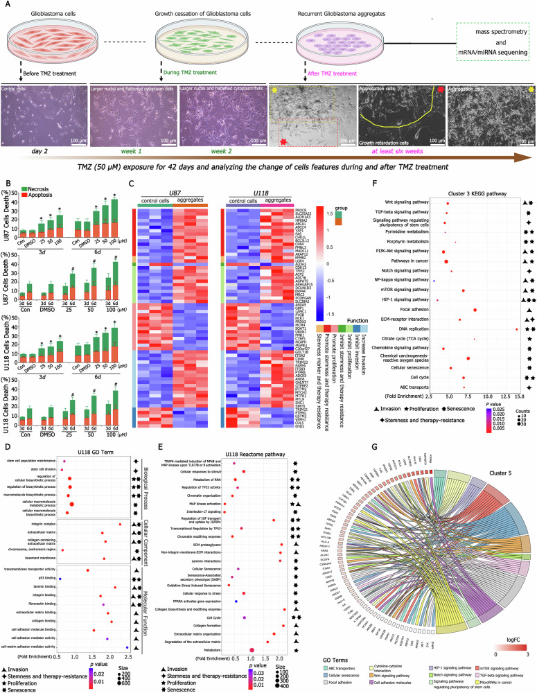
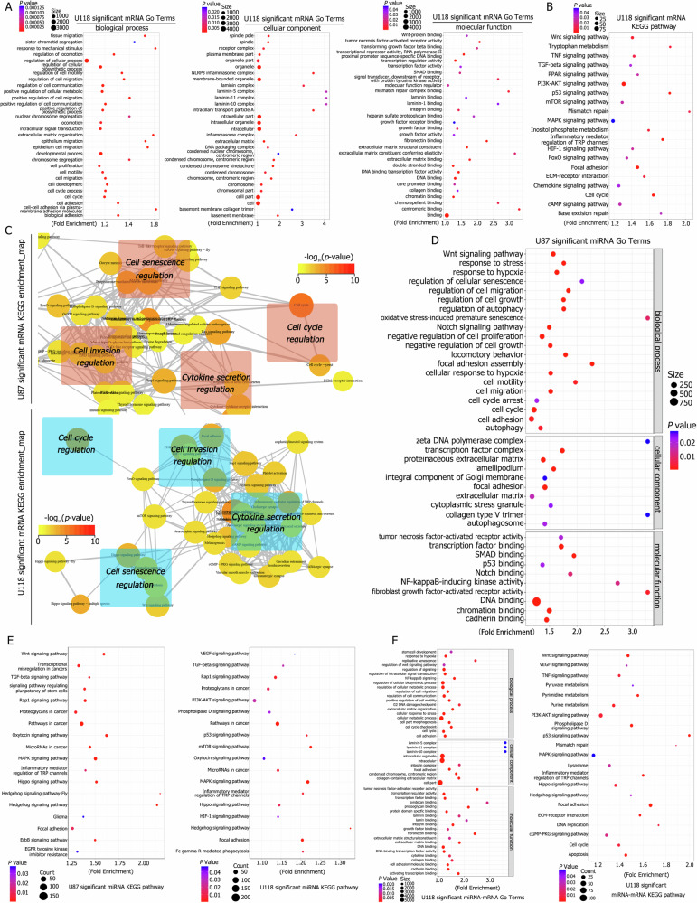
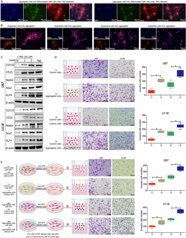
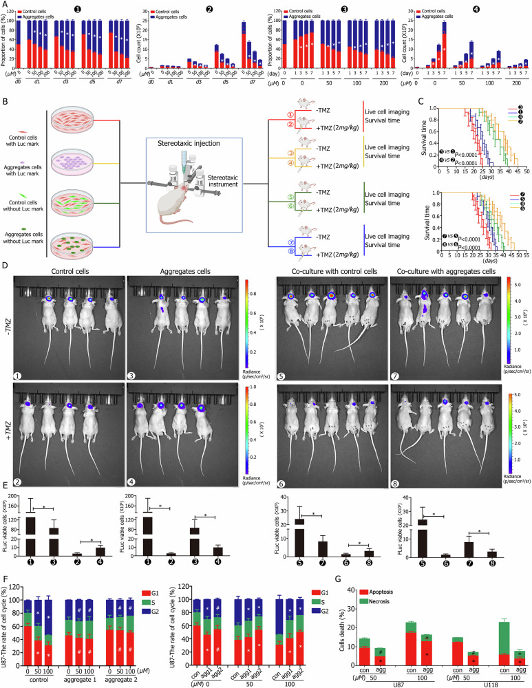
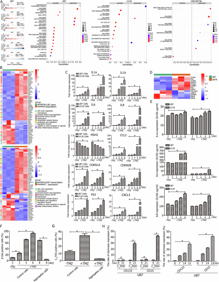
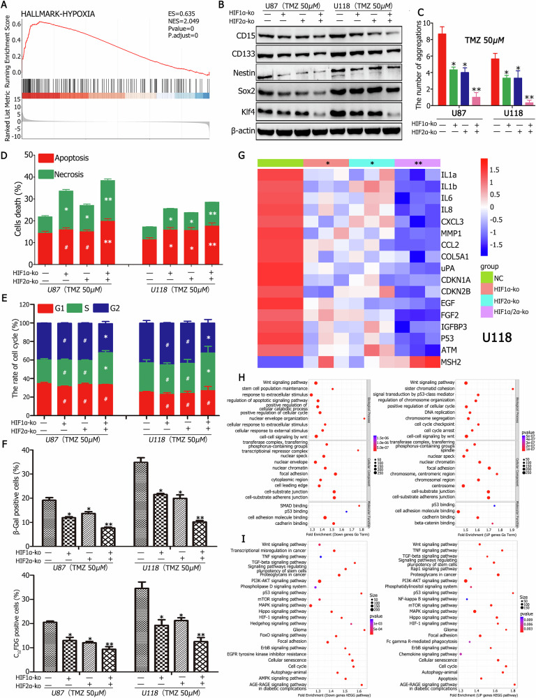
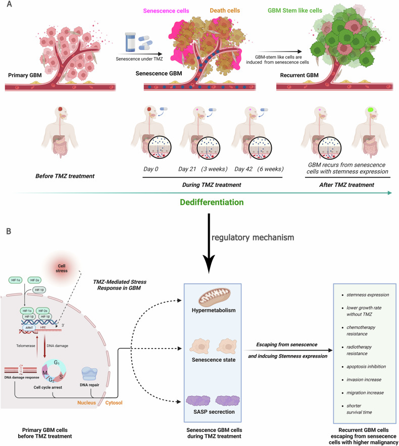
A critical challenge in glioblastoma multiforme (GBM) treatment is that tumors recurring after temozolomide (TMZ) therapy become more malignant, exhibiting increased invasiveness and stemness compared to the primary tumor. However, the underlying mechanisms remain unclear. While the majority of GBM cells are eradicated by TMZ, a subset enters cell cycle arrest, adopts a senescence-associated secretory phenotype (SASP), and activates senescence-related signaling pathways. These cells eventually escape senescence, re-enter the cell cycle, and form aggregates exhibiting stem-like characteristics such as elevated stemness marker expression, enhanced colony formation, increased invasiveness, and resistance to chemotherapy. Furthermore, these aggregates promote the invasion and chemotherapy resistance of surrounding cells. Gene Set Enrichment Analysis (GSEA) and KEGG pathway analysis of miRNA and mRNA sequences revealed activation of hallmark hypoxia and HIF1 signaling pathways. The study demonstrated that HIF1α and HIF2α expression fluctuates during and after TMZ treatment. Knockout of HIF1α and HIF2α in GBM cells exposed to TMZ reduced the formation of senescent cells and stem-like aggregates. These findings challenge the efficacy of TMZ therapy by highlighting its role in inducing the process of cellular senescence, thereby contributing to the enhanced stemness and malignancy of recurrent GBM. The regulatory roles of HIF1α and HIF2α are emphasized, underscoring the necessity of preventing senescent cell formation and inhibiting HIF1α/HIF2α expression to improve therapeutic outcomes.


Conflict of interest statement

Competing interests: The authors declare no conflicts of interest. Ethics: This study was done in compliance with the principles of the Declaration of Helsinki. Ethical related to GBM tissues were approved by the ethics committee of Chongqing General Hospital (No. KY S2022-094-01) and the informed consent was obtained from the patients. All the animal procedures were approved by the ethics committee of ChongQing Medical University (No. IACUC-CQMU-2023-0167).

Figures

Fig. 1
Fig. 1. Activation of senescence- and stemness-associated pathways through proteomic analysis following TMZ treatment.
A TMZ treatment process and associated changes in cellular morphology before, during, and after treatment. GBM cells displayed larger nuclei and a flattened cytoplasm, while new types of GBM aggregation cells formed after six to eight weeks of treatment. B GBM cells were cultured under varying TMZ concentrations (0, 25, 50, and 100 μM) for three to six days. Approximately 20% of GBM cells (including apoptotic and necrotic cells) died after three days of TMZ treatment at 50 μM, with significantly higher death rates compared to the control. The death rate increased proportionally with higher TMZ concentrations and longer exposure duration. C Mass spectrometry analysis indicated that aggregation cells highly expressed proteins associated with promoting stemness, invasion, and therapy resistance, whereas proteins that inhibited these processes or promoted proliferation were less expressed. D GO analysis revealed significant differences in terms related to stemness, invasion, chemotherapy resistance, and senescence between control and treated groups. Key terms included stem cell population maintenance, stem cell division, extracellular matrix, integrin complex, centromeric region, mismatched DNA binding, mismatch repair complex, integrin alpha2-beta1 complex, cell motility, and Notch signaling pathway. E Reactome pathway analysis highlighted the activation of pathways involved in extracellular matrix organization, transcriptional regulation by TP53, senescence-associated secretory phenotype (SASP), cellular senescence, cell cycle regulation, and diseases of mismatch repair (MMR). F, G KEGG pathway analysis identified activation of pathways associated with cellular senescence, focal adhesion, HIF1 signaling, mismatch repair, pluripotency of stem cells, cell cycle regulation, MAPK signaling, and others linked to senescence, stemness, invasion, and chemotherapy resistance. *P < 0.05 were determined using one-way ANOVA and #P < 0.05 were determined using Student’s t-test.
Fig. 2
Fig. 2. mRNA and miRNA sequencing analysis of control and aggregation cells.
A GO analysis of differentially expressed mRNAs demonstrated activation of terms related to stemness, invasion, chemotherapy resistance, and senescence, including cell cycle regulation, positive regulation of locomotion, replicative senescence, cell migration, condensed chromosome, and mismatch repair complex binding. B KEGG pathway analysis of differentially expressed mRNAs revealed significant differences in pathways related to stemness, invasion, and senescence, such as cell cycle, focal adhesion, p53 signaling, mismatch repair, HIF1 signaling, ECM-receptor interaction, TNF signaling, and others. C KEGG pathway enrichment maps showed activation of pathways associated with cellular senescence, invasion, hypoxia response, and stemness, including focal adhesion, HIF1 signaling, PI3K-AKT signaling, and cGMP-PKG signaling. D GO analysis of differentially expressed miRNAs indicated activation of terms associated with stemness, invasion, hypoxia response, and senescence, including response to hypoxia, cell cycle regulation, Notch signaling, cell motility, autophagy, and regulation of cellular senescence. E KEGG pathway analysis of differentially expressed miRNAs revealed activation of pathways such as MAPK signaling, Wnt signaling, regulation of stem cell pluripotency, focal adhesion, HIF1 signaling, and mTOR signaling. F Combined analysis of differentially expressed mRNAs and miRNAs demonstrated significant differences in terms associated with replicative senescence, stem cell development, hypoxia response, cell cycle regulation, focal adhesion, collagen-containing extracellular matrix, integrin binding, and laminin binding. KEGG pathway analysis confirmed activation of processes related to stemness, invasion, hypoxia response, senescence, and cell communication.
Fig. 3
Fig. 3. Characteristics of newly formed aggregation cells, highlighting stemness and invasion capabilities.
A Immunofluorescence analysis showed high expression levels of stemness markers CD133, Nestin, and transcription factors Sox2 and Klf4 in aggregation cells. B Elevated expression levels of CD133, Nestin, Sox2, and Klf4 were observed after two weeks of culturing aggregation cells. C Western blot analysis confirmed increased expression of stemness markers CD133, CD15, Nestin, Sox2, and Klf4 after one to two weeks of TMZ treatment, with higher expression levels in aggregation cells. D Aggregation cells (group ②) demonstrated greater invasive capacity compared to controls (group ①). Co-culturing control and aggregation cells showed a higher number of migrating cells when control cells were co-cultured with aggregation cells (group ④) than with control cells (group ③). E RFP-labeled aggregation cells co-cultured with aggregation cells exhibited the highest invasive capacity (group ④) compared to other groups, in the order: group ④ > group ③ (RFP-aggregation cells co-cultured with control cells) > group ② (RFP-control cells co-cultured with aggregation cells) > group ① (RFP-control cells co-cultured with control cells). *P < 0.05 was determined using Student’s t-test.
Fig. 4
Fig. 4. Aggregation cells exhibit lower proliferation rates but higher chemotherapy resistance.
A Control and aggregation cells were co-cultured at a 1:1 ratio with or without TMZ for one week to assess proliferation. Without TMZ treatment, the proportion of U87 aggregation cells decreased from ~40% to ~30% over seven days (③), with cell counts of 6.12 × 105 and 18.03 × 105 for control cells (④). With TMZ treatment, the proportion of aggregation cells increased with longer exposure times (③) or higher TMZ concentrations (①), showing similar trends in cell counts (② and ④). B Schematic of in vivo experiments involving orthotopic implantation of control or aggregation cells with or without TMZ intraperitoneal injection. CE Without TMZ, control cells (group ①, median survival: 25 days) formed larger tumors but resulted in longer survival compared to aggregation cells (group ③, median survival: 22 days). TMZ treatment reduced tumor volume and extended survival in both groups, with longer survival in control cells (group ②, median survival: 37 days) than aggregation cells (group ④, median survival: 32 days). Mixing control and aggregation cells (1:1 ratio) and orthotopically implanting them showed similar trends, with median survival of 31 vs. 27 days (groups ⑤ vs. ⑦) without TMZ and 39 vs. 34 days (groups ⑥ vs. ⑧) with TMZ. F Cell cycle analysis revealed that control cells predominantly occupied the G1 phase, while aggregation cells were in the G2 + S state. TMZ (50 μM) arrested control cells in the G2 phase, with an increased G2 proportion at 100 μM. Aggregation cells displayed no significant change in the cell cycle proportion with increasing TMZ concentration. G Aggregation cells exhibited lower apoptosis and necrosis rates than control cells under TMZ concentrations of 50 and 100 μM. *P < 0.05 was determined using one-way ANOVA or Student’s t-test, and #P > 0.05 was determined using Student’s t-test. Overall survival was analyzed using the log-rank test.
Fig. 5
Fig. 5. Aggregation cells are formed through cellular senescence under TMZ treatment.
A GSEA analysis indicated upregulation of senescence-associated hallmarks and inhibition of growth-promoting pathways. B The heatmap of U87 overlapping differentially expressed genes (DEGs) with SASP at the mRNA level revealed upregulation of most DEGs, including IL6, IL7, CXCL3, CXCL2, ICAM1, CCL2, CCL3, MMP7, and TIMP1. Conversely, senescence-inhibiting genes such as CDC25B, CDC25C, CDC25A, CDKN2D, MSH6, and MSH5 were downregulated. C RT-qPCR analysis demonstrated significant time-dependent increases in the expression of senescence-promoting genes, including IL1a, IL1b, IL6, IL8, CCL2, CDKN1A, CDKN2B, P53, and CXCL3, while the expression of the senescence-inhibiting gene MSH2 decreased significantly. D Mass spectrometry revealed high expression of senescence-promoting SASP proteins, including ITGA4, MMP15, FN1, IGFB3, and FAS, with a concomitant decrease in senescence-suppressing SASP proteins, MSH2 and MSH6. E ELISA confirmed increased expression of IL1a, IL6, and IL8 in a time-dependent manner following TMZ treatment. F SA-β-Gal staining revealed a significant time-dependent increase in β-Gal-positive cells, and the rate of β-Gal positivity was lower in aggregation cells compared to control cells under TMZ treatment. G C12FDG expression increased approximately threefold after one week of TMZ treatment in CD133CD15 GBM cells, with lower levels of expression in aggregation cells under equivalent TMZ concentrations. H C12FDG-negative and -positive cells were cultured under TMZ for 21 days, showing significantly higher levels of CD133 and CD15 in C12FDG-positive cells over time, while CD133 and CD15 expression was not significant difference in C12FDG-negative cells. *P < 0.05 and #P > 0.05 were determined using Student’s t-test.
Fig. 6
Fig. 6. Regulation of senescence and aggregation stem-like cell formation by HIF1α/HIF2α under TMZ treatment.
A GSEA analysis demonstrated significant upregulation of hypoxia hallmark pathways in newly formed aggregation cells. B HIF1α and HIF2α knockout CD133CD15 cells cultured under TMZ for two months exhibited the lowest expression levels of stemness markers CD133, CD15, Nestin, and transcription factors Sox2 and Klf4. Cells with simultaneous knockout showed the least expression, followed by single knockouts, compared to the control. C HIF1α and HIF2α knockout CD133CD15 cells cultured with TMZ for two months exhibited almost no aggregation formation. Aggregation formation was significantly reduced in single knockouts compared to the control. D Apoptosis and necrosis rates were most significantly increased in cells with simultaneous HIF1α and HIF2α knockouts, followed by single knockouts, compared to the control. E Cell cycle analysis revealed that control cells arrested in the G2/M phase, while HIF1α and HIF2α knockout cells entered the S phase. F The rates of β-Gal-positive and C12FDG-positive cells decreased most significantly in simultaneous HIF1α and HIF2α knockout cells, with intermediate decreases in single knockouts compared to the control. G RT-qPCR analysis showed the lowest expression of SASP factors, including IL1a, IL1b, IL6, IL8, CCL2, and others, in simultaneous knockouts, followed by single knockouts, compared to the control in U118 CD133CD15 cells. H GO analysis of miRNA sequences from simultaneous and single HIF1α and HIF2α knockouts revealed activation of terms associated with senescence and stemness, such as stem cell population maintenance, DNA replication, cell cycle arrest, centrosomes, and p53 binding. I KEGG pathway analysis of miRNA sequences from simultaneous and single HIF1α and HIF2α knockouts indicated activation of pathways associated with senescence and stemness, including cell cycle regulation, cellular senescence, Wnt signaling, regulation of pluripotency in stem cells, focal adhesion, and autophagy. *P < 0.05, **P < 0.01, and #P > 0.05 were determined using Student’s t-test.
Fig. 7
Fig. 7. Dedifferentiation process in differentiated GBM cells under TMZ treatment and its mechanism.
A Differentiated GBM cells exposed to TMZ undergo cellular senescence in addition to cell death. These senescent GBM cells experience growth retardation and hypermetabolism, ultimately dividing into new cells characterized by enhanced stemness, higher invasion potential, reduced proliferation, and increased therapy resistance. This process contributes to the heightened malignancy of recurrent GBM. B HIF1α and HIF2α play critical roles in the formation of senescent cells, promoting hypermetabolism, cell cycle arrest, and DNA damage response. These cells subsequently divide rapidly into more malignant progeny, thereby accelerating GBM recurrence.

Similar articles

References

    1. Chen B, Zhou XX, Yang LT, Zhou HS, Meng M, Wu H, et al. Glioma stem cell signature predicts the prognosis and the response to tumor treating fields treatment. CNS Neurosci Ther. 2022;28:2148–62. - PMC - PubMed
    1. Wu HB, Guo CC, Wang CY, Xu J, Zheng SY, Duan J, et al. Single-cell RNA sequencing reveals tumor heterogeneity, microenvironment, and drug-resistance mechanisms of recurrent glioblastoma. Cancer Sci. 2023;114:2609–21. - PMC - PubMed
    1. Chen C, Jing WQ, Chen Y, Wang GY, Abdalla M, Gao L, et al. Intracavity generation of glioma stem cell-specific CAR macrophages primes locoregional immunity for postoperative glioblastoma therapy. Sci Transl Med. 2022;14:eabn1128. - PubMed
    1. Wang P, Wan WW, Xiong SL, Feng H, Wu N. Cancer stem-like cells can be induced through dedifferentiation under hypoxic conditions in glioma, hepatoma and lung cancer. Cell Death Discov. 2017;3:16105. - PMC - PubMed
    1. Wang P, Yan Q, Liao B, Zhao L, Xiong SL, Wang JW, et al. The HIF1α/HIF2α-miR210-3p network regulates glioblastoma cell proliferation, dedifferentiation and chemoresistance through EGF under hypoxic conditions. Cell Death Dis. 2020;11:992. - PMC - PubMed

MeSH terms

Substances

LinkOut - more resources