. 2025 Apr 19;16(1):319.
doi: 10.1038/s41419-025-07602-3.

Transcription factor JUNB is required for transformation of EpCAM-positive hepatocellular carcinoma (HCC) cells into CD90-positive HCC cells in vitro

Affiliations

Transcription factor JUNB is required for transformation of EpCAM-positive hepatocellular carcinoma (HCC) cells into CD90-positive HCC cells in vitro

Yutaro Kawakami et al. Cell Death Dis. .

Abstract

Hepatocellular carcinoma (HCC) harbors two types of stem cells-epithelial and mesenchymal stem cells. The mechanism by which epithelial EpCAM-positive HCC cells transform into mesenchymal CD90-positive HCC cells remains unclear. On peritumoral fibrotic nodules, epithelial HCC cells form communities with stromal cells, driving tumor growth and malignancy. We aimed to clarify the mechanism by which epithelial cell adhesion molecule (EpCAM)-positive HCC cells contribute to the phenotype of mesenchymal CD90-positive HCC cells that metastasize to distant sites by elucidating the interaction between EpCAM-positive HCC cells and fibroblasts. EpCAM-positive CD90-negative epithelial HCC cells (Huh1, Huh7, and HCC cells) were converted into metastasis-prone CD90-positive HCC cells by co-culture with fibroblasts (Lx-2 and Tig3-20). We identified the transcription factor JUNB as responsible for this altered phenotype. We found that the overexpression of JUNB in CD90-negative epithelial HCC cells resulted in significant transformation to mesenchymal CD90-positive HCC in vitro and in vivo, showing metastatic potential to the lungs. In addition, the JUNB expression in EpCAM-positive hepatoma cells was increased by paracrine stimulation with fibroblast-derived TGFb1. This study unravels the mechanism by which fibroblasts aggravate the malignancy of liver cancer, and the results suggest that JUNB may be a target for treating liver cancer metastasis.


Conflict of interest statement

Competing interests: The authors declare no competing interests. Ethics approval and consent to participate: All animal experiments were approved by the Animal Care and Use Committees of the Institute of Medical, Pharmaceutical, and Health Sciences, Kanazawa University. They were also conducted following the institutional ethical guidelines for animal experiments and the safety guidelines for gene manipulation experiments. The clinical specimens used in this study were performed in accordance with the Declaration of Helsinki, and the protocol was approved by the Ethics Committee of Kanazawa University Graduate School of Medicine. All subjects gave their informed consent for inclusion before they participated in the study, in which the tissue would be used for future study.

Figures
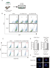
Fig. 1
Fig. 1. Coculture of EpCAM epithelial HCC cells with fibroblasts increases the expression of CD90 and the number of CD90-positive cancer cells in vitro.
A The cell experiment schedule shows the coculture of GFP-labeled EpCAM-positive epithelial HCC cell lines Huh1 and Huh7 with fibroblasts Lx2 or Tig3-20 at a 1:2 ratio. B, C GFP-labeled HCC cells after coculture with fibroblasts were separated using GFP with flow cytometry (B), and the percentage of CD90-positive cells among GFP-positive HCC cells is shown (C). D Expression level of CD90 mRNA after separation of Huh1 and Huh7 cells cocultured with fibroblasts Lx2 or Tig3-20. E Expression of CD90 evaluated using fluorescent immunostaining after separation of Huh1 and Huh7 cells cocultured with fibroblasts Tig3-20. Data are presented as the mean (SD) (N = 3) and were analyzed using the one-way ANOVA. ***P < 0.001.
Fig. 2
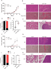
Fig. 2. EpCAM-positive epithelial HCC cells metastasize at a high rate when mixed with fibroblasts and transplanted into immunodeficient mice.
AE Tumor growth curve (A), HE-stained images of subcutaneous tumors and Sirius red-stained images showing fibrosis (B), and HE-stained images of the lungs after subcutaneous implantation of a mixture of Huh1 and Tig3-20 in an immunodeficient model NOD-SCID mouse (N = 5) (C), probability of metastasis to the lungs (D), semi-quantified graph showing the degree of fibrosis in the tumor nodule of a subcutaneous tumor (E). FJ Tumor growth curve, HE-stained images of subcutaneous tumors, Sirius red-stained images, HE-stained images of the lungs after subcutaneous implantation of a mixture of Huh7 and Tig3-20 in a NOD-SCID mouse (N = 5), and probability of metastasis to the lungs. Semiquantified graph showing the degree of fibrosis in the nodules of a subcutaneous tumor. Data in A, E, F, and J are presented as mean (SD) (N = 5) and were analyzed using the Mann–Whitney U-test. *P < 0.05. In D and I, data were analyzed using the χ2 test. ***P < 0.001.
Fig. 3
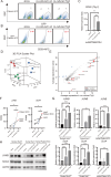
Fig. 3. Induced of SOX4 and JUNB in epithelial HCC cells by fibroblasts.
AC Human HCC cells isolated from surgical specimens were labeled with GFP and cocultured with fibroblasts. After coculture, cells were separated using GFP, and the percentage of CD90-positive cells is shown, along with the expression level of CD90 mRNA. D, E HCC cells Huh1, Huh7, and patient HCC were isolated with or without coculture with fibroblasts Lx-2 or Tig3-20. The isolated HCC cells were subjected to RNA-seq analysis, and the PCA diagram and scatter plot are shown. FH mRNA levels of JUNB and SOX4 assessed using RNA-seq, and mRNA and protein levels of JUNB and SOX4 in Huh1, Huh7, and patient HCC cocultured with fibroblasts. In C, F, and G, data are presented as the mean (SD) (n = 3) and were analyzed using the one-way ANOVA. *P < 0.05, **P < 0.01, ***P < 0.001. In D, data statistics were calculated using Huh1 (N = 1), Huh7 (N = 1), and patient HCC (N = 1) as three samples in total.
Fig. 4
Fig. 4. High JUNB expression in epithelial HCC cells transforms them into mesenchymal CD90-positive HCC cells in vitro.
AC Protein levels of SOX4 and JUNB after transfection of epithelial hepatoma cells Huh1 (A), Huh7 (B), and patient HCC (C). D Flow cytometry analysis shows the conversion rate of epithelial hepatoma cells Huh1, Huh7, and patient HCC transfected with SOX4 and JUNB to CD90-positive cells. EH Graph of invasion rate and HE-stained images of membrane filters of epithelial hepatoma cells Huh1, Huh7, and patient HCC transfected with JUNB determined using invasion assay. In EG, data are presented as the mean (SD) (n = 3) and were analyzed using the one-way ANOVA. **P < 0.01, ***P < 0.001.
Fig. 5
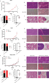
Fig. 5. JUNB overexpression in epithelial HCC cells induces lung metastasis in vivo.
AE JUNB-overexpressing Huh1 cells were subcutaneously transplanted into an immunodeficient NOD-SCID mouse model (n = 5). The tumor growth curve after transplantation, HE-stained images of the subcutaneous tumor, Sirius red-stained image, positivity rate for fibrosis, the probability of metastasis to the lungs, and HE-stained images of the lungs are shown. FJ The tumor growth curve after subcutaneous transplantation of JUNB-overexpressing Huh7 into NOD-SCID mice (n = 5). HE-stained images of subcutaneous tumor, Sirius Red stain image, and positivity rate for fibrosis, probability of metastasis to the lungs, and HE-stained images of the lungs are shown. KO Tumor growth curve after subcutaneous transplantation of JUNB-overexpressing patient HCC into NOD-SCID mice (n = 5), HE-stained images of the subcutaneous tumor, Sirius Red stain image and positivity rate for fibrosis, probability of metastasis to the lungs, and HE-stained images of the lungs are shown. A, E, F, J, K, and O, data are presented as the mean (SD) (n = 5) and were analyzed using the Mann–Whitney U-test. *P < 0.05, *P < 0.01. In D, H, and L, data are analyzed using the χ2 test. *P < 0.05.
Fig. 6
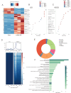
Fig. 6. Analysis of gene expression and pathways directly altered in JUNB-transfected epithelial hepatoma cells.
AC Heat map of 2957 genes with P < 0.05 identified using RNA-seq analysis of JUNB-overexpressing Huh1 (n = 3), GO enrichment analysis, and KEGG pathway analysis. DF Diagram showing the increased JUNB-binding peak due to overexpression of JUNB, distribution diagram of JUNB-binding to gDNA (n = 1), and graph depicting the results of GO analysis of genes bound to gDNA based on integrating RNA-seq and ChIP-seq data.
Fig. 7
Fig. 7. Stimulation with TGFb1 increases the expression of JUNB in epithelial HCC cells and promotes the rate of JUNB-mediated transformation into CD90-positive cells.
A, B mRNA and protein levels of JUNB and SOX4, induced in a concentration-dependent manner, 24 h after the addition of TGFb1 to the epithelial HCC cell lines Huh7, Huh1, and patient HCC. C Activation of total AKT/pAKT (Ser473), total SMAD3/pSMAD3 (Ser423/425), and total JNK/pJNK (Thr183/Tyl185), 24 h after the addition of 2.5 ng and 5 ng of TGFb1 recombinant protein to the epithelial HCC cell lines Huh7, Huh1, and patient HCC overexpressing JUNB. In A, data are presented as the mean (SD) (n = 3) and were analyzed using the one-way ANOVA. *P < 0.05, **P < 0.01, ***P < 0.001.
Fig. 8
Fig. 8. Primary tumors of human HCC with high JUNB expression have a high probability of metastasizing to the lungs.
A Diagram depicting the comparison of the expression of JUNB in the primary human HCC cases depending on the presence (n = 14) or absence (n = 11) of metastasis to the lungs (GSE40367). B Immunohistochemical staining images showing the localization of JUNB expression in the primary human HCC. C Diagram showing comparison of the expression of JUNB determined using immunohistochemical staining of the primary human HCC specimens according to the presence (N = 9) or absence (n = 4) of lung metastasis. In A, data are presented as the mean (SD) and were analyzed using the Mann–Whitney U-test. *P < 0.05. In C, data were analyzed using the χ2 test. *P < 0.05.

Similar articles

References

    1. Sung H, Ferlay J, Siegel RL, Laversanne M, Soerjomataram I, Jemal A, et al. Global cancer statistics 2020: GLOBOCAN estimates of incidence and mortality worldwide for 36 cancers in 185 countries. CA Cancer J Clin. 2021;71:209–49. 10.3322/caac.21660. - PubMed
    1. Villanueva A. Hepatocellular carcinoma. N Engl J Med. 2019;380:1450–62. 10.1056/NEJMra1713263. - PubMed
    1. Yamashita T, Ji J, Budhu A, Forgues M, Yang W, Wang HY, et al. EpCAM-positive hepatocellular carcinoma cells are tumor-initiating cells with stem/progenitor cell features. Gastroenterology. 2009;136:1012–24. 10.1053/j.gastro.2008.12.004. - PMC - PubMed
    1. Yamashita T, Honda M, Nakamoto Y, Baba M, Nio K, Hara Y, et al. Discrete nature of EpCAM+ and CD90+ cancer stem cells in human hepatocellular carcinoma. Hepatology. 2013;57:1484–97. 10.1002/hep.26168. - PMC - PubMed
    1. Tabrizian P, Jibara G, Shrager B, Schwartz M, Roayaie S. Recurrence of hepatocellular cancer after resection: patterns, treatments, and prognosis. Ann Surg. 2015;261:947–55. 10.1097/SLA.0000000000000710. - PubMed

MeSH terms