. 2025 Jun:83:103615.
doi: 10.1016/j.redox.2025.103615. Epub 2025 Mar 25.

GPX4 deficiency-induced ferroptosis drives endometrial epithelial fibrosis in polycystic ovary syndrome

Affiliations

GPX4 deficiency-induced ferroptosis drives endometrial epithelial fibrosis in polycystic ovary syndrome

Zhenhong Ye et al. Redox Biol. 2025 Jun.

Abstract

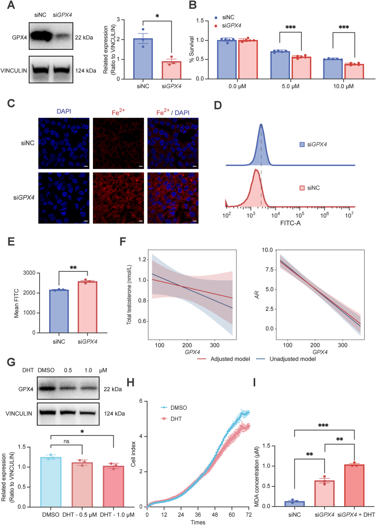
The increased risk of infertility and endometrial lesions (such as endometrial hyperplasia or cancer) in polycystic ovary syndrome (PCOS) are closely associated with the lack of cyclical transformation in the endometrium. However, the underlying mechanisms remain incompletely understood. Though integrating single-cell RNA-sequencing, transcriptomics, and metabolomics analysis, we found that glutathione (GSH) metabolism disorder and the overactivation of ferroptosis, triggered by glutathione peroxidase 4 (GPX4) deficiency in endometrial epithelial cells, were the consequences of the prolonged endometrial proliferative phase in PCOS. This change may collectively contribute to some extent to decidualization failure. We further performed GSVA analysis and determined that the negative correlation between ferroptosis and fibrosis-related pathway was the most significant. Therefore, we first confirmed the presence of fibrosis in the proliferative endometrium of PCOS and PCOS-like mouse uteri. Additionally, by establishing endometrial organoids (EEOs) models and in vitro cell line models, we demonstrated that GPX4 deficiency contributed to extracellular matrix remodeling and excessive collagen deposition, via activating the TGF-β1/Smad2/3 pathway, which ultimately accelerated fibrosis. GSH intervention to the EEOs of PCOS could alleviate their fibrotic phenotypes at different stages. These findings may serve as a promising therapeutic target for PCOS-related endometrial dysfunction, as well as valuable strategies for improving PCOS-related adverse pregnancy outcomes.

Keywords: Endometrial fibrosis; Female infertility; Ferroptosis; Organoid; Polycystic ovary syndrome.


Conflict of interest statement

Declaration of competing interest The authors declare no conflict of interest.

Figures

Image 1
Graphical abstract
Fig. 1
Fig. 1
Ferroptosis participated the periodic transformation of the normal endometrium during menstrual cycles. (A) Activation of seven cell types along the ferroptosis pathway. (B) Volcano plot illustrated differentially expressed genes between proliferative and secretory unciliated epithelium. Up-regulated genes were shown in red (p < 0.05); down-regulated genes were marked in blue (p < 0.05); non-significant genes were indicated in gray (p > 0.05). (C) GSEA analysis showed the differences in ferroptosis pathway between proliferative and secretory unciliated epithelium based on the WP database. (D) Volcano plot illustrated differentially expressed genes between the proliferative and secretory endometrium in each cell type. Up-regulated genes were shown in red (p < 0.05); down-regulated genes were marked in blue (p < 0.05). (E and F) KEGG pathway enrichment analysis of up-regulated (E) and down-regulated differential genes (F) in unciliated epithelium.
Fig. 2
Fig. 2
The combined analysis of transcriptomics and scRNA-seq elucidated the key molecules and pathways influenced the endometrial dysfunction of PCOS. (A) Volcano plot of endometrial transcriptomics illustrated differentially expressed genes between PCOS and non-PCOS patients. Up-regulated genes were shown in red (p < 0.05); down-regulated genes were marked in blue (p < 0.05); non-significant genes were indicated in gray (p > 0.05). (B and C) KEGG pathway analysis of up-regulated (B) and down-regulated differential genes (C) in endometrium compared PCOS to non-PCOS patients. (D) Intersection of transcriptomics genes and scRNA-seq genes of ferroptosis pathway and GSH metabolism. (E) Distribution of GPX4 in unciliated epithelium of the proliferative and secretory phases. (F and G) qPCR verified the expression of GPX4 in endometrial epithelial cell (EEC) (F) and endometrial stromal cell (ESC) (G), respectively. (H) Western blot detected the expression of GPX4 in EEC. (I and J) Immunohistochemical staining of GPX4 (I) and 4-HNE (J) expression in endometrium from each group. Data are presented as mean ± SEM. Statistical analysis was performed using independent sample t-test or Mann-Whitney U test. ∗p < 0.05, ∗∗p < 0.01, ∗∗∗p < 0.001.
Fig. 3
Fig. 3
GPX4 was a key molecule of ferroptosis in endometrial epithelial cells. (A) Left: The protein levels of GPX4 in siGPX4 ishikawa cells detected by western blotting. Right: relative band intensities analyzed by ImageJ. (B) Cell viability of siGPX4 and siNC ishikawa cells treated with erastin for 48 h measure by CCK8. (C) Intracellular Fe2+ level was detected by FerroOrange assay of siGPX4 cells. Scale bar, 75 μm. (D and E) Flow cytometry analyzed ROS level of siGPX4 cells. (F) Correlation analysis of the gene expression of GPX4 with total testosterone levels and AR expression. Unadjusted model was shown in blue; adjusted model for age and BMI was shown in red. (G) Western blot detected the expression of GPX4 in ishikawa cells treated with different concentration of DHT. (H) Cell viability of ishikawa cells treated with DHT (1 μM). (I) MDA contents of siGPX4 ishikawa cells treated with DHT for 48 h. Error bars, mean ± SEM. Statistical analysis was performed using independent sample t-test. ∗p < 0.05, ∗∗p < 0.01, ∗∗∗p < 0.001.
Fig. 4
Fig. 4
Metabolomics revealed abnormal GSH metabolism of proliferative endometrium in PCOS. (A and D) OPLS-DA analysis of untargeted metabolomics (A) and targeted metabolomics (D) in the endometrium of PCOS and non-PCOS patients. Blue and red circles represent the non-PCOS and PCOS group, respectively. (B and E) Volcano plot of untargeted metabolomics (B) and targeted metabolomics (E) illustrated differential metabolites between PCOS and non-PCOS patients. (C and F) KEGG pathway analysis of untargeted metabolomics (C) and targeted metabolomics (F) of differential metabolites in endometrium compared PCOS to non-PCOS patients. (G) Heatmap shown the levels of top 20 differential metabolites from each sample of PCOS and non-PCOS patients. (H) GSH content in untargeted metabolomics. (I) A schematic diagram of GSH metabolism and ferroptosis. Cys: cysteine; Glu: glutamic acid; γ-GCS: γ-glutamylcysteine; Gly: glycine; GSS: glutathione synthetase; GSH: glutathione; GSSG: oxidized glutathione. Created with BioRender.com (https://biorender.com/).
Fig. 5
Fig. 5
GPX4 deficiency contribute to fibrosis of endometrial epithelial cells. (A) GSVA score of distinct biological processes and GSVA score partial correlation in non-PCOS and PCOS groups. (B and D) Masson trichrome staining images of non-PCOS and PCOS endometrium (B) and control and DHT uteri (D). Scale bar, 250 μm (left) and 50 (right) μm. (C and E) Quantitative statistical graph of Masson trichrome staining scores in two groups. (F) The mRNA expression of ECM-related genes of siGPX4 ishikawa cells using qPCR. (G) GSEA enrichment analysis of TGF beta pathway in endometrium. (H) The mRNA expression of TGFB1 of siGPX4 ishikawa cells using qPCR. (I) Western blotting detected the phosphorylation level of Smad2/3 in siGPX4 ishikawa cells treated with TGF-β1. Data are presented as mean ± SEM. Statistical analysis was performed using independent sample t-test. ∗p < 0.05, ∗∗p < 0.01, ∗∗∗p < 0.001.
Fig. 6
Fig. 6
GPX4 deficiency triggered the fibrotic phenotype of endometrial epithelial organoids. (A) Scheme for stimulation of endometrial organoids. Organoids were passaged and plated on day 0 (d0) in expansion medium (ExM). On d4, ExM was changed to different medium for 8 d. ExM: EEOs established in ExM continuously cultured. Prol: EEOs were primed with E2 to mimic proliferative endometrium. Sec: EEOs were primed with P4 + cAMP to mimic secretory endometrium. Mid-sec: EEOs were primed with differentiation medium (P4 + cAMP + hCG + PRL + hPL) to mimic endometrium during pregnancy. Created with BioRender.com (https://biorender.com/). (B) Typical bright field images of organoids with different generations (upper) and cultivation days (lower). (C) The protein expression of fibronectin, GPX4, Smad2/3 and phosphorylated Smad2/3 in EEOs from non-PCOS patient (donor 1) treated with or without FIN56. (D) IF staining of EEOs for fibronectin between non-PCOS patient (upper) and non-PCOS patient treated with FIN56 (lower). Scale bar, 20 μm. (E) IF staining of organoid for phosphorylated Smad2/3 between non-PCOS patient (upper) and non-PCOS patient treated with FIN56 (lower). Scale bar, 20 μm.
Fig. 7
Fig. 7
Supplementing GSH rescued the fibrotic phenotype of endometrial epithelial organoids. (A and B) IF staining of organoids for fibronectin from non-PCOS (donor 2, A) and PCOS (donor 3, B) patient. Scale bar, 20 μm. (C) IF staining of organoids for fibronectin from PCOS (donor 3) patient treated with GSH. Scale bar, 20 μm. (D) Schematic diagram depicting proposed mechanism that ferroptosis induced by GPX4 deficiency, promoted endometrial epithelial fibrosis via the TGF-β1/Smad2/3 pathway. Cys: cysteine; Glu: glutamic acid; γ-GCS: γ-glutamylcysteine; Gly: glycine; GSS: glutathione synthetase; GSH: glutathione; GSSG: oxidized glutathione. Created with BioRender.com (https://biorender.com/).

Similar articles

Cited by

References

    1. Yang R., Li Q., Zhou Z., Qian W., Zhang J., Wu Z., Jin L., Wu X., Zhang C., Zheng B., Tan J., Hao G., Li S., Tian T., Hao Y., Zheng D., Wang Y., Norman R.J., Li R., Liu P., Qiao J. Lancet Reg Health West Pac. 2022;25 doi: 10.1016/j.lanwpc.2022.100494. - DOI - PMC - PubMed
    1. Escobar-Morreale H.F. Nat. Rev. Endocrinol. 2018;14(5):270. doi: 10.1038/nrendo.2018.24. - DOI - PubMed
    1. Goodarzi M.O., Dumesic D.A., Chazenbalk G., Azziz R. Nat. Rev. Endocrinol. 2011;7(4):219. doi: 10.1038/nrendo.2010.217. - DOI - PubMed
    1. Palomba S., de Wilde M.A., Falbo A., Koster M.P., La Sala G.B., Fauser B.C. Hum. Reprod. Update. 2015;21(5):575. doi: 10.1093/humupd/dmv029. - DOI - PubMed
    1. Liu S., Mo M., Xiao S., Li L., Hu X., Hong L., Wang L., Lian R., Huang C., Zeng Y., Diao L. Front. Endocrinol. 2020;11 doi: 10.3389/fendo.2020.575337. - DOI - PMC - PubMed

MeSH terms

Substances

LinkOut - more resources