. 2025 Apr 20;16(1):194.
doi: 10.1186/s13287-025-04313-6.

Identifying TNFSF4low-MSCs superiorly treating idiopathic pulmonary fibrosis through Tregs differentiation modulation

Affiliations

Identifying TNFSF4low-MSCs superiorly treating idiopathic pulmonary fibrosis through Tregs differentiation modulation

Yuanyuan Xie et al. Stem Cell Res Ther. .

Abstract

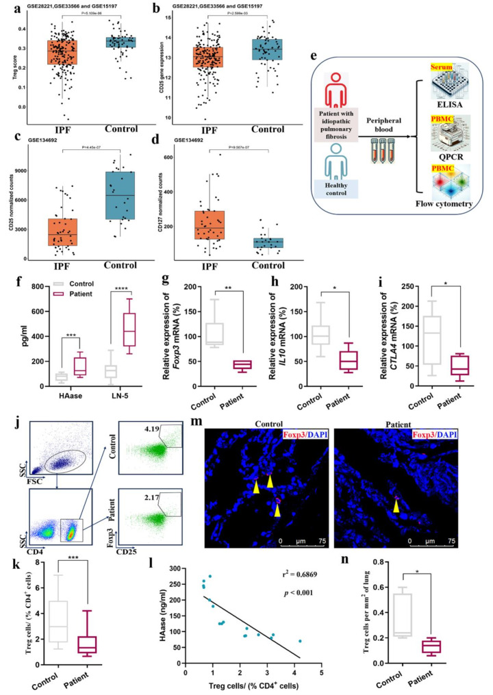
Background: Idiopathic pulmonary fibrosis is a progressive lung disorder, presenting clinically with symptoms such as shortness of breath and hypoxemia. Despite its severe prognosis and limited treatment options, the pathogenesis of idiopathic pulmonary fibrosis remains poorly understood. This study aims to investigate the therapeutic potential of mesenchymal stromal cells in treating idiopathic pulmonary fibrosis, focusing on their ability to modulate regulatory T cells through the low tumor necrosis factor superfamily member 4 (TNFSF4) pathway. The goal is to identify mesenchymal stromal cells subtypes with optimal immunomodulatory effects to enhance regulatory T cells functions and ameliorate fibrosis.

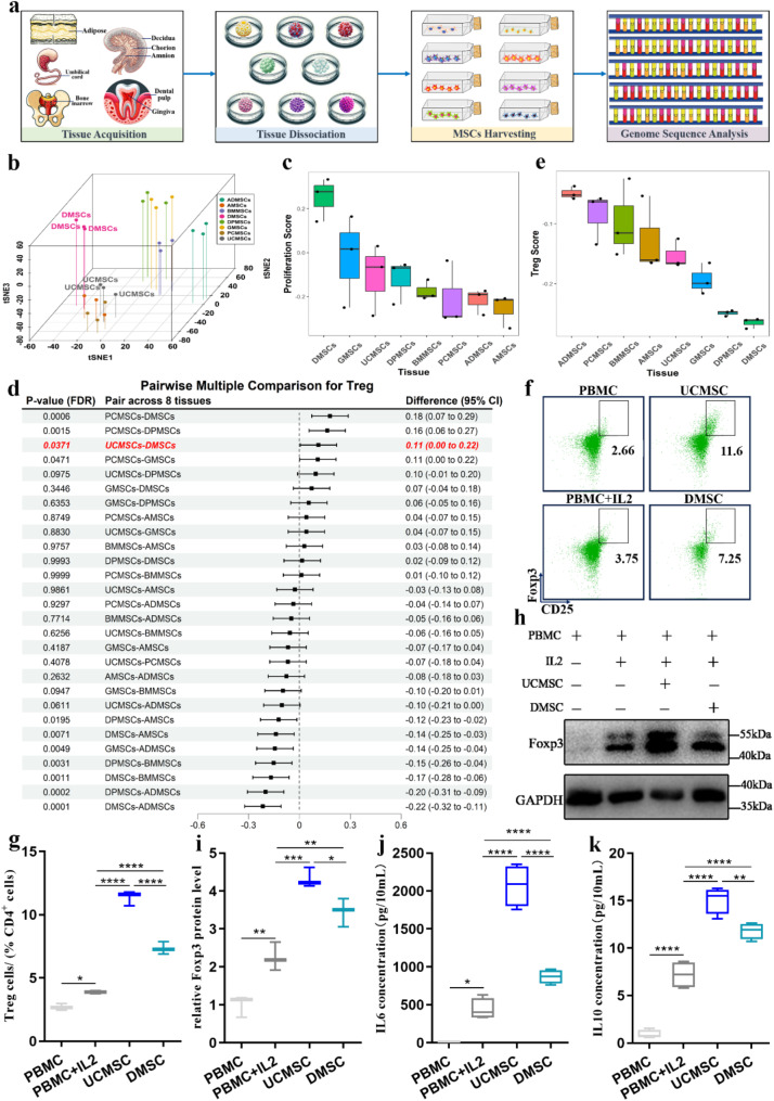
Methods: We identified the immune characteristics of idiopathic pulmonary fibrosis by mining and analyzing multiple public datasets and detecting regulatory T cells in the blood and lung tissues of idiopathic pulmonary fibrosis patients. An extensive examination followed, including assessing the impact of mesenchymal stromal cells on regulatory T cells proportions in peripheral blood and lung tissue, and exploring the specific role of TNFSF4 expression in regulatory T cells modulation. Whole-genome sequencing and cluster analysis were used to identify mesenchymal stromal cells subtypes with low TNFSF4 expression.

Results: Mesenchymal stromal cells characterized by TNFSF4 expression (TNFSF4low-MSCs) demonstrated enhanced ability to regulate regulatory T cells subpopulations and exhibited pronounced anti-fibrotic effects in the bleomycin-induced idiopathic pulmonary fibrosis mouse model. These mesenchymal stromal cells increased regulatory T cells proportions, reduced lung fibrosis, and improved survival rates. TNFSF4-tumor necrosis factor receptor superfamily member 4 (TNFRSF4) signaling was identified as a critical pathway influencing regulatory T cells generation and function.

Conclusions: Our findings underscore the pivotal role of TNFSF4 in mesenchymal stromal cells mediated regulatory T cells modulation and highlight the therapeutic potential of selecting mesenchymal stromal cells subtypes based on their TNFSF4 expression for treating idiopathic pulmonary fibrosis. This approach may offer a novel avenue for the development of targeted therapies aimed at modulating immune responses and ameliorating fibrosis in idiopathic pulmonary fibrosis.

Trial registration: Our study involved collecting 10 mL of peripheral blood from idiopathic pulmonary fibrosis patients, and the Medical Ethics Committee of Nanjing Drum Tower Hospital approved our study protocol with the approval number 2023-675-01.

Keywords: Fibrosis therapy; Idiopathic pulmonary fibrosis; Immunomodulation; Mesenchymal stromal cells; Regulatory T cells; TNFSF4.


Conflict of interest statement

Declarations. Ethics approval and consent to participate: Ethnic approval was granted by the Medical Ethics Committees of Nanjing Drum Tower Hospital (Project title: Utilization of Clinical Patient Samples (Tissue/Blood/Body Fluids) and Aborted Fetal Tissue to Extract Stromal Cells for Basic and Clinical Research in Regenerative Medicine and Treatment of Clinical Diseases; Approval No. 2017-161-08; Date of approval: 2017-11-30. Project title: TNFSF4(low)-MSCs Improve IPF by Regulating Tregs Subtypes; Approval No. 2023-675-01; Date of approval: 2023-03-20). All experiments involving mice were approved by the Ethics Committee of Nanjing Drum Tower Hospital (Project title: Research on the Mechanism of MSCs in Treating Pulmonary Fibrosis; Approval No. DWSY-22047178; Date of approval: 2022-04-07). All participants provided written informed consent before enrollment in the study. Consent for publication: Not applicable. Competing interests: The authors declare that they have no competing interest.

Figures

Fig. 1
Fig. 1
UCMSCs with stronger Tregs-regulatory capacities have superior IPFtherapeutic effects compared to DMSCs. (a) Flowchart of UCMSCs/DMSCs treatment of BLM-induced IPF mice. (b-d)Representative immunohistochemistry images for a-SMA,Col I and quantification; scale bar, 20um. (e) TheHydroxyproline levels in the lungs. (f) The Collagen content in thelungs using Sircol® soluble collagen assay kit. (g-h) The Tregs (CD4+CD25+Foxp3+) levels in the blood of BLM-induced IPF mice treatedwith UC-Mix and D-Mix, determined by FCM. (i-j) The Tregs (CD4+CD25+Foxp3+)levels in the thoracic lymph nodes of BLM-inducedIPF mice treated with UC-Mix and D-Mix, determined by FCM. (k-l) The Tregs(CD4+CD25+Foxp3+) levels in the spleen ofBLM-induced IPF mice treated with UC-Mix and D-Mix, determined by FCM. (m) Flowchartof PC61 antibody administration in BLM-induced IPF micetreated with UC-Mix. PC61 mAb (anti-murine CD25 ratIgG1) was used to deplete Tregs. (n-q) The levels of Tregs in the blood and lymph nodes of mice afterblockade with the PC61 antibody, as determined by FCM. (r-s) The effectof PC61 antibody blockade on the efficacy of UC-Mix treatment in IPF mice vialung hydroxyproline and collagen levels. (UC-Mix/D-Mix: Clinical-gradeUCMSCs/DMSCs derived from three donors were mixed in a 1:1:1 ratio to eliminateindividual variations.) 3
Fig. 2
Fig. 2
Tregs in PB and lung tissue were significantly lower than IPF patients. (a) We evaluated immune infiltration using gene expression profiles from the GSE28221, GSE33566, and GSE15197 datasets from the GEO database, which include IPF samples and healthy controls. The results showed a significant decrease in Tregs scores in IPF compared to healthy controls. (b-c) The expression of the CD25/IL2RA gene was significantly decreased in IPF compared to healthy controls in both array and sequencing data. (d) The Tregs-associated negative marker gene CD127 showed a significant increase in IPF patients in the sequencing data. (e) PB was collected from healthy individuals and patients with IPF. The fibrosis-related proteins in serum were analyzed by ELISA. The expression of relevant genes in PBMCs was analyzed using qPCR. Additionally, CD4+ T cells in PBMCs was analyzed using FCM. (f) The protein expression levels of HAase and LN-5 in PB were analyzed by ELISA. (g-i) QPCR was used to analyze the relevant gene expression of the Tregs, including IL-10, Foxp3 and CTLA4. (j-k) Flow cytometric analysis was performed on PBMCs to determine the percentages of Tregs (CD4+ CD25+ Foxp3+). Contour plots showing the cell populations from the indicated gates. (l) Correlation analysis between the expression of HAase and the proportions of Tregs. (m-n) By using immunofluorescence to stain human lung tissue paraffin sections with Foxp3 and DAPI, Tregs number in the lung tissues of healthy individuals and IPF patients was counted, followed by statistical analysis between the two groups
Fig. 3
Fig. 3
Bioinformatics reveals tissue heterogeneity of MSCs in regulating Tregs differentiation capability. (a) Schematic diagram of the acquisition and detection of MSCs from eight tissue sources. (b) The cluster analysis, t-distributed stochastic neighbor embedding analysis of gene expression of 24 samples from eight tissues, supported the heterogeneity of mesenchymal stromal cell. (c) Box plot of proliferation comparison of mesenchymal stromal cell across eight tissues. (d) Forest plot of multiple comparisons across eight tissues for Tregs. (e) Box plot of Tregs-infiltrated fraction comparison of mesenchymal stromal cell across eight tissues. (f-g) UCMSC and DMSC were co-cultured with PBMCs induced by IL-2, and the percentage of Tregs was determined by flow cytometric analysis. (h-i) The protein levels of the Tregs marker Foxp3 were detected by western blotting and quantified in each group. (j-k) The levels of Tregs related cytokines IL10 and IL6 in the supernatants of each group were determined by CBA kit. Images of unedited full blots in Figure S8
Fig. 4
Fig. 4
Bioinformatics data analysis shows that the gene most associated with the heterogeneity of MSCs in regulating Tregs differentiation is TNFSF4. (a) Bubble plot showing the GO enrichment analysis of immune-related significant pathways of DEGs between UCMSCs and DMSCs, especially enriched in plenty of T cell related pathways. (b) Gene expression heatmap showing the expression levels of 21 Tregs-related genes between UCMSCs and DMSCs. The color of each square indicates the scaled expression level of each gene. The columns show each sample, while the rows show each DEGs related to Tregs. (c) Bar plot indicates the Tregs-related DEGs between UCMSCs and DMSCs. There was significant difference for TNFSF4 with the maximal foldchange. Compared to DMSCs, the orange represents Tregs-related genes that are upregulated in UCMSCs, while the blue represents those that are downregulated. (d) The expression levels of TNFSF4 in UC-Mix and D-Mix were determined by qPCR. (e-f) The proportion of TNFSF4+ cells in the UC-Mix and D-Mix were determined by flow cytometric analysis. (g-h) Protein levels of TNFSF4 in UC-Mix and D-Mix were determined by western blotting and quantified. Images of unedited full blots in Figure S9
Fig. 5
Fig. 5
Gene editing technology demonstrate that the most significant gene associated with the heterogeneity of MSCs in regulating Tregs differentiation is TNFSF4. (a) Using virus plasmid transfection for gene knockout technology, the TNFSF4 gene in D-Mix cells was knocked out, and the stable cell line after puromycin selection was named D-Mix-sh-TNFSF4. The control group D-Mix cells were transfected with only the empty vector virus plasmid, and the stable cell line after selection was named D-Mix-sh-LV. QPCR to determine the mRNA expression of TNFSF4 in D-Mix-sh-TNFSF4 and D-Mix-sh-LV. (b) The proportion of TNFSF4+ MSCs in D-Mix-sh-TNFSF4 by flow cytometric analysis. (c-d) Protein levels of TNFSF4 in D-Mix-sh-TNFSF4 were determined by western blotting and quantified. (e) D-Mix-sh-TNFSF4 were co-cultured with PBMCs induced by IL-2, and the percentage of Tregs was determined by flow cytometric analysis. (f-g) The levels of Tregs related cytokines IL10 and IL6 in the supernatants of each group were determined by CBA kit. (h-i) The protein levels of the Tregs marker Foxp3 were detected by western blotting and quantified in each group. (j-s) Through virus plasmid transfection and gene overexpression technology, the TNFSF4 gene was overexpressed in UC-Mix cells, and the stable cell line after puromycin selection was named UC-Mix-TNFSF4. For the control group UC-Mix cells, only the empty vector virus plasmid was transfected, and the stable cell line after selection was named UC-Mix-LV. The experiments described in this figure, i-t, were conducted on both UC-Mix-TNFSF4 and UC-Mix-LV cell lines. Images of unedited full blots in Figure S10
Fig. 6
Fig. 6
MSCs regulate the differentiation, subtype distribution, and proliferation of Tregs subpopulations through the TNFSF4-TNFRSF4 axis. (a-b) Flow cytometric analysis of Tregs after addition of Oxelumab (anti-TNFSF4), Telazorlimab (anti-TNFSF4) and Amlitelimab (TNFRSF4) to the D-Mix and PBMCs co-culture systems. Contour plots showing the cell populations from the indicated gates. The percentage of Tregs (CD4+ CD25+ Foxp3+). (c-d) UC-Mix/D-Mix were co-cultured with purified CD4+ T cells induced by IL2. The proportion of activated Tregs (CD4+ CD25+ CD38+ HLA-DR+) was determined by FCM. (e-g) The relative mRNA expression levels of Tregs subtype-related genes (Foxp3, GTIR and Helios) in each group were determined by qPCR. (h-i) The expression levels of Tregs subtype-associated proteins (Foxp3, GTIR and Helios) in each group were determined by western blotting. (j-k) UC-Mix/D-Mix were co-cultured with purified CD4+ T cells induced by IL2 and the percentage of each Tregs subtype was determined by FCM. The samples were analyzed for the expression of CD4, CD25, CD45RA and CCR7. Contour plots showing the cell populations from the indicated gates. The proportions of TregCM, TregE, TregEM and Tregnaive were counted. (l-m) FCM was used to analyze the proliferation of Tregs in each group. Representative FCM gating strategy correspond to Tregs (CD4+ CD25+ Foxp3+), and proliferated Tregs gating was further stratified by the expression of Ki67. The proportions of Ki67 Tregs were counted. Images of unedited full blots in Figure S11

Similar articles

References

    1. du Bois RM. Idiopathic pulmonary fibrosis: present Understanding and future options. Eur Respiratory Review: Official J Eur Respiratory Soc. 2011;20(121):132–3. - PMC - PubMed
    1. Raghu G, Remy-Jardin M, Myers JL, Richeldi L, Ryerson CJ, Lederer DJ, et al. Diagnosis of idiopathic pulmonary fibrosis. An official ATS/ERS/JRS/ALAT clinical practice guideline. Am J Respir Crit Care Med. 2018;198(5):e44–68. - PubMed
    1. Richeldi L, Collard HR, Jones MG. Idiopathic pulmonary fibrosis. Lancet. 2017;389(10082):1941–52. - PubMed
    1. George PM, Patterson CM, Reed AK, Thillai M. Lung transplantation for idiopathic pulmonary fibrosis. Lancet Respir Med. 2019;7(3):271–82. - PubMed
    1. Tzouvelekis A, Toonkel R, Karampitsakos T, Medapalli K, Ninou I, Aidinis V, et al. Mesenchymal stem cells for the treatment of idiopathic pulmonary fibrosis. Front Med (Lausanne). 2018;5:142. - PMC - PubMed

MeSH terms

LinkOut - more resources