

Relationship between temporomandibular joint space and articular disc displacement
- PMID: 40254585
- PMCID: PMC12010630
- DOI: 10.1186/s12903-025-05991-7
Relationship between temporomandibular joint space and articular disc displacement
Abstract
Objective: Analyse the correlation between the changes in joint space of TMJ and the displacement and degree of articular disc for clinical diagnosis.
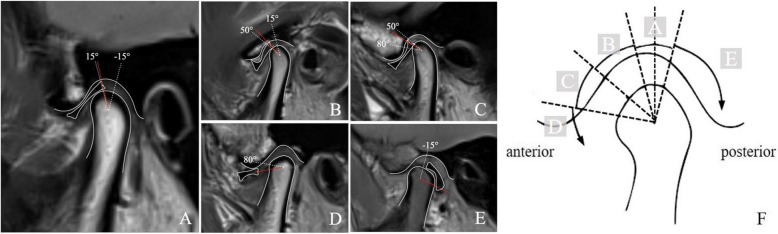
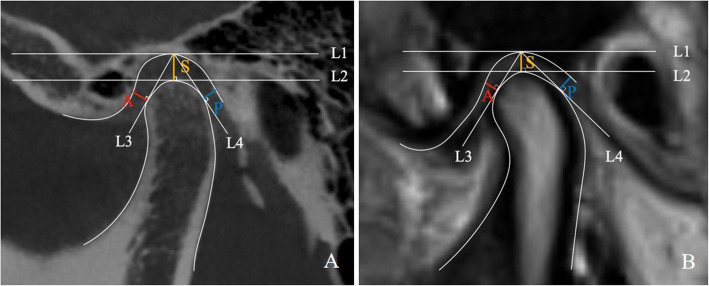
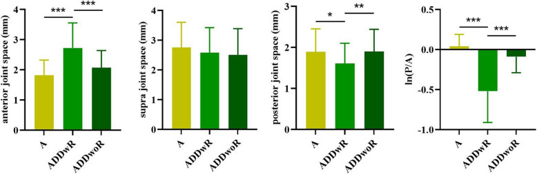
Methods: Two hundred sixteen TMJs of 108 temporomandibular disorders (TMD) patients with clinical symptoms and MRI examination were included in the study. 30 of these patients had undergone CBCT before MRI. According to the degree of articular disc displacement, the 216 joints are divided into five groups. Group A: no disc displacement (40 cases); group B: mild anterior disc displacement (44 cases); group C: moderate anterior disc displacement (36 cases); group D: severe anterior disc displacement (52 cases); group E: posterior displacement (44 cases). The 132 sides of these anteriorly displaced discs (ADD) were further divided into two groups, anterior disc displacement with reduction (ADDwR) and anterior disc displacement without reduction (ADDwoR). We analysed the concordance of the joint space measured by MRI and CBCT, and explored the relationship between joint space, ln(P/A) values and joint disc displacement.
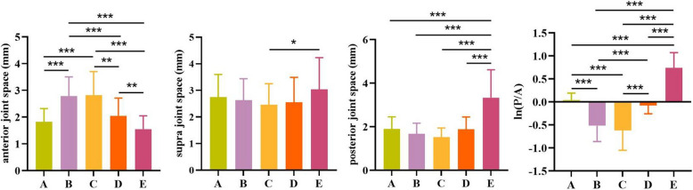
Results: There was no statistically significant difference between the joint spaces measured by CBCT and MRI (P > 0.05). The anterior joint space in group B (2.7 ± 0.72 mm) and C (2.82 ± 0.88 mm) was larger than group A (1.82 ± 0.50 mm) (P < 0.05), and ln(P/A) value in group B (-0.52 ± 0.34) and C (-0.62 ± 0.43) was smaller than group A (0.04 ± 0.15) (P < 0.05). The posterior joint space (3.33 ± 1.28 mm) and ln(P/A) value (0.74 ± 0.33) in group E was larger than group A (P < 0.05). There was no significant difference in the anterior, superior and posterior joint space and ln(P/A) value between group D and A (P > 0.05). The ADDwR group had a larger anterior joint space (2.72 ± 0.83 mm) than group A (P < 0.05), while having a smaller posterior joint space (1.61 ± 0.49 mm) and ln(P/A) value (-0.52 ± 0.39 mm) (P < 0.05). Compared with group A, there was no significant difference in the anterior joint space and ln(P/A) value in the ADDwoR group(P > 0.05).
Conclusion: There is no significant change in anterior, supra, and posterior joint space in severe anterior disc displacement. The anterior joint space increases in mild to moderate anterior disc displacement, but does not change in severe anterior disc displacement-the posterior joint space increases when the joint disc is displaced posteriorly. The position of the joint disc cannot be accurately inferred by observing the joint space through CBCT, and a combination of MRI and clinical examination is required to make a definitive judgement.
Keywords: ADD; Joint space; MRI; TMD.
© 2025. The Author(s).
Conflict of interest statement
Declarations. Ethics approval and consent to participate: The experimental protocol was established according to the ethical guidelines of the Helsinki Declaration and was approved by the Human Ethics Commit tee of Stomatological Hospital of Zhejiang Chinese Medical University(No.ZCMUHSIRB - 2024101015). Written informed consent was obtained from individual or guardian participants. Consent for publication: Not applicable. Competing interests: The authors declare no competing interests.
Figures
References
MeSH terms
Grants and funding
LinkOut - more resources
Full Text Sources
Medical
