

Real-Time Identification of Lymph Vessels Using Indocyanine Green in a Patient With Chylothorax Associated With Lymphangioleiomyomatosis
- PMID: 40254818
- PMCID: PMC12010055
- DOI: 10.1111/ases.70067
Real-Time Identification of Lymph Vessels Using Indocyanine Green in a Patient With Chylothorax Associated With Lymphangioleiomyomatosis
Abstract
Introduction: Lymphangioleiomyomatosis (LAM) is often complicated by chylothorax and may require surgical intervention; however, the treatment is complicated because of difficulties in identifying the location of the fistula intraoperatively. This is the first report to identify the site of a chyle fistula associated with LAM in real time during surgery by using indocyanine green (ICG) lymphangiography.
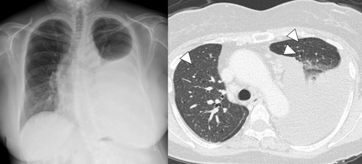
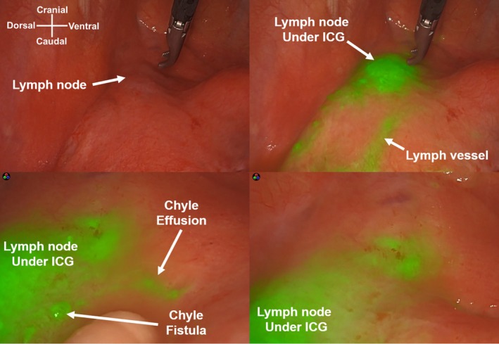
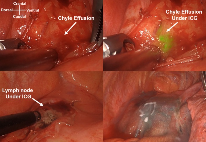
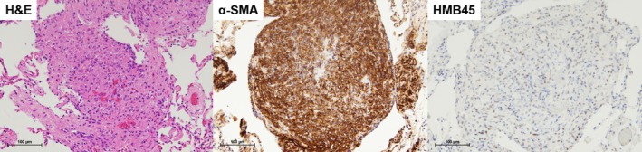
Materials and surgical technique: A 56-year-old woman received a diagnosis of a treatment-resistant left chylothorax associated with LAM. To identify the chyle fistula during surgery, 1 mL of ICG (2.5 mg) was injected into both inguinal lymph nodes under ultrasound guidance after anesthesia, with 1 mL per side for a total of 5 mg of ICG. We performed video-assisted thoracic surgery and observed near-infrared light acquisition and overlay technology using Stryker. Approximately 1 h after administration, fluorescence was observed in the anterior mediastinal lymph nodes, and a chyle fistula was observed around them. Although we attempted ligation of the lymph trunk, the surgical procedure damaged well-developed lymph vessels. The damaged area and anterior mediastinal lymph nodes, including the surrounding lymph vessels, were incinerated using soft coagulation and covered with polyglycolic acid sheets and fibrin glue. Consequently, the amount of chylous effusion decreased.
Discussion: The use of ICG allowed visualization of the lymphatic pathway and location of the chyle fistula in real time during surgery, enabling precise local treatment to reduce chyle effusion.
Keywords: chylothorax; indocyanine green; lymphangioleiomyomatosis.
Asian Journal of Endoscopic Surgery© 2025 The Author(s). Asian Journal of Endoscopic Surgery published by Asia Endosurgery Task Force and Japan Society of Endoscopic Surgery and John Wiley & Sons Australia, Ltd.
Conflict of interest statement
The authors declare no conflicts of interest.
Figures
References
-
- Ryu J. H., Doerr C. H., Fisher S. D., Olson E. J., and Sahn S. A., “Chylothorax in Lymphangioleiomyomatosis,” Chest 123 (2003): 623–627. - PubMed
-
- Thammineedi S. R., Patnaik S. C., Reddy P., Shukla S., Vashist Y. K., and Nusrath S., “Impact of Fluorescent Thoracic Duct Lymphography via Intranodal Approach in Minimal Access Esophageal Cancer Surgery,” Langenbeck's Archives of Surgery 408 (2023): 426. - PubMed
-
- Northrup H., Krueger D. A., and International Tuberous Sclerosis Complex Consensus Group , “Tuberous Sclerosis Complex Diagnostic Criteria Update: Recommendations of the 2012 International Tuberous Sclerosis Complex Consensus Conference,” Pediatric Neurology 49, no. 4 (2013): 243–254, 10.1016/j.pediatrneurol.2013.08.001. - DOI - PMC - PubMed
Publication types
MeSH terms
Substances
LinkOut - more resources
Full Text Sources
Medical
