. 2025 Jun;21(23):e2501789.
doi: 10.1002/smll.202501789. Epub 2025 Apr 21.

Fusogenic Nanoreactor-Based Detection of Extracellular Vesicle-derived miRNAs for Diagnosing Atherosclerosis

Affiliations

Fusogenic Nanoreactor-Based Detection of Extracellular Vesicle-derived miRNAs for Diagnosing Atherosclerosis

Jiyoon Lee et al. Small. 2025 Jun.

Abstract

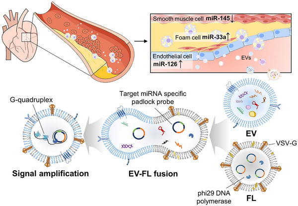
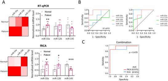
Extracellular vesicle (EV) microRNAs (miRNAs) are critical liquid-biopsy biomarkers that facilitate noninvasive clinical diagnosis and disease monitoring. However, conventional methods for detecting these miRNAs require EV lysis, which is expensive, labor-intensive, and time-consuming. Inspired by natural viral infection mechanisms, a novel strategy is developed for detecting EV miRNAs in situ via vesicle fusion mediated by viral fusion proteins. A padlock probe encapsulated within fusogenic liposomes is activated by target miRNAs, thereby initiating a highly sensitive and specific rolling circle amplification (RCA) reaction. Three EV miRNAs associated with atherosclerosis are successfully analyzed using this method, thereby enabling clear differentiation of healthy and diseased mice at several disease stages. Overall, the developed platform offers a simple approach for detecting EV miRNAs and demonstrates significant potential for broad use in applications involving disease diagnosis and monitoring.

Keywords: atherosclerosis diagnosis; extracellular vesicle; fusogenic nanoreactor; microrna; rolling circle amplification.


Conflict of interest statement

The authors declare no conflict of interest.

Figures

Scheme 1
Scheme 1
Fusogenic nanoreactor‐based EV‐miRNA detection method for diagnosing atherosclerosis. We developed a novel vesicle fusion‐based platform for direct detection of EV miRNAs, enabling highly specific and sensitive disease stage discrimination. By engineering FLs carrying VSV‐G and reagents for RCA, we achieved target‐specific signal generation within intact EVs without RNA extraction or reverse transcription. This platform detects three miRNAs (miR‐33a, miR‐126, and miR‐145) via RCA‐triggered G‐quadruplex formation, allowing fluorescence‐based quantification. The system was validated using cell‐derived, mouse, and human serum EVs, exhibiting robust diagnostic performance for atherosclerosis – an asymptomatic, artery‐narrowing disease with limited blood‐based diagnostic options. By capturing EVs released from plaque, our method offers a minimally invasive alternative. The method simplifies the workflow, reduces the sample loss and cost, and maintains high analytical accuracy through direct in situ detection of EV miRNAs.
Figure 1
Figure 1
Characterizing FLs and HEK293T‐cell‐derived EVs. A) Schematic depicting FL production. B) Size‐distribution profiles for liposomes with (FL) and without VSV‐G (L). C) Zeta potentials of FLs; n = 5–6, Student's t‐test. D) Cryo‐TEM image of FLs. Scale bar: 100 nm. E) Schematic depicting EV production. F) Size‐distribution profile for EVs. G) Western‐blot analysis of EV marker proteins (CD9, CD63, CD81, Alix) and a non‐EV protein (calnexin). Cell lysate and EV samples were loaded for comparison. H) TEM image of EVs. Scale bar: 100 nm. All values are means ± SDs: ****p < 0.0001.
Figure 2
Figure 2
Evaluating FL and EV membrane fusion using a FRET‐based assay. A) Schematic depicting the FRET‐based detection of FL/EV membrane fusion. FLs are labeled with a donor–acceptor FRET pair, with fusion leading to a reduction in FRET. B) Emission spectra of the donor fluorophore and C) corresponding FRET ratios at various pH values; n = 5, Student's t‐test. D) FRET spectra after a 2‐h fusion reaction and E) corresponding FRET ratios that compare fusion efficiencies using different fusion methods; n = 3. F) Size‐distribution profiles of L, FLs, and FL‐EV mixtures after fusion reactions. All values are means ± SDs: *p < 0.05.
Figure 3
Figure 3
Sensitivity and selectivity of the RCA‐based miRNA detection method. A) Schematic depicting the three EV‐associated miRNA targets (miR‐33a‐5p, miR‐126‐3p, and miR‐145‐5p) selected for diagnosing atherosclerosis. B) Emission spectra and C) gel electropherograms of RCA reactions performed in the presence (w/) or absence (w/o) of the three target miRNAs. In (B), solid lines represent reactions containing the respective target miRNA, and dashed lines indicate reactions conducted without the target miRNA. D) Reaction‐time‐dependent RCA reaction intensities; n = 3. E) Selectivity of each padlock probe for its corresponding miRNA target. F) Target miRNA‐concentration‐dependent RCA reaction intensities; n = 3. All values are means ± SDs.
Figure 4
Figure 4
Feasibility of fRCA for analyzing EV miRNA in situ. A,C,E) Schematic depicting the analysis steps and B,D,F) normalized detection‐signal data for the three EV‐miRNA‐detection methods using: a commercially available PCR kit for RNA extraction and reverse transcription (A–B, RT‐qPCR), an RCA reaction performed after extracting the RNA from the EVs (C–D, RCA), and a membrane‐fusion‐based RCA reaction without RNA isolation (E–F, fRCA). Note that RCA reactions were conducted under two different time conditions: 2 h and overnight (O/N), n = 3–4. All values are means ± SDs: ****p < 0.0001.
Figure 5
Figure 5
In vivo detection of EV miRNAs via fRCA during atherosclerosis diagnosis. A–E) Male ApoE −/− mice were fed a normal chow diet for 10, 20, or 40–44 weeks (n = 5, 5, and 11, respectively). A) Schematic depicting the feeding protocol and time points for serum isolation at each stage. B) Representative photographic images of atherosclerotic plaques on the aortic arch. Scale bars: 0.2 cm. Red triangles indicate plaque lesions at respective stages. C) Representative en‐face images of Oil‐Red‐O‐stained whole aortas at different time points. D) Quantifying plaque areas as percentages of the total aorta. Each data point represents an individual mouse; one‐way ANOVA. E) Serum levels of T‐chol, LDL‐C, and HDL‐C in normal C57BL/6J control (Ctrl) mice (n = 5; 10 weeks old) and ApoE −/− mice at 10, 20, and 40 weeks (n = 5 for each group). Each data point represents an individual mouse; one‐way ANOVA. F) Detecting EV miRNAs using the RT‐qPCR (top) and fRCA (bottom) methods. Each data point represents an individual mouse. G) RT‐qPCR/fRCA correlation analysis for miR‐33a, miR‐126 and miR‐145; Pearson correlation analysis. H) ROC curves and AUC analyses for miR‐33a, miR‐126, and miR‐145 RT‐qPCR data (left) and fRCA data (right). I) ROC curves and AUC analyses of the logistic regression model based on the combination of miR‐33a, miR‐126, and miR‐145, for RT‐qPCR data (blue) and fRCA data (red). All values are means ± SDs: **p < 0.01, ***p < 0.001, and ****p < 0.0001. Statistical significance was analyzed relative to the control group (Ctrl). T‐chol, total cholesterol; LDL‐C, low‐density lipoprotein cholesterol; HDL‐C, high‐density lipoprotein cholesterol; ANOVA, analysis of variance; W, weeks.
Figure 6
Figure 6
Detecting EV miRNAs via fRCA during atherosclerosis diagnosis in humans. A) Detecting EV miRNAs using the RT‐qPCR (top) and fRCA (bottom) methods. Each data point represents an individual. B) ROC curves and AUC analyses for miR‐33a, miR‐126, and miR‐145 RT‐qPCR data (left) and fRCA data (right). C) ROC curves and AUC analyses of the logistic regression model based on the combination of miR‐33a, miR‐126, and miR‐145, for RT‐qPCR data (blue) and fRCA data (red). All values are means ± SDs: **p < 0.01, ***p < 0.001, and ****p < 0.0001. Statistical significance was analyzed relative to the normal group. Normal, healthy individual; Patient, patient with coronary artery disease.

Similar articles

References

    1. a) Tuttolomondo A., Di Raimondo D., Pecoraro R., Arnao V., Pinto A., Licata G., Curr. Pharm. Des. 2012, 18, 4266; - PubMed
    2. b) Malekmohammad K., Bezsonov E. E., Rafieian‐Kopaei M., Front. Cardiovasc. Med. 2021, 8, 707529. - PMC - PubMed
    1. a) Ibanez B., Badimon J. J., Garcia M. J., Am. J. Med. 2009, 122, S15 ; - PubMed
    2. b) Penalva R. A., Huoya M. D. O., Correia L. C. L., Feitosa G. S., Ladeia A. M. T., Arq. Bras. Cardiol. 2008, 90, 24. - PubMed
    1. a) Badimon L., Suades R., Vilella‐Figuerola A., Crespo J., Vilahur G., Escate R., Padro T., Chiva‐Blanch G., Antioxid. Redox. Signal 2020, 33, 645; - PubMed
    2. b) Chiva‐Blanch G., Padro T., Alonso R., Crespo J., Perez de Isla L., Mata P., Badimon L., Arterioscler Thromb. Vasc. Biol. 2019, 39, 945. - PubMed
    1. a) Gurunathan S., Kang M. H., Qasim M., Khan K., Kim J. H., Int. J. Nanomed. 2021, 16, 3357; - PMC - PubMed
    2. b) Simeone P., Bologna G., Lanuti P., Pierdomenico L., Guagnano M. T., Pieragostino D., Del Boccio P., Vergara D., Marchisio M., Miscia S., Mariani‐Costantini R., Int. J. Mol. Sci. 2020, 21, 2514; - PMC - PubMed
    3. c) Palviainen M., Saraswat M., Varga Z., Kitka D., Neuvonen M., Puhka M., Joenvaara S., Renkonen R., Nieuwland R., Takatalo M., Siljander P. R. M., PLoS One 2020, 15, 0236439; - PMC - PubMed
    4. d) Revenfeld A. L., Baek R., Nielsen M. H., Stensballe A., Varming K., Jorgensen M., Clin. Ther. 2014, 36, 830. - PubMed
    1. a) Price N. L., Rotllan N., Canfran‐Duque A., Zhang X., Pati P., Arias N., Moen J., Mayr M., Ford D. A., Baldan A., Suarez Y., Fernandez‐Hernando C., Cell Rep. 2017, 21, 1317; - PMC - PubMed
    2. b) Martinez‐Arroyo O., Ortega A., Flores‐Chova A., Sanchez‐Garcia B., Garcia‐Garcia A. B., Chaves F. J., Martin‐Escudero J. C., Forner M. J., Redon J., Cortes R., Eur. J. Intern. Med. 2023, 113, 49; - PMC - PubMed
    3. c) Leistner D. M., Boeckel J. N., Reis S. M., Thome C. E., De Rosa R., Keller T., Palapies L., Fichtlscherer S., Dimmeler S., Zeiher A. M., Eur. Heart J. 2016, 37, 1738. - PubMed

LinkOut - more resources