. 2025 May 9;11(5):1064-1077.
doi: 10.1021/acsinfecdis.4c00631. Epub 2025 Apr 21.

Anti-SARS-CoV-2 Small Molecule Targeting of Oxysterol-Binding Protein (OSBP) Activates Cellular Antiviral Innate Immunity

Affiliations

Anti-SARS-CoV-2 Small Molecule Targeting of Oxysterol-Binding Protein (OSBP) Activates Cellular Antiviral Innate Immunity

Bharathiraja Subramaniyan et al. ACS Infect Dis. .

Abstract

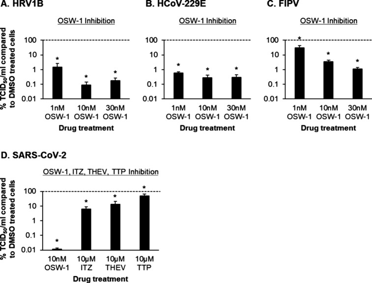
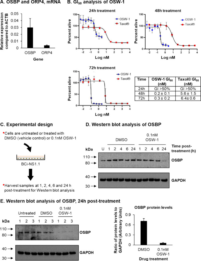
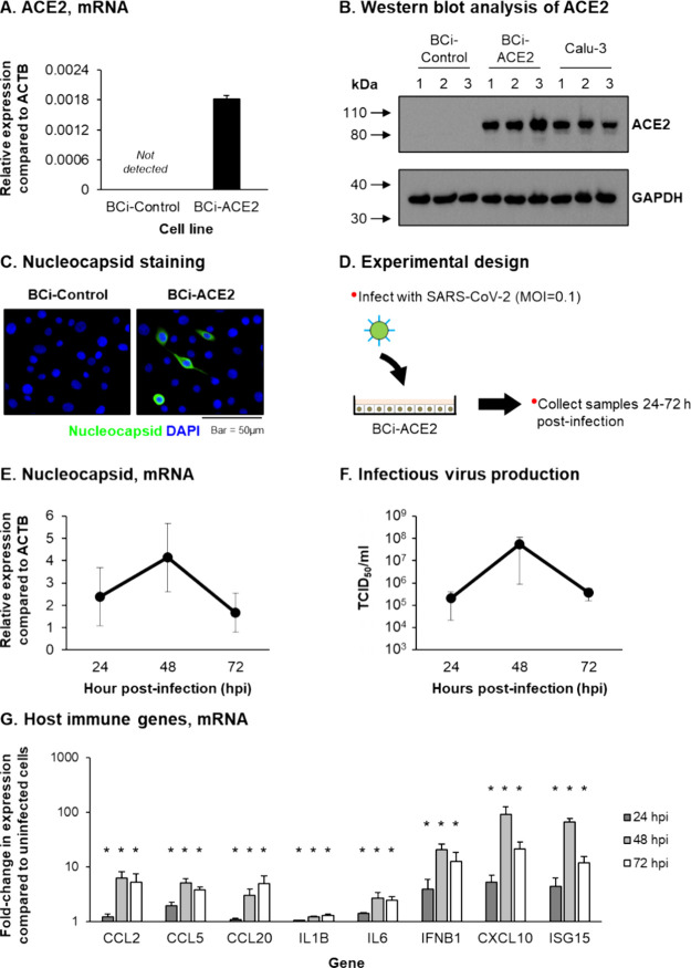
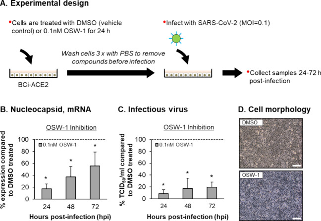
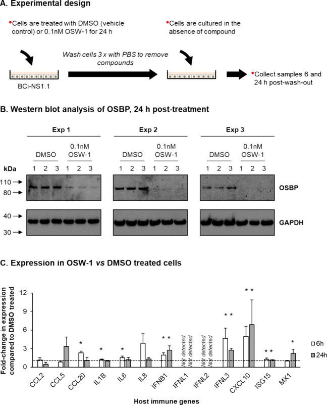
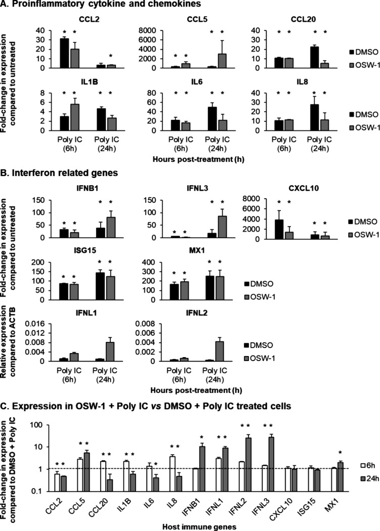
Human oxysterol-binding protein (OSBP) is a potentially druggable mediator in the replication of a broad spectrum of positive-sense (+) single-stranded RNA (ssRNA) viruses, including members of the Picornaviridae, Flaviviridae, and Coronaviridae. OSBP is a cytoplasmic lipid transporting protein capable of moving cholesterol and phosphoinositides between the endoplasmic reticulum (ER) and Golgi, and the ER and lysosome. Several structurally diverse antiviral compounds have been reported to function through targeting OSBP, including the natural product compound OSW-1. Our prior work shows that transient OSW-1 treatment induces a reduction in OSBP protein levels over multiple successive cell generations (i.e., multigenerational), with no apparent cellular toxicity, and the OSW-1-induced reduction of OSBP has antiviral activity against multiple (+)ssRNA viruses. This study extends these findings and establishes that OSW-1 has in vitro antiviral activity against multiple pathogenic (+)ssRNA viruses, including human rhinovirus (HRV1B), the feline coronavirus peritonitis virus (FIPV), human coronavirus 229E (HCoV-229E), and severe acute respiratory syndrome coronavirus 2 (SARS-CoV-2). We also demonstrate that OSW-1 treatment in human airway epithelial cells alters the expression of multiple antiviral innate immune mediators, including the interferon (IFN) related genes IFNB1, IFNL3, CXCL10, ISG15, and MX1. Furthermore, OSW-1 enhances the induction of specific components of type I and III IFN antiviral responses triggered by the RNA viral mimetic polyinosinic-polycytidylic acid (Poly IC). In summary, this study further demonstrates the importance of OSBP in (+)ssRNA virus replication and presents OSBP as a potential regulator of cellular antiviral innate immune responses.

Keywords: OSW-1; SARS-CoV-2; antiviral compounds; innate antiviral immune response; oxysterol-binding protein (OSBP); rhinovirus.


Conflict of interest statement

The authors declare no competing financial interest.

Figures

Figure 1
Figure 1
OSW-1 treatment inhibits replication of HRV1B, HCoV-229E, FIPV and SARS-CoV-2. (A) HRV1B-infected H1-HeLa cells treated with DMSO or OSW-1 from n = 3 independent experiments. (B) HCoV-229E infected MRC-5 cells treated with DMSO or OSW-1 from n = 4 independent experiments. (C) FIPV-infected CRFK cells treated with DMSO or OSW-1 from n = 3 independent experiments. (D) SARS-CoV-2-infected Vero E6 cells treated with DMSO, OSW-1, ITZ, THEV or TTP from n = 3 independent experiments. For A–D, the data are presented as percentage inhibition of virus replication by OSW-1, ITZ, THEV or TTP. For A–D, the bars represent the mean percentage of TCID50/mL in OSW-1, ITZ, THEV or TTP treated compared to DMSO treated cells, and the error bars indicate the standard error of the mean (SEM). * p ≤ 0.05.
Figure 2
Figure 2
Treatment of BCi-NS1.1 cells with OSW-1 leads to a knockdown of OSBP protein levels. (A) qPCR of OSBP and ORP4 gene expression in BCi-NS1.1 cells. Bars represent mean expression in n = 3 replicates from a single experiment. Error bars indicate the SEM (B) CellTiter-Blue assay to determine the GI50 of OSW-1 and Taxol in BCi-NS1.1 cells at 24–72 h (h) post-treatment. Graphs are representative of one experiment with the mean value from n = 3 replicates shown for each concentration. Error bars indicate the standard deviation (SD). The table displays the mean and SD calculated from n ≥ 3 experiments for each compound and time point. (C) Experimental design for treatment of BCi-NS1.1 cells with OSW-1. (D) Western blot analysis of OSBP and GAPDH in whole cell lysates of BCi-NS1.1 cells either untreated or treated with DMSO or 0.1 nM OSW-1 for 1, 2, 4, 6, and 24 h. (E) Western blot analysis of OSBP and GAPDH in whole cell lysates of BCi-NS1.1 cells either untreated or treated with DMSO or 0.1 nM OSW-1 for 24 h. OSBP protein levels are shown with a representative Western blot shown as well as quantified values from three independent experiments, normalized to GAPDH protein levels for each treatment group. Bar represents mean levels from n = 3 replicates and error bars the SEM.
Figure 3
Figure 3
ACE2 expressing BCi-NS1.1 cells supports infection and replication of SARS-CoV-2. (A) qPCR of ACE2 gene expression in BCi-Control and BCi-ACE2 cells. Bars represent mean expression in n = 3 replicates from a single experiment. Error bars indicate the SEM (B) Western blot analysis of ACE2 and GAPDH in whole cell lysates of BCi-Control, BCi-ACE2 and Calu-3 cells. Data shown from a single experiment with n = 3 replicates per cell line. (C) Co-immunofluorescent staining of SARS-CoV-2 nucleocapsid (green) and cell nuclei (blue, DAPI) in BCi-Control and BCi-ACE2 cells infected with SARS-CoV-2. (D) Experimental design for infection of BCi-ACE2 cells with SARS-CoV-2. (E) qPCR of SARS-CoV-2 nucleocapsid gene expression at 24, 48, and 72 h post infection (hpi) in cultures of SARS-CoV-2 infected BCi-ACE2 cells. The circles represent the mean expression from n = 3 independent experiments and the error bars indicate the SEM (F) Infectious virus titer (TCID50/mL) at 24, 48, and 72 hpi in cultures of SARS-CoV-2 infected BCi-ACE2 cells. The circles represent the mean TCID50/mL from n = 3 independent experiments and the error bars indicate the SEM (G) qPCR of host immune gene expression (CCL2, CCL5, CCL20, IL1B, IL6, IFNB1, CXCL10 and ISG15) at 24 (dark gray bars), 48 (light gray bars) and 72 (white bars) hpi in cultures of SARS-CoV-2 infected BCi-ACE2 cells. Bars represent mean fold-change expression compared to uninfected cells in n = 3 independent experiments and error bars indicate the SEM * p ≤ 0.05.
Figure 4
Figure 4
OSW-1 treatment inhibits the replication of SARS-CoV-2. (A) Experimental design for SARS-CoV-2 infection of BCi-ACE2 cells pretreated with DMSO or 0.1 nM OSW-1. (B) qPCR of SARS-CoV-2 nucleocapsid gene expression at 24, 48, and 72 h post infection (hpi) in cultures of SARS-CoV-2 infected BCi-ACE2 cells either pretreated with DMSO or 0.1 nM OSW-1 from n = 3 independent experiments. The data are presented as percentage inhibition of virus replication by OSW-1 The bars represent the mean percentage of expression in OSW-1 treated compared to DMSO treated cells and the error bars indicate the SEM (C) Infectious virus titer (TCID50/mL) at 24, 48, and 72 hpi in cultures of SARS-CoV-2 infected BCi-ACE2 cells either pretreated with DMSO or 0.1 nM OSW-1 from n = 3 independent experiments. The data are presented as percentage inhibition of virus replication by OSW-1. The bars represent the mean percentage of TCID50/mL in OSW-1 treated compared to DMSO treated cells and the error bars indicate the SEM * p ≤ 0.05. (D) Morphology of DMSO and OSW-1 treated BCi-ACE2 cells at 72 hpi with SARS-CoV-2. Bar = 50 μm.
Figure 5
Figure 5
OSW-1 treatment leads to activation of the antiviral innate immune response. (A) Experimental design for the treatment of BCi-NS1.1 cells with DMSO or 0.1 nM OSW-1. (B) Western blot analysis of OSBP and GAPDH in whole cell lysates of BCi-NS1.1 cells either treated with DMSO or 0.1 nM OSW-1 for 24 h. Data shown from a n = 3 experiments with n = 3 replicates per treatment. (C) qPCR of host immune gene expression (CCL2, CCL5, CCL20, IL1B, IL6, IL8, IFNB1, CXCL10, and ISG15) at 6 (white bars) and 24 (light gray bars) hour (h) postwashout of DMSO/OSW-1. Each bar represents mean fold-change in expression compared to DMSO treated cells in n = 3 independent experiments and error bars indicate the SEM * p ≤ 0.05.
Figure 6
Figure 6
Poly IC stimulation of BCi-NS1.1 following OSW-1 treatment leads to induction of a potent innate immune response. (A) qPCR of proinflammatory cytokine and chemokine gene expression (CCL2, CCL5, CCL20, IL1B, IL6 and IL8) at 6 and 24 h (h) poststimulation with Poly IC (10 μg/mL). (B) qPCR of IFN related genes expression (IFNB1, IFNL1, IFNL2, IFNL3, CXCL10, ISG15 and MX1) at 6 and 24 h (h) poststimulation with Poly IC (10 μg/mL). For both A and B, black bars represent DMSO treated group and dark gray bars represent the OSW-1 treated group. Each bar represents mean fold-change in expression compared to untreated (non-Poly IC) cells in n = 3 independent experiments and error bars indicate the SEM. Due to the lack of expression of IFNL1 and IFNL2 in the untreated control group, the data are presented as relative expression compared to ACTB. * p ≤ 0.05 compared to the untreated control group. (C) qPCR of host immune gene expression (CCL2, CCL5, CCL20, IL1B, IL6, IL8, IFNB1, IFNL1, IFNL2, IFNL3, CXCL10, ISG15 and MX1) in OSW-1 + Poly IC treated cells at 6 (white bars) and 24 (light gray bars) hour (h) poststimulation with Poly IC (10 μg/mL). Each bar represents mean fold-change in expression compared to DMSO + Poly IC treated cells in n = 3 independent experiments and error bars indicate the SEM * p ≤ 0.05.

Similar articles

References

    1. von Delft A.; Hall M. D.; Kwong A. D.; Purcell L. A.; Saikatendu K. S.; Schmitz U.; Tallarico J. A.; Lee A. A. Accelerating Antiviral Drug Discovery: Lessons from COVID-19. Nat. Rev. Drug Discovery 2023, 22 (7), 585–603. 10.1038/s41573-023-00692-8. - DOI - PMC - PubMed
    1. Roberts B. L.; Severance Z. C.; Bensen R. C.; Le-McClain A. T.; Malinky C. A.; Mettenbrink E. M.; Nuñez J. I.; Reddig W. J.; Blewett E. L.; Burgett A. W. G. Differing Activities of Oxysterol-Binding Protein (OSBP) Targeting Anti-Viral Compounds. Antiviral Res. 2019, 170, 10454810.1016/j.antiviral.2019.104548. - DOI - PMC - PubMed
    1. Roberts B. L.; Severance Z. C.; Bensen R. C.; Le A. T.; Kothapalli N. R.; Nuñez J. I.; Ma H.; Wu S.; Standke S. J.; Yang Z.; Reddig W. J.; Blewett E. L.; Burgett A. W. G. Transient Compound Treatment Induces a Multigenerational Reduction of Oxysterol-Binding Protein (OSBP) Levels and Prophylactic Antiviral Activity. ACS Chem. Biol. 2019, 14 (2), 276–287. 10.1021/acschembio.8b00984. - DOI - PMC - PubMed
    1. Kobayashi J.; Arita M.; Sakai S.; Kojima H.; Senda M.; Senda T.; Hanada K.; Kato R. Ligand Recognition by the Lipid Transfer Domain of Human OSBP Is Important for Enterovirus Replication. ACS Infect. Dis. 2022, 8 (6), 1161–1170. 10.1021/acsinfecdis.2c00108. - DOI - PubMed
    1. Albulescu L.; Strating J. R. P. M.; Thibaut H. J.; Van Der Linden L.; Shair M. D.; Neyts J.; Van Kuppeveld F. J. M. Broad-Range Inhibition of Enterovirus Replication by OSW-1, a Natural Compound Targeting OSBP. Antiviral Res. 2015, 117, 110–114. 10.1016/j.antiviral.2015.02.013. - DOI - PubMed

LinkOut - more resources