

An unusual and incidental diagnosis of acute promyelocytic leukaemia through eye casualty
- PMID: 40255332
- PMCID: PMC12008112
- DOI: 10.1093/jscr/rjaf228
An unusual and incidental diagnosis of acute promyelocytic leukaemia through eye casualty
Abstract
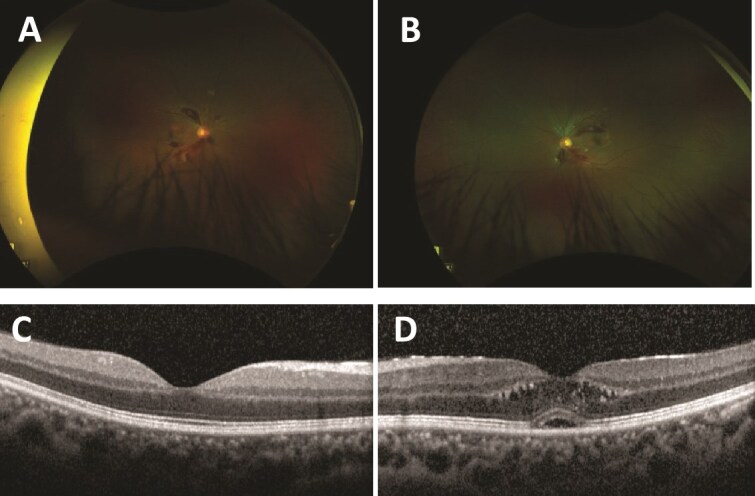
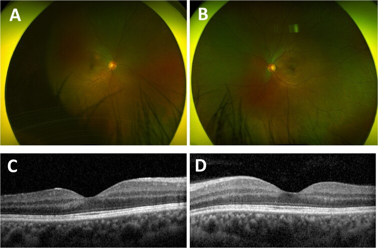
This case study reports a young man with hypertension who presented to eye casualty and was incidentally diagnosed with acute promyelocytic leukaemia (APML). Dilated fundus examination showed multiple retinal flame haemorrhages from the optic discs with scattered exudates bilaterally. Optical coherence tomography showed left cystoid macular oedema. He had preserved vision bilaterally and was systemically well with no constitutional symptoms. This case report is the first of our knowledge that reports on this unusual clinical presentation of APML. The patient successfully completed 29 days of induction all-trans-retinoic acid/arsenic trioxide therapy with repeat bone marrow biopsy showing haematological remission. Ophthalmic review 2 months later revealed resolution of retinal haemorrhages and cystoid macular oedema. Retinal haemorrhages may be the initial and only manifestation of APML. A full blood count should be considered in patients who present to eye casualty with haemorrhagic retinopathy of unknown aetiology.
Keywords: APML; acute promyelocytic leukaemia; leukaemic retinopathy; retinal haemorrhages.
Published by Oxford University Press and JSCR Publishing Ltd. © The Author(s) 2025.
Conflict of interest statement
None declared.
Figures
Similar articles
-
Acute promyelocytic leukaemia presenting as necrotising fasciitis of the perineum (Fournier gangrene).BMJ Case Rep. 2018 Dec 7;11(1):e226837. doi: 10.1136/bcr-2018-226837. BMJ Case Rep. 2018. PMID: 30567203 Free PMC article.
-
Molecular remission without blood product support using all-trans retinoic acid (ATRA) induction and combined arsenic trioxide/ATRA consolidation in a Jehovah's Witness with de novo acute promyelocytic leukaemia.Br J Haematol. 2000 Dec;111(4):1103-5. doi: 10.1046/j.1365-2141.2000.02480.x. Br J Haematol. 2000. PMID: 11167746
-
Isolated Ocular Manifestations in Chronic Myeloid Leukaemia.Cureus. 2021 Nov 10;13(11):e19450. doi: 10.7759/cureus.19450. eCollection 2021 Nov. Cureus. 2021. PMID: 34912600 Free PMC article.
-
Recent advances in acute promyelocytic leukaemia.F1000Res. 2017 Jul 28;6:1273. doi: 10.12688/f1000research.10736.1. eCollection 2017. F1000Res. 2017. PMID: 28794865 Free PMC article. Review.
-
Acute promyelocytic leukaemia:a review.Expert Opin Pharmacother. 2003 Aug;4(8):1379-92. doi: 10.1517/14656566.4.8.1379. Expert Opin Pharmacother. 2003. PMID: 12877645 Review.
References
Publication types
LinkOut - more resources
Full Text Sources
