. 2025 Apr 4:16:1548548.
doi: 10.3389/fimmu.2025.1548548. eCollection 2025.

CD47 regulates antigen modulation and red blood cell clearance following an incompatible transfusion

Affiliations

CD47 regulates antigen modulation and red blood cell clearance following an incompatible transfusion

Ryan P Jajosky et al. Front Immunol. .

Abstract

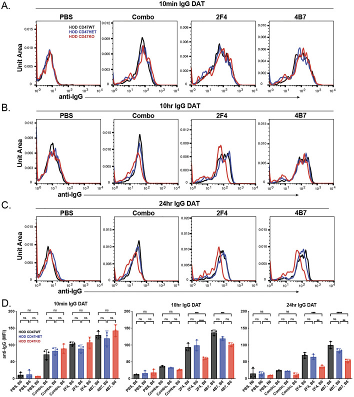
Red blood cell (RBC) alloantibodies can result in the rapid removal of incompatible RBCs following transfusion. However, antibody-mediated clearance of RBCs is not the inevitable outcome of an incompatible transfusion. Antibody engagement can also result in the modulation of the target antigen, often rendering RBCs resistant to antibody-mediated removal. Despite this, the factors that regulate antibody-induced RBC removal or antigen modulation remain incompletely understood. Given the ability of CD47 to regulate RBC survival in general, we examined the possible role of CD47 in governing antibody-mediated RBC clearance and antigen modulation. This was achieved by crossing the well-established HEL-OVA-Duffy (HOD) mouse model with CD47 knockout (KO) mice to generate offspring that express the HOD antigen and either WT (HOD CD47 WT), heterozygote (HOD CD47 HET) or KO (HOD CD47 KO) levels of CD47. Using the commonly employed anti-HEL immunization model, our results demonstrate that while antibody engagement of HOD CD47 WT RBCs resulted in rapid antigen modulation in the absence of detectable RBC clearance, antibody binding to HOD CD47 HET RBCs did result in detectable RBC removal despite similar rates and overall levels of antigen modulation. In contrast, despite accelerated clearance of HOD CD47 KO RBCs in the absence of anti-HEL antibodies, the rate of RBC removal and antigen modulation was enhanced in the presence of anti-HEL antibodies. Taken together, these results suggest a role for CD47 in regulating the overall consequence of an incompatible RBC transfusion.

Keywords: alloimmunization; antibodies; antigen modulation; incompatible transfusion; red blood cell.


Conflict of interest statement

The authors declare that the research was conducted in the absence of any commercial or financial relationships that could be construed as a potential conflict of interest.

Figures

Figure 1
Figure 1
CD47 knockout and heterozygous HOD RBCs possess reduced levels of CD47 with comparable HOD antigen levels. (A) Depictions of distinct HOD CD47 RBC populations used in this model. (B) Protocol for detecting HEL antigen on the surface of HOD CD47 RBCs. (C) Examination of HEL antigen expression on HOD CD47 WT, HOD CD47 heterozygous (HET) or HOD CD47 knockout (KO) shown by dot plots examining anti-HEL antibody (2F4 or 4B7) engagement by side scatter (SSC). (D) Evaluation of anti-HEL antibody binding over a range of concentrations toward HOD CD47 WT, HOD CD47 HET and HOD CD47 KO RBCs by histogram. (E) Quantification of histogram mean fluorescent intensity (MFI) values using a range of concentrations of anti-HEL antibodies individually or in combination toward HOD CD47 RBCs WT, HOD CD47 HET and HOD CD47 KO RBCs. (F) Histogram analysis of CD47 expression on HOD CD47 WT, HOD CD47 HET and HOD CD47 KO RBCs. (G) Quantification of histogram MFI values of CD47 antigen expression on HOD CD47 WT, HOD CD47 HET and HOD CD47 KO RBCs. ****p < 0.0001.
Figure 2
Figure 2
Anti-HEL antibodies enhance clearance of HOD CD47 heterozygous and knockout RBCs, while failing to impact HOD CD47 WT RBC removal. (A) Depiction of the experimental design using HOD CD47 WT, HOD CD47 heterozygous (HET) or HOD CD47 knockout (KO) RBCs transfused into non-immunized (PBS) or recipients immunized with one (2F4 or 4B7) or a combination (2F4 and 4B7) of anti-HEL antibodies. (B) Flow cytometry gating strategy used to detect DiI labeled HOD CD47 WT, HOD CD47 HET and HOD CD47 KO RBCs or DiO labeled B6 RBCs following transfusion into non-immunized or anti-HEL immunized recipients. (C) Survival of transfused HOD CD47 WT, HOD CD47 HET and HOD CD47 KO RBCs over time in comparison to B6 RBCs in the same recipient. (D) Normalized survival of transfused HOD CD47 WT, HOD CD47 HET and HOD CD47 KO RBCs over time in immunized recipients in comparison to the survival observed following transfusion of each population into non-immunized (PBS) recipients evaluated in parallel. Normalized survival for HOD CD47 KO not calculated for time points at which PBS controls reach 0% survival. (E) Bar graph representation of normalized survival of transfused HOD CD47 WT, HOD CD47 HET and HOD CD47 KO RBCs 4 hours following transfusion. ns = not significant. *p<0.05, **p < 0.01, ***p < 0.001.
Figure 3
Figure 3
The levels of anti-HEL antibodies decrease over time following transfusion of HOD CD47 WT, heterozygous and knockout RBCs into immunized recipients. Detection of antibody levels on the surface of HOD CD47 WT, HOD CD47 heterozygous (HET) or HOD CD47 knockout (KO) RBCs following transfusion into non-immunized (PBS) or recipients immunized with a single (2F4 or 4B7) or combination (combo - 2F4 and 4B7) of anti-HEL antibodies shown as histograms at 10 minutes (A), 10 hours (B), and 24 hours (C) following transfusion. (D) Quantification of histogram MFI values of antibody bound on HOD CD47 RBCs WT, HOD CD47 HET and HOD CD47 KO RBCs at 10 minutes, 10 hours, and 24 hours post-transfusion. ns = not significant. **p<0.01, ***p < 0.001, ****p < 0.0001.
Figure 4
Figure 4
Anti-HEL antibodies reduce HOD antigen levels on HOD CD47 WT, heterozygous and knockout RBCs. Detection of HOD antigen levels on the surface of HOD CD47 WT, HOD CD47 heterozygous (HET) or HOD CD47 knockout (KO) RBCs following transfusion into non-immunized (PBS) or recipients immunized with a single (2F4 or 4B7) or combination (combo - 2F4 and 4B7) of anti-HEL antibodies shown as histograms at 10 minutes (A), 10 hours (B), and 24 hours (C) following transfusion. (D) Quantificaiton of histogram MFI values of HOD antigen on HOD CD47 RBCs WT, HOD CD47 HET and HOD CD47 KO RBCs at 10 minutes, 10 hours, and 24 hours post-transfusion. ns = not significant. *p<0.05, **p<0.01.
Figure 5
Figure 5
Enhanced antibody-mediated clearance of HOD CD47 heterozygous and knockout RBCs requires Fcγ receptors. (A) Depiction of the experimental design using HOD CD47 WT, HOD CD47 heterozygous (HET) or HOD CD47 knockout (KO) RBCs transfused into WT or Fcγ receptor (FcγR) KO recipients that were either non-immunized (PBS) or immunized with a single (2F4 or 4B7) or combination (combo - 2F4 and 4B7) of anti-HEL antibodies. (B) Survival of transfused HOD CD47 WT, HOD CD47 HET and HOD CD47 KO RBCs over time in comparison to B6 RBCs in the same recipient following transfusion into either WT or FcγR KO recipients that were either non-immunized (PBS) or immunized with a single (2F4 or 4B7) or combination (combo - 2F4 and 4B7) of anti-HEL antibodies. (C) Normalized survival of transfused HOD CD47 WT, HOD CD47 HET and HOD CD47 KO RBCs over time in comparison to B6 RBCs in the same recipient following transfusion into either WT or FcγR KO recipients that were either non-immunized (PBS) or immunized with a single (2F4 or 4B7) or combination (combo - 2F4 and 4B7) of anti-HEL antibodies. Normalized survival for HOD CD47 KO not calculated for time points at which PBS controls reach 0% survival. (D) Bar graph representation of normalized survival of transfused HOD CD47 WT, HOD CD47 HET and HOD CD47 KO RBCs in comparison to B6 RBCs in the same recipient following transfusion into either WT or FcγR KO recipients that were either non-immunized (PBS) or immunized with a single (2F4 or 4B7) or combination (combo - 2F4 and 4B7) of anti-HEL antibodies 4 hours following transfusion. ns = not significant. *p<0.05, ***p < 0.001.
Figure 6
Figure 6
The levels of bound anti-HEL antibodies decrease over time following transfusion of HOD CD47 WT, heterozygous and knockout RBCs into WT and Fcγ receptor immunized recipients. Detection of antibody levels on the surface of HOD CD47 WT, HOD CD47 heterozygous (HET) or HOD CD47 knockout (KO) RBCs following transfusion into WT or Fcγ receptor (FcγR) KO recipients that were either non-immunized (PBS) or immunized with a combination (combo - 2F4 and 4B7) of anti-HEL antibodies shown as histograms at 10 minutes (A), 10 hours (B), and 24 hours (C) following transfusion. (D) Quantification of histogram mean fluorescence intensity (MFI) values of antibody bound on HOD CD47 WT, HOD CD47 HET and HOD CD47 KO RBCs following transfusion into either WT or FcγR KO recipients that were either non-immunized (PBS) or immunized with a combination (combo - 2F4 and 4B7) of anti-HEL antibodies, 10 minutes, 10 hours, and 24 hours post-transfusion. ns = not significant. *p<0.05, ***p < 0.001.
Figure 7
Figure 7
Anti-HEL antibodies reduce levels of HOD without changing Ter119 levels or inducing complement deposition. (A) HOD antigen levels on the surface of HOD CD47 WT, HOD CD47 heterozygous (HET) or HOD CD47 knockout (KO) RBCs following transfusion into WT or Fcγ receptor (FcγR) KO recipients that were either non-immunized (PBS) or immunized with a combination (combo - 2F4 and 4B7) of anti-HEL antibodies shown as histograms at 10 minutes and 10 hours post-transfusion. (B) Levels of Ter119 on the surface of HOD CD47 WT, HOD CD47 HET or HOD CD47 KO RBCs following transfusion into WT or FcγR KO recipients that were either non-immunized (PBS) or immunized with a combination (combo - 2F4 and 4B7) of anti-HEL antibodies shown as histograms at 1 hour and 24 hours post-transfusion. (C) Measurement of C3 deposition on the surface of HOD CD47 WT, HOD CD47 HET or HOD CD47 KO RBCs following transfusion into WT or FcγR KO recipients that were either non-immunized (PBS) or immunized with a combination (combo - 2F4 and 4B7) of anti-HEL antibodies shown as histograms 24 hours post-transfusion. ns = not significant.

Similar articles

References

    1. Yazdanbakhsh K, Ware RE, Noizat-Pirenne F. Red blood cell alloimmunization in sickle cell disease: pathophysiology, risk factors, and transfusion management. Blood. (2012) 120:528–37. doi: 10.1182/blood-2011-11-327361 - DOI - PMC - PubMed
    1. Pirenne F, Floch A, Diop S. Alloimmunisation against red blood cells in sickle cell disease: transfusion challenges in high-income and low-income countries. Lancet Haematol. (2023) 10:e468–76. doi: 10.1016/S2352-3026(23)00066-2 - DOI - PubMed
    1. Arthur CM, Stowell SR. The development and consequences of red blood cell alloimmunization. Annu Rev Pathol. (2023) 18:537–64. doi: 10.1146/annurev-pathol-042320-110411 - DOI - PMC - PubMed
    1. Yazdanbakhsh K. Mechanisms of sickle cell alloimmunization. Transfus Clin Biol. (2015) 22:178–81. doi: 10.1016/j.tracli.2015.05.005 - DOI - PMC - PubMed
    1. Jajosky RP, Wu SC, Zheng L, Jajosky AN, Jajosky PG, Josephson CD, et al. . ABO blood group antigens and differential glycan expression: Perspective on the evolution of common human enzyme deficiencies. iScience. (2023) 26:105798. doi: 10.1016/j.isci.2022.105798 - DOI - PMC - PubMed

LinkOut - more resources