

Application of a novel thermal/pH-responsive antibacterial paeoniflorin hydrogel crosslinked with amino acids for accelerated diabetic foot ulcers healing
- PMID: 40255581
- PMCID: PMC12008599
- DOI: 10.1016/j.mtbio.2025.101736
Application of a novel thermal/pH-responsive antibacterial paeoniflorin hydrogel crosslinked with amino acids for accelerated diabetic foot ulcers healing
Abstract
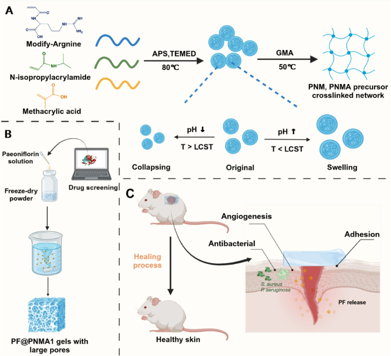
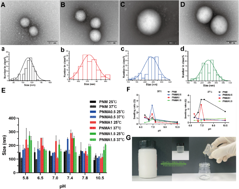
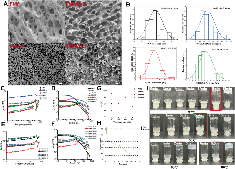
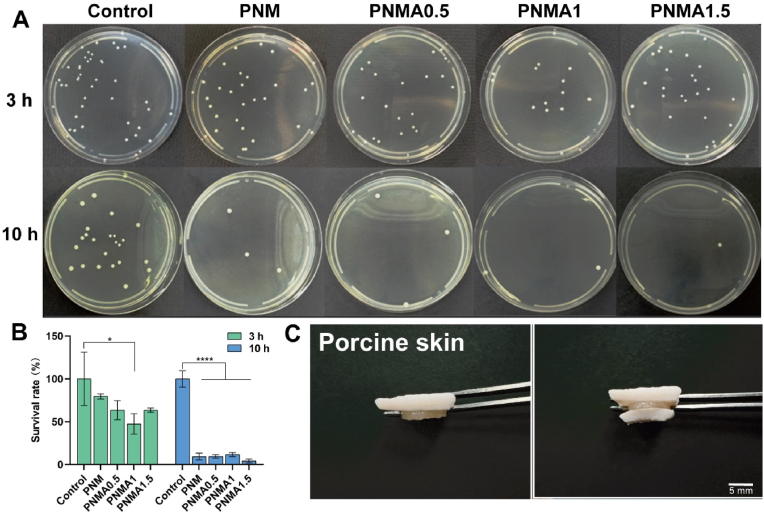
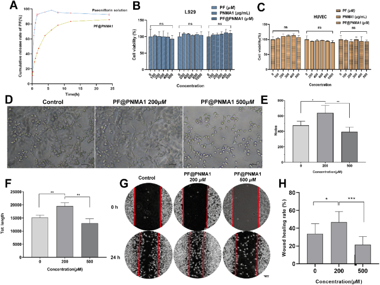
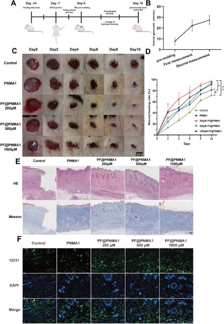
Diabetic foot ulcers (DFUs), a severe and common complication of diabetes, present significant treatment challenges due to the limitations of conventional dressings, such as poor mechanical properties, bioactivity, and limited functionality, which hinder fast and effective wound healing. To address these issues, we developed a novel natural amino acid-based hydrogel loaded with paeoniflorin (PF@PNMA1) and comprehensively evaluated its properties and functions. The nanogel particles (NGs) were synthesized via emulsion polymerization using N-isopropylacrylamide (NIPAM), methacrylic acid (MAA), and chemically modified arginine (MArg). The poly(NIPAM-co-MAA) (PNM) and poly(NIPAM-co-MAA-co-MArg) (PNMA) gels were prepared by functionalizing the NGs with glycidyl methacrylate (GMA). The different concentrations of amino acids were added to explore the optimal mechanical properties of the gel. Through the rheological measurement, we found that PNMA1 gel has good ductile properties with a critical strain up to about 63 %. At the same time, we also verified its antibacterial activity and found that the viability of bacteria decreased to 47.46 % after 3 h. Preliminary tests using network pharmacology and molecular docking confirmed the therapeutic potential of PF for DFUs. The PF@PNMA1 gel demonstrated excellent biocompatibility, and in vivo experiments revealed its effectiveness in promoting angiogenesis and wound healing. After 10 days, the wound healing rate was 25.6 % higher than that of the control group. The PF@PNMA1 shows great potential as an effective therapy for DFUs treatment.
Keywords: Amino acids; Crosslinker; Diabetic foot ulcers; Paeoniflorin; Wound dressing.
© 2025 The Authors. Published by Elsevier Ltd.
Conflict of interest statement
The authors declare that they have no known competing financial interests or personal relationships that could have appeared to influence the work reported in this paper.
Figures

Similar articles
-
Glucose-responsive multifunctional metal-organic drug-loaded hydrogel for diabetic wound healing.Acta Biomater. 2022 Mar 1;140:206-218. doi: 10.1016/j.actbio.2021.11.043. Epub 2021 Dec 5. Acta Biomater. 2022. PMID: 34879294
-
Dual-functional hybrid quaternized chitosan/Mg/alginate dressing with antibacterial and angiogenic potential for diabetic wound healing.J Orthop Translat. 2021 Aug 12;30:6-15. doi: 10.1016/j.jot.2021.07.006. eCollection 2021 Sep. J Orthop Translat. 2021. PMID: 34466384 Free PMC article.
-
The role of gel wound dressings loaded with stem cells in the treatment of diabetic foot ulcers.Am J Transl Res. 2021 Dec 15;13(12):13261-13272. eCollection 2021. Am J Transl Res. 2021. PMID: 35035674 Free PMC article. Review.
-
Novel Diabetic Foot Wound Dressing Based on Multifunctional Hydrogels with Extensive Temperature-Tolerant, Durable, Adhesive, and Intrinsic Antibacterial Properties.ACS Appl Mater Interfaces. 2021 Jun 16;13(23):26770-26781. doi: 10.1021/acsami.1c05514. Epub 2021 Jun 7. ACS Appl Mater Interfaces. 2021. PMID: 34096258
-
Research advances in smart responsive-hydrogel dressings with potential clinical diabetic wound healing properties.Mil Med Res. 2023 Aug 23;10(1):37. doi: 10.1186/s40779-023-00473-9. Mil Med Res. 2023. PMID: 37608335 Free PMC article. Review.
Cited by
-
Exo-hydrogel therapy: a revolutionary approach to managing diabetic complications.J Nanobiotechnology. 2025 Aug 11;23(1):558. doi: 10.1186/s12951-025-03621-6. J Nanobiotechnology. 2025. PMID: 40790200 Free PMC article. Review.
References
-
- Theocharidis G., Yuk H., Roh H., Wang L., Mezghani I., Wu J., Kafanas A., Contreras M., Sumpio B., Li Z., Wang E., Chen L., Guo C., Jayaswal N., Katopodi X., Kalavros N., Nabzdyk C., Vlachos I., Veves A., Zhao X. A strain-programmed patch for the healing of diabetic wounds. Nat Biomed Eng. 2022;6(10):1118–1133. - PubMed
-
- Boyko E.J., Zelnick L.R., Braffett B.H., Pop-Busui R., Cowie C.C., Lorenzi G.M., Gubitosi-Klug R., Zinman B., de Boer I.H. Risk of foot ulcer and lower-extremity amputation among participants in the diabetes control and complications trial/epidemiology of diabetes interventions and complications study. Diabetes Care. 2022;45(2):357–364. - PMC - PubMed
LinkOut - more resources
Full Text Sources
