. 2025 Apr 5:32:101734.
doi: 10.1016/j.mtbio.2025.101734. eCollection 2025 Jun.

Development of a decellularized extracellular matrix-derived wet adhesive for sustained drug delivery and enhanced wound healing

Affiliations

Development of a decellularized extracellular matrix-derived wet adhesive for sustained drug delivery and enhanced wound healing

Xinming Wang et al. Mater Today Bio. .

Abstract

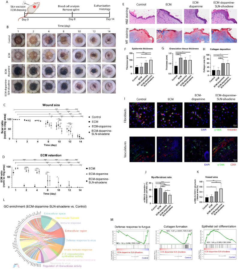
Complete tissue recovery following traumatic injury remains a major clinical challenge. While tissue adhesives show promise for managing traumatic injuries, developing materials with robust wet adhesion and high biocompatibility remains difficult. Decellularized extracellular matrix (ECM)-derived materials are widely utilized in tissue engineering due to their superior biocompatibility and bioactivity. In this study, a wet adhesive is developed by functionalizing ECM with dopamine. The resulting ECM-dopamine exhibits strong wet adhesion and excellent biocompatibility. Furthermore, ECM-dopamine can be engineered into a drug delivery platform for small agents and macromolecules. Solid lipid nanoparticles (SLNs) are incorporated into ECM-dopamine to enable sustained release of small molecules. The ECM-dopamine-SLN system ensures sustained drug release for at least one week upon adhesion to target tissues. ECM-dopamine-SLN loaded with antimicrobials accelerates wound healing and promotes angiogenesis by modulating the inflammatory response in a mouse skin excision model. Additionally, ECM-dopamine can deliver bioactive macromolecules to injured tissue. ECM-dopamine loaded with insulin-like growth factor-1 promotes skeletal muscle regeneration in a mouse volumetric muscle loss model, likely through the modulation of M2-like macrophage polarization. The dual functionality of ECM-dopamine as both a wet adhesive and a drug delivery platform offers significant potential for regenerative medicine applications.

Keywords: Drug delivery; Extracellular matrix derived-material; Volumetric muscle loss; Wet adhesion; Wound healing.


Conflict of interest statement

The authors declare that they have no known competing financial interests or personal relationships that could have appeared to influence the work reported in this paper.

Figures

Image 1
Graphical abstract
Fig. 1
Fig. 1
ECM-dopamine fabrication and characterization. (A) Schematic representation of the experimental design. Dopamine molecules were conjugated to ECM via EDC/NHS chemistry. ECM-dopamine exhibited strong wet adhesion to tissue. Drugs incorporated into ECM-dopamine were capable of sustained release at the wound site. (B) The ECM solution turned from opaque white to dark brown following dopamine modification. ECM-dopamine formed an aerogel-like substrate after lyophilization. (C) FTIR spectroscopy revealed additional peaks around 2350 cm−1 and 1740 cm−1 in ECM-dopamine. (D) XRD analysis showed a sharp peak indicative of crystalline microstructure in ECM samples, while ECM-dopamine exhibited an amorphous microstructure. (E) Optical microscopy revealed entangled fibers in dry ECM-dopamine. Upon rehydration, ECM-dopamine remained intact, while ECM partially dissolved. (F) SEM showed a similar microstructure for both ECM and ECM-dopamine, with ECM-dopamine primarily consisting of fibers. (G) ECM-dopamine showed a broader distribution of fiber diameters compared to ECM. The average fiber diameter of ECM-dopamine was about 1.7 μm, which is significantly thicker than that of ECM. (H) Tissue-to-tissue adhesion in the air and underwater was also examined. Porcine muscle, fat, and skin were adhered to the finger using ECM-dopamine in both conditions. (I) The release rate of exogenous proteins from ECM-dopamine was assessed using BSA, with ECM-dopamine (without BSA) as the control. ECM-dopamine released proteins at a slower rate compared to ECM within 24 h. (J) Over a period from day 3 to day 9 in an infinite dilution experiment, ECM-dopamine released significantly more proteins compared to ECM. (n = 3 in panel I and J. T-test applied for panel G. ∗∗∗∗p < 0.0001. Data are presented as mean ± SD.). (For interpretation of the references to color in this figure legend, the reader is referred to the Web version of this article.)
Fig. 2
Fig. 2
ECM-dopamine revealed strong wet adhesion and good cell compatibility. (A) The adhesive force of ECM-dopamine was examined underwater. Dopamine molecules were conjugated to ECM via different amounts of EDC/NHS. (B) The shear strength was first measured. Increasing the concentration of EDC/NHS resulted in reduced adhesive strength. (C) Next, the pull strength was measured. A negative correlation was observed between the amount of EDC/NHS used and the adhesive strength. (D) Young's modulus was measured using atomic force microscopy. (E) ECM-dopamine exhibited a lower Young's modulus compared to ECM. (F) Dopamine modification increased the adhesive force by approximately 8 times. (G) Lyophilized ECM-dopamine can be degraded by collagenase or pepsin within 24 h under physiological conditions. (H) Protein release during collagenase-induced ECM-dopamine degradation was examined. A relatively consistent protein release was observed over 8 h. (I) The hemocompatibility of ECM-dopamine was examined using fresh sheep blood. ECM-dopamine exhibited similar (J) hemolytic and (K) hemostatic effects on the blood compared to the negative controls. (L) Next, cell compatibility was assessed using L929 cells. Large clusters of L929 cells were observed growing on ECM-dopamine. (M) Cells were cultured in the presence of ECM-dopamine for 2 days and imaged via fluorescent microscopy. (N) No significant difference in cell density was observed between the control and the ECM-dopamine group. (n = 3, One-way ANOVA and Tukey's test applied for panel B, C, and K, t-test applied for panel E, F, and N. ∗p < 0.05, ∗∗p < 0.01, ∗∗∗p < 0.001, ∗∗∗∗p < 0.0001. Data are presented as mean ± SD.)
Fig. 3
Fig. 3
ECM-dopamine-SLN facilitates sustained release of drugs into the target organ. (A) SLNs were prepared by oil-in-water emulsion and sonification of the drug-solid lipids mixture at 60 °C. FTIR spectroscopy indicated amplified peaks at 2920 cm−1, 1735 cm−1, and 940 cm−1 in ECM-dopamine-SLN samples. (B) The incorporation of SLNs did not alter the meshwork structure of ECM-dopamine; however, SLNs appeared as 300 nm white dots adhered to the ECM-dopamine. (C) SLNs containing fluorescein were utilized to assess the SLN release kinetics. ECM-dopamine-SLN slowly released FITC into the environment over 24 h. (D) A sustained release of FITC was observed over 14 days in an infinite dilution experiment. (E) The release rate of SLNs was modulated by genipin crosslinking. Lyophilized ECM-dopamine-SLN containing FITC was treated with 0.5 %, 0.05 %, or 0.005 % genipin buffer before measuring the FITC release kinetics. The FITC release rate was inversely correlated with genipin concentration. (F) SLN retention was investigated in vivo, with ECM-dopamine-SLN containing biotin-PEG-Cy5 adhered to the liver surface. The distribution of biotin-PEG-Cy5 in the body was examined via live imaging on days 1, 3, and 7 post implantation, along with an assessment of major organ distribution on day 7. (G) The ECM-dopamine-SLN group had higher total flux compared to the SLNs group on days 3 and day 7. In addition, the Cy5 signal faded slower in the ECM-dopamine-SLN group. (H) Biotin-PEG-Cy5 primarily accumulated in the liver in both SLNs and ECM-dopamine-SLN groups, with the liver in the ECM-dopamine-SLN group exhibiting the highest total flux. (n = 3, two-way ANOVA and Tukey's test applied, ∗p < 0.05, ∗∗p < 0.01, ∗∗∗p < 0.001, ∗∗∗∗p < 0.0001. Data are presented as mean ± SD.)
Fig. 4
Fig. 4
ECM-dopamine-SLN loaded with antimicrobial promoted skin wound healing. (A) Experimental design. A 10 mm silicone ring was used to splint the wound after skin excision. ECM, ECM-dopamine, or ECM-dopamine-SLN-silvadene was applied 6 h after excision. Silicone rings were removed, and blood cell counts were analyzed on day 8. All mice were euthanized on day 14 for histological analysis. (B) Wound healing was regularly monitored through imaging. (C) Wound size over 14 days. ECM-dopamine and ECM-dopamine-SLN-silvadene treatments reduced wound sizes on day 8 compared to day 1. After silicone ring removal, the ECM-dopamine-SLN-silvadene group demonstrated significantly accelerated wound healing compared to other groups. (D) ECM retention was also examined. Non-modified ECM was largely absent by day 2 and barely detectable thereafter, while ECM-dopamine and ECM-dopamine-SLN-silvadene remained visible throughout the entire healing process. (E) Hematoxylin and eosin staining, as well as Masson's trichrome staining were performed to evaluate wound healing process. (F) ECM-dopamine increased epithelial thickness compared to the control, with further improvements observed in the ECM-dopamine-SLN-silvadene group. (G) ECM and its derivatives increased granulation tissue thickness in comparison to the control. (H) Collagen deposition, as indicated by MT staining, was notably higher in ECM-dopamine groups (with or without silvadene) compared to both the ECM-only and control groups. (I) Myofibroblasts and vasculatures were examined via immunostaining. (J) The ECM-dopamine-SLN-silvadene group exhibited the lowest myofibroblast to fibroblast ratio. Besides, ECM-dopamine lowered the myofibroblast ratio compared to the control and ECM-only treatments. (K) Both ECM-dopamine and ECM-dopamine-SLN-silvadene increased vessel area compared to the control, with ECM-dopamine-SLN-silvadene treatment showing a greater vessel area than ECM treatment. (L) mRNA sequencing revealed that ECM-dopamine-SLN-silvadene altered the transcription of genes related to extracellular matrix remodeling and inflammation, as shown by Gene Ontology analysis. (M) Gene Set Enrichment Analysis demonstrated that ECM-dopamine-SLN-silvadene suppressed the transcription of genes associated with fungus defense and collagen production. In contrast, ECM-dopamine-SLN-silvadene facilitated the transcription of genes related to epithelial cell differentiation. (n = 6 in panel C and D, two-way ANOVA and Tukey's test applied. n = 6 in panel F, G, H, J and K, one-way ANOVA and Tukey's test applied. ∗p < 0.05, ∗∗p < 0.01, ∗∗∗p < 0.001, ∗∗∗∗p < 0.0001. Data are presented as mean ± SD.)
Fig. 5
Fig. 5
Blood cell analysis on day 8 post-skin injury. (A) Red blood cells and platelets were analyzed via hydrodynamically focused impedance measurements. (B) White blood cells were analyzed using fluorescent flow cytometry. ECM treatments did not affect (C) white blood cell and (D) red blood cell counts. (E) Platelet counts were elevated in both ECM-dopamine-SLN-silvadene and ECM-only groups compared to the control. (F) The platelet-large cell ratio decreased in these same groups relative to the control. (G) Neutrophil, (H) lymphocyte, and (I) eosinophil counts did not differ significantly across the treatment groups. (J) However, ECM-dopamine-SLN-silvadene increased monocyte count compared to the control and ECM-only groups. (n = 6 in panel C, D, E, F, G, H, I and J, one-way ANOVA and Tukey's test applied, ∗p < 0.05, ∗∗p < 0.01, ∗∗∗p < 0.001. Data are presented as mean ± SD.). (For interpretation of the references to color in this figure legend, the reader is referred to the Web version of this article.)
Fig. 6
Fig. 6
ECM-dopamine loaded with insulin-like growth factor improved skeletal muscle repair. (A) Experimental design. Volumetric muscle loss was established by surgical ablation of tibialis anterior muscle using a 3 mm biopsy punch. ECM samples were implanted into the defect. (B) The grip strength of rear limbs was measured using a grip strength meter. (C) Volumetric muscle loss resulted in significantly reduced peak force on day 3. ECM-dopamine-IGF treatment promoted force recovery compared to the control on day 7 after surgery. (D) Mice gait was evaluated using a video-based gait analysis system. (E) ECM-dopamine and ECM-dopamine-IGF treatments significantly increased rear right foot pressure compared to the control on day 7. (F) Myocyte diameter and scar formation on day 21 post-surgery were examined using H&E staining and MT staining. (G) Both ECM and ECM-dopamine treatments increased myocyte diameter in the injury area compared to the control, with ECM-dopamine-IGF group exhibiting the highest myocyte diameter. (H) Fibrotic area was significantly reduced in the ECM-dopamine group compared to the control. (I) The population of various immune cells was analyzed using flow cytometry. (J) ECM and ECM-dopamine-IGF treatments promoted the T cell number compared to the control. (K) The percentage of T cells within CD45+ immune cells was lower in ECM-dopamine and ECM-dopamine-IGF groups than in the other groups. (L) Both ECM-dopamine and ECM-dopamine-IGF treatments increased the number of M2-like macrophages compared to other treatments. (M) Only ECM-dopamine group exhibited a higher percentage of M2-like macrophages compared to the control. (n = 5 in panel E, G, H, J, K, L and M, one-way ANOVA and Tukey's test applied, n = 5 in panel C, two-way ANOVA and Tukey's test applied. ∗p < 0.05, ∗∗p < 0.01, ∗∗∗p < 0.001, ∗∗∗∗p < 0.0001. Data are presented as mean ± SD.)

Similar articles

Cited by

References

    1. Prevaldi C., Paolillo C., Locatelli C., Ricci G., Catena F., Ansaloni L., Cervellin G. Management of traumatic wounds in the emergency department: position paper from the academy of emergency medicine and Care (AcEMC) and the world society of emergency surgery (WSES) World J. Emerg. Surg. Jun. 2016;11(1):1–6. doi: 10.1186/S13017-016-0084-3/TABLES/4. - DOI - PMC - PubMed
    1. Nair A., Dahiya A., Yadav P., Sharma N., Butola B.S. Materials for the management of traumatic Wounds: a descriptive review. Eur. Polym. J. Jan. 2025;222 doi: 10.1016/J.EURPOLYMJ.2023.112475. - DOI
    1. Moriscot A., Miyabara E.H., Langeani B., Belli A., Egginton S., Bowen T.S. Firearms-related skeletal muscle trauma: pathophysiology and novel approaches for regeneration. npj Regenerative Medicine 2021. Mar. 2021;6(1):1–17. doi: 10.1038/s41536-021-00127-1. 6:1. - DOI - PMC - PubMed
    1. Fan H., Gong J.P. Bioinspired underwater adhesives. Adv. Mater. Nov. 2021;33(44) doi: 10.1002/adma.202102983. - DOI - PubMed
    1. Frantzis P. Durability of adhesive joints made underwater. J. Mater. Civ. Eng. Oct. 2008;20(10):635–639. doi: 10.1061/(ASCE)0899-1561(2008)20:10(635). - DOI

LinkOut - more resources