. 2025 Apr 15:20:4777-4802.
doi: 10.2147/IJN.S508171. eCollection 2025.

Gold and Silver Nanoparticles Efficiently Modulate the Crosstalk Between Macrophages and Cancer Cells

Affiliations

Gold and Silver Nanoparticles Efficiently Modulate the Crosstalk Between Macrophages and Cancer Cells

Dóra Izabella Adamecz et al. Int J Nanomedicine. .

Abstract

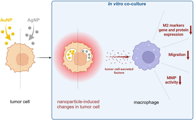
Background: Macrophages, polarized into pro-inflammatory M1 or anti-inflammatory M2 states, are essential cellular elements of innate immunity. In the tumor microenvironment, owing to a paracrine manipulative program by cancerous cells, tumor-associated macrophages (TAMs) evolve, which can shift between M1-like and M2-like phenotypes. Since it is fairly unknown how the promising anticancer agents, silver (AgNPs) and gold nanoparticles (AuNPs) affect the bidirectional communication and reprogramming in the tumor stroma, we examined the behavior, the tumor-supporting functions, and the expression of polarization and functional marker genes of TAMs to reveal how these are modulated upon interaction with nanoparticle-exposed cancer cells.

Methods: We established co-cultures of murine immortalized J774 or primary bone marrow-derived macrophages with 4T1 breast cancer cells treated with AuNPs or AgNPs or with none of the nanoparticles. We assessed the expression of macrophage polarization and functional markers using RT-qPCR and Proteome Profiler Array and evaluated macrophage migration and matrix metalloproteinase activity by specific assays.

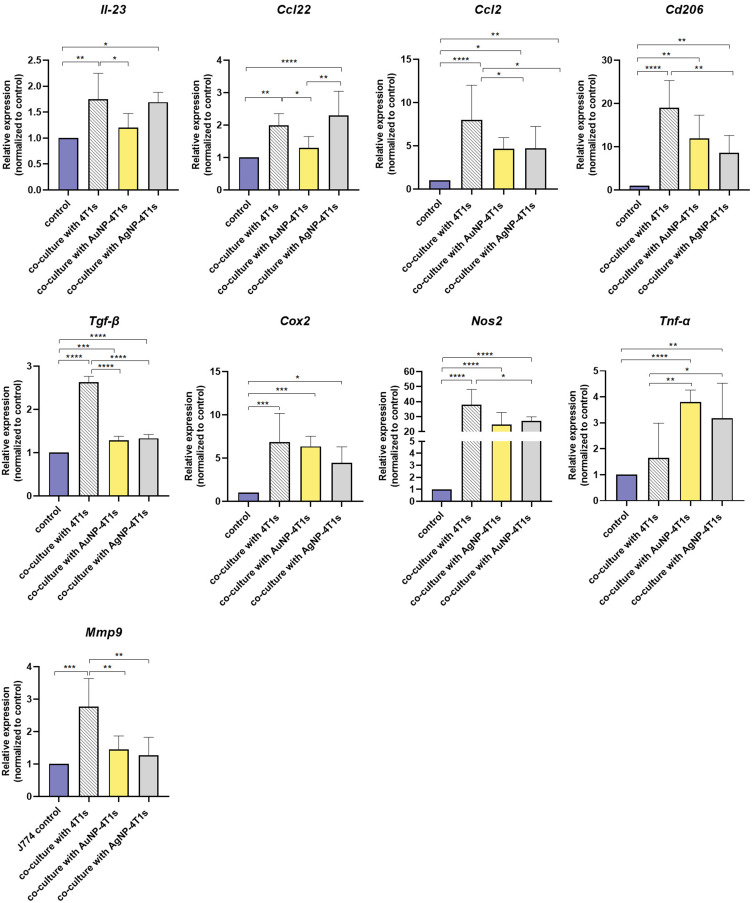
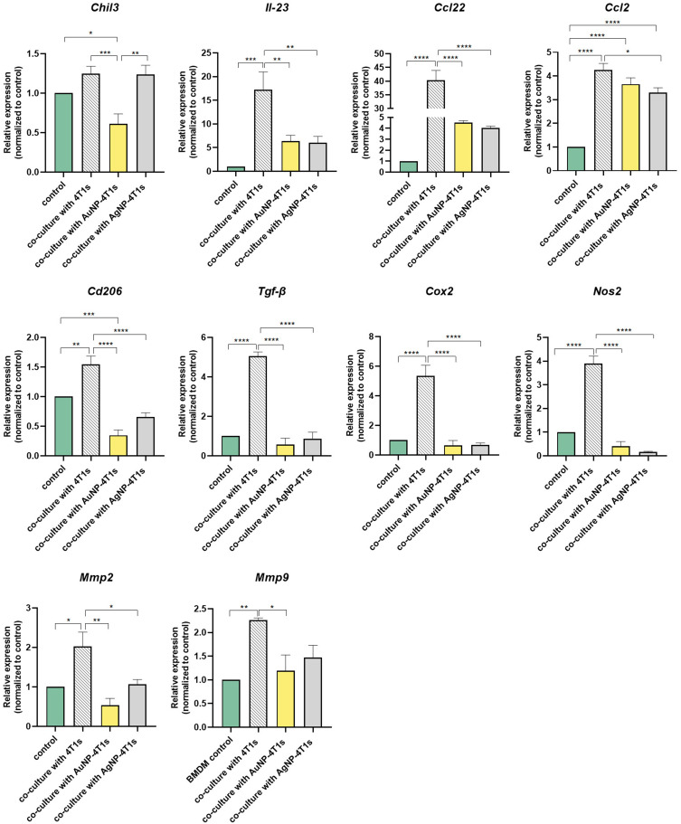
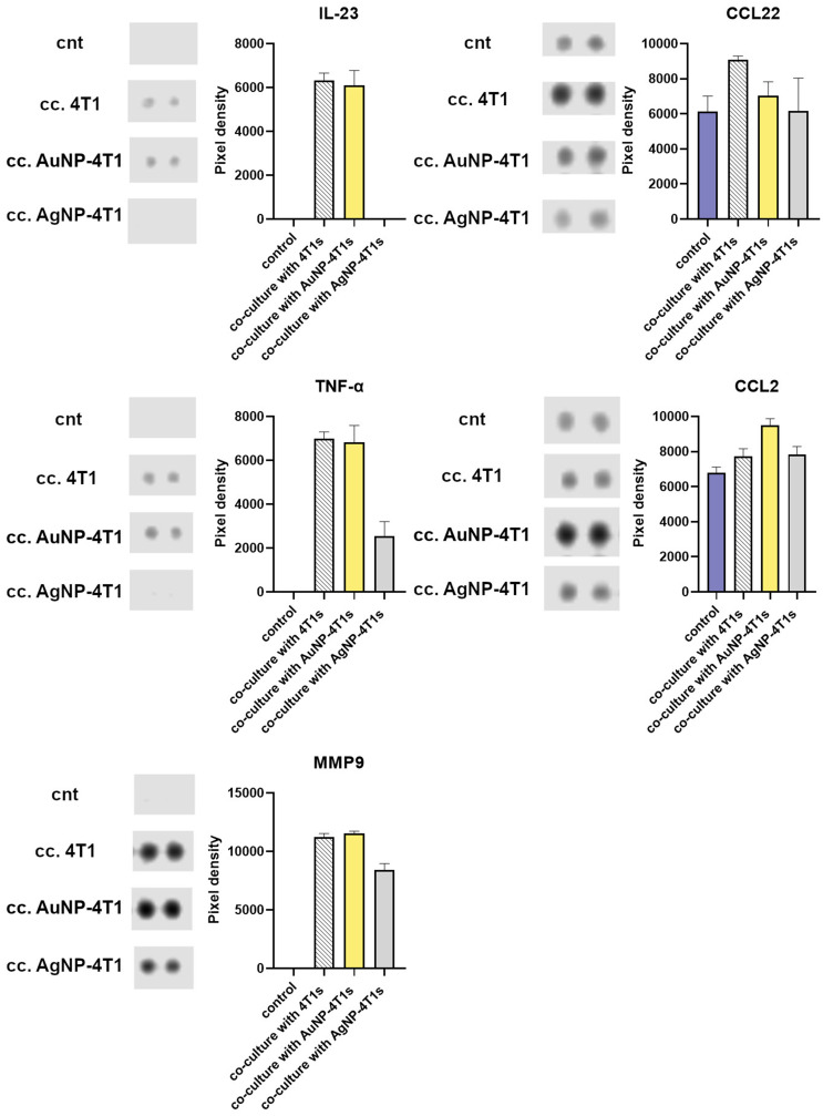
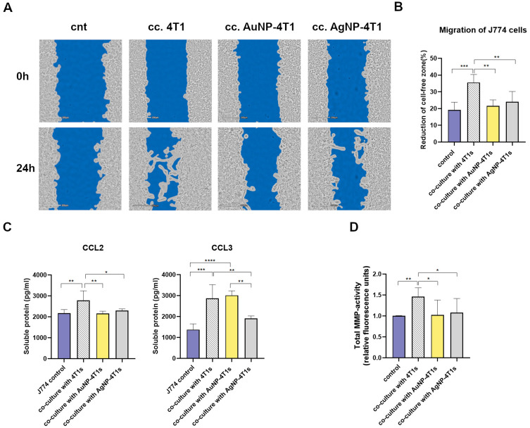
Results: Protein and mRNA levels of most examined factors - except tumor necrosis factor-alpha - such as C-C-motif chemokine ligands 2 and 22, interleukin-23, inducible nitric oxide synthase, cyclooxygenase-2, the macrophage mannose receptor CD206, transforming growth factor-beta, and chitinase-like-3 protein decreased, and the expression of polarization markers revealed a shift towards M1-like phenotype in macrophages co-cultured with AgNP- or AuNP-treated 4T1 cells. Both nanoparticle treatments reduced the levels and activity of cell migration-related factors, such as C-C motif chemokine ligand 3, matrix metalloproteinases, and suppressed macrophage migration.

Conclusion: Both AuNPs and AgNPs showed a remarkable ability to influence macrophage-cancer cell communication, suppressed indirectly M2-like TAM polarization, and perturbed the migration behavior of TAMs that is critical for tumor invasion, indicating modulated immunological functions and debilitated cancer-promoting capabilities of TAMs in this microenvironment.

Keywords: macrophage-tumor cell co-culture; metal nanoparticles; paracrine; tumor microenvironment; tumor-associated macrophage.


Conflict of interest statement

The authors declare that the research was conducted in the absence of any commercial or financial relationships that could be construed as a potential conflict of interest.

Figures

None
Graphical abstract
Figure 1
Figure 1
Physicochemical characterization of the synthesized gold (AuNPs) and silver (AgNPs) nanoparticles. (A) Representative transmission electron microscopic images of AuNPs and AgNPs, (B) size distribution by dynamic light scattering of metal nanoparticles, and (C) ultraviolet-visible spectra obtained of AuNPs and AgNP samples.
Figure 2
Figure 2
Cytotoxic effects and internalization of gold (AuNPs) and silver (AgNPs) nanoparticles by 4T1 breast cancer cells. (A) The cytotoxicity of metal nanoparticles on 4T1 cells was assessed using a viability assay, and (B) internalization of AuNPs and AgNPs in 4T1 cancer cells was verified by transmission electron microscopic analysis (AuNP: left panels, AgNP: right panels). (**P=0.0099, ****P<0.0001, One-way ANOVA; Dunnett’s multiple comparisons test, values represent mean ± standard deviation (SD)).
Figure 3
Figure 3
Relative expression levels of various polarization and functional marker genes in J774 macrophages. Gene expression was examined in monocultured J774 cells (J774 control), as well as in J774 cells co-cultured with untreated, or gold (AuNP)-, or silver (AgNP) nanoparticle-treated 4T1 breast cancer cells, determined by RT-qPCR (control: monoculture of J774 cells, co-culture with 4T1s: J774 cells co-cultured with untreated 4T1 cells, co-culture with AuNP-treated 4T1s: J774 cells co-cultured with AuNP-treated 4T1 cells, co-culture with AgNP-4T1s: J774 cells co-cultured with AgNP-treated 4T1 cells).
Figure 4
Figure 4
Relative expression levels of various polarization and functional marker genes in primary bone-marrow-derived macrophage (BMDM) cells maintained in monoculture (control) or in co-culture with untreated, gold (AuNP)-, or silver (AgNP) nanoparticle-treated 4T1breast cancer cells determined by RT-qPCR (control: monoculture of BMDM cells, co-culture with 4T1s: BMDM cells co-cultured with untreated 4T1 cells, co-culture with AuNP-treated 4T1s: BMDM cells co-cultured with AuNP-treated 4T1 cells, co-culture with AgNP-4T1s: BMDM cells co-cultured with AgNP-treated 4T1 cells).
Figure 5
Figure 5
Relative amounts of selected cytokines, chemokines, and soluble factors secreted by J774 macrophages upon exposure to cancer cells and metal nanoparticles. The relative quantity of these proteins was assessed in J774 supernatants collected from macrophage monocultures or from co-cultures with 4T1 breast cancer cells by Proteome Profiler Array. Representative pictures and quantification of target dots based on chemiluminescence intensity are presented. Values represent mean ± standard deviation (SD) calculated from the intensity of two dots/target. (cnt: monoculture of J774s control, cc. 4T1: J774s co-cultured for 48 hours with untreated 4T1 cells, cc. AuNP-4T1: J774s co-cultured for 48 hours with AuNP-treated 4T1 cells, cc. AgNP-4T1: J774s co-cultured for 48 hours with AgNP-treated 4T1 cells).
Figure 6
Figure 6
Relative amounts of selected cytokines, chemokines, and soluble factors secreted by primary bone-marrow-derived macrophages upon exposure to cancer cells and metal nanoparticles. The relative quantity of these proteins was assessed in primary bone-marrow-derived macrophage (BMDM) supernatants collected from macrophage monocultures or from co-cultures with 4T1 breast cancer cells by Proteome Profiler Array. Representative pictures and quantification of target dots based on chemiluminescence intensity are presented. Values represent mean ± standard deviation (SD) calculated from the intensity of two dots/target. (cnt: monoculture of BMDMs control, cc. 4T1: BMDMs co-cultured for 48 hours with untreated 4T1 cells, cc. AuNP-4T1: BMDMs co-cultured for 48 hours with AuNP-treated 4T1 cells, cc. AgNP-4T1: BMDMs co-cultured for 48 hours with AgNP-treated 4T1 cells).
Figure 7
Figure 7
The presence of cancer cells and also nanoparticles affects the migratory capacity of J774 macrophages. (A) Representative images and (B) the quantified migratory capacity of J774 cells after 24 h co-culturing with 4T1 breast cancer cells and nanoparticle treatments. (C) Quantity of chemokines C-C motif chemokine ligand 2 (CCL2) and C-C motif chemokine ligand 3 (CCL3) measured by Enzyme-Linked Immunosorbent Assay (ELISA) from supernatants of J774 cells co-cultured for 24 hours with untreated or metal nanoparticle-treated 4T1 cancer cells. (D) The activity of J774-secreted matrix metalloproteinase (MMP) enzymes was detected by Total MMP activity assay from 24 h co-culture supernatants (control: monoculture of J774 cells, co-culture with 4T1s: J774 cells co-cultured for 24 hours with untreated 4T1 cells, co-culture with AuNP-treated 4T1s: J774 cells co-cultured for 24 hours with AuNP-treated 4T1 cells, co-culture with AgNP-4T1s: J774 cells co-cultured for 24 hours with AgNP-treated 4T1 cells). (*P<0.0370, **P<0.0097, ***P<0.0004, ****P<0.0001, One-way ANOVA; Tukey’s multiple comparisons test, values represent mean ± standard deviation (SD)).

Similar articles

References

    1. Classen A, Lloberas J, Celada A. Macrophage activation: classical versus alternative. Methods mol Biol. 2009;531:29–43. doi:10.1007/978-1-59745-396-7_3 - DOI - PubMed
    1. Shapouri-Moghaddam A, Mohammadian S, Vazini H, et al. Macrophage plasticity, polarization, and function in health and disease. J Cell Physiol. 2018;233(9):6425–6440. doi:10.1002/jcp.26429 - DOI - PubMed
    1. Wang L, Zhang S, Wu H, Rong X, Guo J. M2b macrophage polarization and its roles in diseases. J Leukoc Biol. 2019;106(2):345–358. doi:10.1002/JLB.3RU1018-378RR - DOI - PMC - PubMed
    1. Murray PJ, Allen JE, Biswas SK, et al. Macrophage activation and polarization: nomenclature and experimental guidelines. Immunity. 2014;41(1):14–20. doi:10.1016/j.immuni.2014.06.008 - DOI - PMC - PubMed
    1. Gao J, Liang Y, Wang L. Shaping Polarization Of Tumor-Associated Macrophages In Cancer Immunotherapy. Front Immunol. 2022;13:888713. doi:10.3389/fimmu.2022.888713 - DOI - PMC - PubMed

MeSH terms

LinkOut - more resources