Case Reports
. 2025 Mar 19;17(3):e80849.
doi: 10.7759/cureus.80849. eCollection 2025 Mar.

Segond Fracture: From X-ray to Surgical Treatment

Affiliations
Case Reports

Segond Fracture: From X-ray to Surgical Treatment

Hamzah Adwan et al. Cureus. .

Abstract

Segond fracture is an avulsion fracture of the lateral side of the tibial plateau. In most cases, this fracture is associated with serious injuries to the knee such as a rupture of the anterior cruciate ligament (ACL). This highlights the importance of recognizing and diagnosing such fractures on X-ray images followed by the use of computed tomography (CT) and magnetic resonance imaging (MRI), in order to accurately diagnose potential additional injuries of the knee joint. This report shows relevant images as well as the outcome of a 59-year-old woman with a right-sided Segond fracture.

Keywords: arthroscopy; ct; mri; segond fracture; trauma surgery; x-ray.


Conflict of interest statement

Human subjects: Consent for treatment and open access publication was obtained or waived by all participants in this study. Conflicts of interest: In compliance with the ICMJE uniform disclosure form, all authors declare the following: Payment/services info: All authors have declared that no financial support was received from any organization for the submitted work. Financial relationships: All authors have declared that they have no financial relationships at present or within the previous three years with any organizations that might have an interest in the submitted work. Other relationships: All authors have declared that there are no other relationships or activities that could appear to have influenced the submitted work.

Figures

Figure 1
Figure 1. (A) Anterior-posterior X-ray view of the right knee shows an avulsion fracture of the lateral tibial plateau (Segond fracture) with minimal displacement (arrow).
Figure 2
Figure 2. (A) Coronal CT image shows the Segond fracture with a craniocaudal size of 16 mm (arrow). (B) Axial CT image in bone window shows the Segond fracture with a maximum anterior-posterior size of approximately 20 mm as well as cortical incongruence of the dorsolateral tibia plateau as an impression fracture (arrows). The Segond fracture has a minimal lateral displacement. Additional fractures of the knee were ruled out. (C) The axial CT image in soft tissue window shows hemarthrosis in the knee joint (arrows).
CT: computed tomography
Figure 3
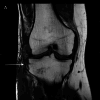
Figure 3. (A) Coronal T1-weighted image shows the Segond fracture (arrow).
Figure 4
Figure 4. (A) Sagittal fat-suppressed proton density-weighted image and (B) paracoronal T2-weighted image show a total rupture of the ACL (arrows). (C) Coronal fat-suppressed proton density-weighted image demonstrates a partial rupture of the MCL (arrow). (D) Axial fat-suppressed proton density-weighted image shows avulsion of the anterolateral capsular structures involving the anterolateral ligament as well as a posterolateral impression fracture of the tibial plateau (arrows). No injuries to the meniscus were observed.
ACL: anterior cruciate ligament, MCL: medial collateral ligament
Figure 5
Figure 5. (A-D) The patient initially underwent an arthroscopy of the knee joint, which confirmed the complete rupture of the ACL. The stump of the ACL was then resected. Chondromalacia grade 1 in the notch as well as medial femoral chondromalacia grade 3 were also seen in the arthroscopy.
ACL: anterior cruciate ligament
Figure 6
Figure 6. After arthroscopy, the Segond fracture was fixated using a screw: (A) initial intraoperative anterior-posterior view before screwing the Segond fracture and (B) intraoperative anterior-posterior view showing the screw with a length of 6.4 cm and diameter of 0.4 cm. (C) The intraoperative image show the result in lateral view.
Figure 7
Figure 7. (A and B) Knee after wound closure.

Similar articles

References

    1. The segond fracture: a narrative review of the anatomy, biomechanics and clinical implications. Phua SK, Lim JY, Li T, Ho SW. J Clin Orthop Trauma. 2023;38:102127. - PMC - PubMed
    1. Achtnich AE, Akoto R, Barié A, et al. AGA Committee Knee Ligament; 2016. Diagnosis of the knee ligament system (Book in German)
    1. Skinner EJ, Davis DD, Varacallo MA. Treasure Island, FL: StatPearls Publishing; 2024. Segond fracture. - PubMed
    1. Avulsion fracture of the medial collateral ligament association with Segond fracture. Albtoush OM, Horger M, Springer F, Fritz J. Clin Imaging. 2019;53:32–34. - PubMed
    1. The Segond fracture of the proximal tibia: a small avulsion that reflects major ligamentous damage. Goldman AB, Pavlov H, Rubenstein D. AJR Am J Roentgenol. 1988;151:1163–1167. - PubMed

Publication types

LinkOut - more resources