

Endoscope-assisted brain tumor removal overcomes the restriction of using intraoperative open magnetic resonance imaging in the suboccipital approach
- PMID: 40256004
- PMCID: PMC12003996
- DOI: 10.18999/nagjms.87.1.168
Endoscope-assisted brain tumor removal overcomes the restriction of using intraoperative open magnetic resonance imaging in the suboccipital approach
Abstract
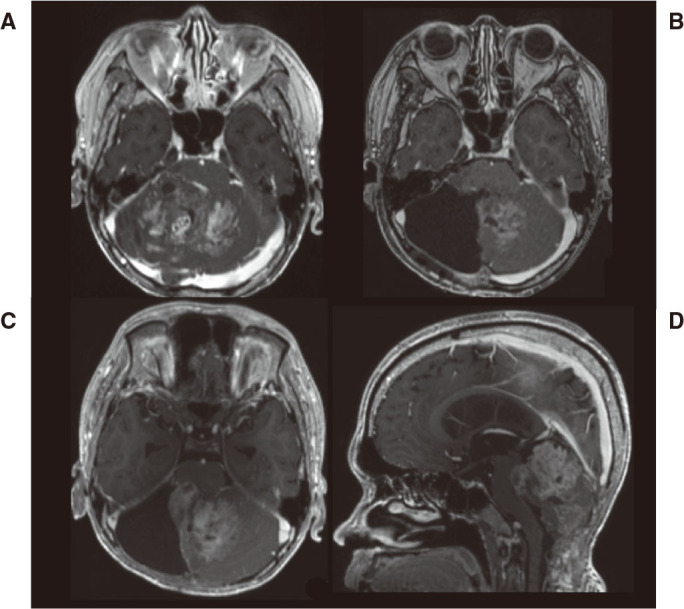
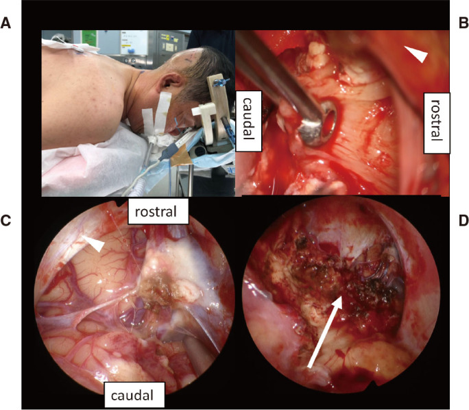
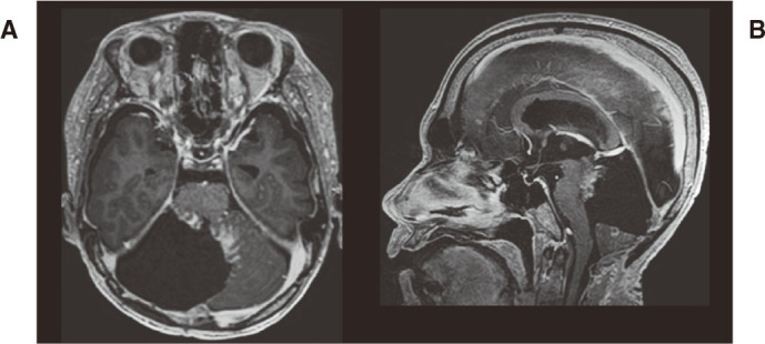
Intraoperative magnetic resonance imaging (iMRI) plays a crucial role in improving the precision of brain tumor surgeries. However, the use of iMRI can impose certain limitations on intraoperative head positioning. In regular microscopic surgery, head positioning is of utmost importance because an appropriate surgical field is important for the efficacy and safety of surgery. Therefore, in cases where adequate head positioning is difficult, usually, iMRI will not be utilized. Herein, we report an adult case of cerebellar astrocytoma whose tumor extended to the culmen of the cerebellum. Upon surgery via the suboccipital approach, the positional limitations imposed by iMRI led to an insufficient vertex-down position and limited surgical field, which hampered the removal of the upper portion of the tumor. However, this concern could be overcome when used in combination with an endoscope. The potential of iMRI applications is anticipated to be enhanced by overcoming positional limitations through combined endoscopic surgery. The use of multimodality in surgery is an optimal example of how surgical support equipment can also improve surgical outcomes. Here, we report on the new possibilities offered by multimodality.
Keywords: endoscope; intraoperative MRI; multimodality; positioning.
Conflict of interest statement
The authors declare that they have no competing interests.
Figures
Similar articles
-
Use of an ultra-low field intraoperative MRI system for pediatric brain tumor cases: initial experience with 'PoleStar N20'.Turk Neurosurg. 2012;22(2):218-25. doi: 10.5137/1019-5149.JTN.5615-11.0. Turk Neurosurg. 2012. PMID: 22437297
-
Safety, Utility, and Clinical Results of Continuous Intraoperative Electrophysiologic Monitoring in 1.5T iMRI-Guided Surgery.World Neurosurg. 2017 Oct;106:198-205. doi: 10.1016/j.wneu.2017.06.054. Epub 2017 Jun 15. World Neurosurg. 2017. PMID: 28624561
-
Intraoperative visualisation of functional structures facilitates safe frameless stereotactic biopsy in the motor eloquent regions of the brain.Br J Neurosurg. 2018 Aug;32(4):372-380. doi: 10.1080/02688697.2017.1416059. Epub 2017 Dec 20. Br J Neurosurg. 2018. PMID: 29260585
-
[Image-guided Brain Tumor Surgery with Intraoperative MRI].No Shinkei Geka. 2025 Mar;53(2):241-252. doi: 10.11477/mf.030126030530020241. No Shinkei Geka. 2025. PMID: 40155328 Review. Japanese.
-
Optimizing brain tumor resection. Midfield interventional MR imaging.Neuroimaging Clin N Am. 2001 Nov;11(4):659-72. Neuroimaging Clin N Am. 2001. PMID: 11995421 Review.
References
Publication types
MeSH terms
LinkOut - more resources
Full Text Sources
Medical