

Added value of diffusion-weighted magnetic resonance imaging in the diagnosis of recurrent cholangiocarcinoma
- PMID: 40256005
- PMCID: PMC12003994
- DOI: 10.18999/nagjms.87.1.22
Added value of diffusion-weighted magnetic resonance imaging in the diagnosis of recurrent cholangiocarcinoma
Abstract
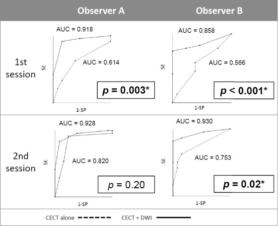
Distinguishing recurrent cholangiocarcinoma lesions from postoperative fibrosis or biliojejunostomy lesions using contrast-enhanced computed tomography (CECT) alone is challenging. This study examined the value of adding diffusion-weighted magnetic resonance imaging (DWI) to CECT for the detection of cholangiocarcinoma recurrence. This single-institution retrospective analysis included 33 patients who underwent cholangiocarcinoma resection between January 2016 and December 2020. Of the patients, 20 were in the recurrence group and 13 were in the non-recurrence group. Two observers independently reviewed the CECT images and subsequently reviewed the combined CECT and DWI images (b-value, 1000 s/mm2), with each image reviewed twice. The diagnostic performance was evaluated using receiver operating characteristic (ROC) curve analysis. Kappa statistics were used to evaluate agreement. The diagnostic performance (area under the ROC curve [AUC]) of both observers improved after the addition of DWI; the AUC improved from 0.614 to 0.918 (P = 0.003) in the first session and from 0.820 to 0.928 (P = 0.20) in the second session for Observer A, whereas it improved from 0.566 to 0.858 (P < 0.001) in the first session and from 0.753 to 0.930 (P = 0.02) in the second session for Observer B. The intraobserver and interobserver agreements improved after the addition of DWI; the kappa value improved from 0.586 to 0.656 for Observer A, from 0.371 to 0.838 for Observer B, from 0.308 to 0.766 in the first session, and from 0.464 to 0.620 in the second session. Adding DWI to CECT improves the detection of cholangiocarcinoma recurrence compared to CECT alone.
Keywords: bile ducts; cholangiocarcinoma; computed tomography; diffusion-weighted imaging; magnetic resonance imaging.
Conflict of interest statement
Yasuo Takehara is a faculty member of the Nagoya University, Department of Fundamental Development for Advanced Low Invasive Diagnostic Imaging, which is endowed by a private company, HIMEDIC, Inc. The other authors have no relevant financial or non-financial interests to disclose.
Figures


References
MeSH terms
LinkOut - more resources
Full Text Sources
Medical