

Can a black pigmented lesion of the oral cavity predict future development of melanoma- Report of a case and review of literature
- PMID: 40256074
- PMCID: PMC12007754
- DOI: 10.4103/jfmpc.jfmpc_1672_24
Can a black pigmented lesion of the oral cavity predict future development of melanoma- Report of a case and review of literature
Abstract
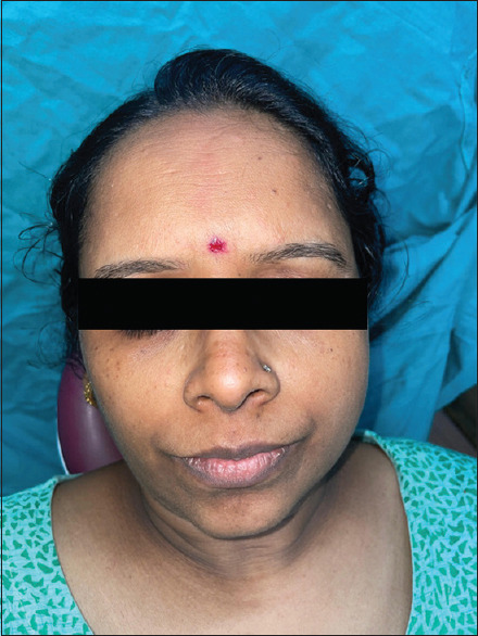
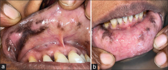
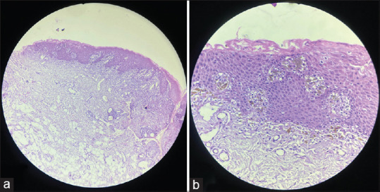
Oral melanotic macules and primary oral malignant melanoma are distinct pigmented lesions that can present clinical challenges in diagnosis. While melanotic macules are generally benign, quite a few instances of progression to melanoma have been reported. We present the case of a 42-year-old female with gradually enlarging painless pigmented macules on her lips, diagnosed as oral melanotic macules after histopathological evaluation. Although no signs of melanocytic hyperplasia or dysplasia were found, the patient was closely monitored due to the lesion's increasing size over 3 months. This case highlights the importance of distinguishing between benign pigmented lesions, which have a propensity to undergo malignant transformation from those that show no such tendency. Presences of histopathological features like atypical melanocytes and melanocytic hyperplasia/dysplasia in benign pigmented lesions are important markers for malignant transformation. Close clinical follow-up is essential to ensure timely intervention if the lesion exhibits suspicious changes.
Keywords: Malignant transformation; melanocytic dysplasia; melanoma; melanotic macule; peutz-jeghers syndrome; premalignant lesion.
Copyright: © 2025 Journal of Family Medicine and Primary Care.
Conflict of interest statement
There are no conflicts of interest.
Figures
Similar articles
-
Pigmented lesion with characteristics of malignancy: a case report.Gen Dent. 2013 Sep-Oct;61(6):e2-5. Gen Dent. 2013. PMID: 24064172
-
Melanotic Macule in Conjunction with a Giant Cell Fibroma.J Contemp Dent Pract. 2017 Oct 1;18(10):981-985. doi: 10.5005/jp-journals-10024-2160. J Contemp Dent Pract. 2017. PMID: 28989141
-
Transformation of a benign oral pigmentation to primary oral melanoma.Oral Surg Oral Med Oral Pathol Oral Radiol Endod. 2005 Oct;100(4):454-9. doi: 10.1016/j.tripleo.2005.01.018. Oral Surg Oral Med Oral Pathol Oral Radiol Endod. 2005. PMID: 16182166
-
Role of In Vivo Reflectance Confocal Microscopy in the Analysis of Melanocytic Lesions.Acta Dermatovenerol Croat. 2018 Apr;26(1):64-67. Acta Dermatovenerol Croat. 2018. PMID: 29782304 Review.
-
Oral pigmented lesions: Clinicopathologic features and review of the literature.Med Oral Patol Oral Cir Bucal. 2012 Nov 1;17(6):e919-24. doi: 10.4317/medoral.17679. Med Oral Patol Oral Cir Bucal. 2012. PMID: 22549672 Free PMC article. Review.
References
-
- Sung H, Ferlay J, Siegel RL, Laversanne M, Soerjomataram I, Jemal A, et al. Global cancer statistics 2020: GLOBOCAN estimates of incidence and mortality worldwide for 36 cancers in 185 countries. CA Cancer J Clin. 2021;71:209–49. - PubMed
-
- Kahn MA, Weathers DR, Hoffman JG. Transformation of a benign oral pigmentation to primary oral melanoma. Oral Surg Oral Med Oral Pathol Oral Radiol Endod. 2005;100:454–9. - PubMed
-
- Kaugars GE, Heise AP, Riley WT, Abbey LM, Svirsky JA. Oral melanotic macules: A review of 353 cases. Oral Surg Oral Med Oral Pathol. 1993;76:59–61. - PubMed
-
- Gupta G, Williams RE, Mackie RM. The labial melanotic macule: A review of 79 cases. Br J Dermatol. 1997;136:772–5. - PubMed
-
- Mukherjee D. Pigmented lesions of the oral cavity –A brief review. J Dent Res Rev. 2020;7:228–31.
Publication types
LinkOut - more resources
Full Text Sources