. 2025 Apr 2;10(14):14487-14495.
doi: 10.1021/acsomega.5c01028. eCollection 2025 Apr 15.

On Levodopa Interactions with Brain Disease Amyloidogenic Proteins at the Nanoscale

Affiliations

On Levodopa Interactions with Brain Disease Amyloidogenic Proteins at the Nanoscale

Talia Bergaglio et al. ACS Omega. .

Abstract

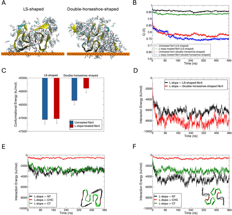
The cerebral accumulation of α-synuclein (α-Syn) and amyloid β-1-42 (Aβ-42) proteins is known to play a key role in the pathology of Parkinson's disease (PD). Currently, levodopa (L-dopa) is the first-line dopamine replacement therapy for treating bradykinetic symptoms (i.e., difficulty initiating physical movements), which become visible in PD patients. Using atomic force microscopy, we evidence at nanometer length scales the differential effects of L-dopa on the morphology of α-Syn and Aβ-42 protein fibrils. L-dopa treatment was observed to reduce the length and diameter of both types of protein fibrils, with a stark reduction mainly observed for Aβ-42 fibrils in physiological buffer solution and human cerebrospinal fluid. The insights gained on Aβ-42 fibril disassembly from the label-free nanoscale imaging experiments are substantiated by using atomic-scale molecular dynamics simulations. Our results indicate L-dopa-driven reversal of amyloidogenic protein aggregation, which might provide leads for designing chemical effector-mediated disassembly of insoluble protein aggregates.


Conflict of interest statement

The authors declare no competing financial interest.

Figures

Figure 1
Figure 1
Study rationale and pathological protein structure. (A) Schematic detailing the differences between normal synapse, neurocognitive disorder synapse, and levodopa-treated synapse. The objects shown are not to scale. Understanding the impact of levodopa on protein aggregates such as alpha-synuclein (B, protein data bank identifier: 2NOA) and amyloid beta (1–42) fibril (C, protein data bank identifier: 5OQV) implicated in neurodegenerative disorders such as Parkinson’s and Alzheimer’s disease using nanoscale imaging is the focus of the present study.
Figure 2
Figure 2
Nanoscale imaging of untreated and L-dopa-treated α-Syn in buffer salt solution. (A) AFM height map of untreated α-Syn protein fibrillar aggregates deposited on a gold surface. (B) AFM height image of α-Syn protein monomers incubated with 100 μM L-dopa and deposited on a gold surface. (C) Size distribution of spherical particles of 10.53 ± 1.96 nm, based on AFM height data. (D, E) Plot of the mean α-Syn fibril length and height values obtained in untreated samples (coded in black) and samples incubated with L-dopa (coded in red). Error bars indicate the standard deviation from the mean. (F) Persistence length of untreated α-Syn fibrils (hollow sphere trace) and L-dopa-treated (red trace) α-Syn fibrils. The worm-like chain model (WLC) is plotted as a dark gray line.
Figure 3
Figure 3
Characterization of untreated and L-dopa-treated Aβ-42 in physiological buffer. (A) Large-area AFM image showing untreated Aβ-42 fibrils. (B) AFM image showing L-dopa-treated Aβ-42 fibrils of diverse lengths formed after incubating Aβ-42 peptides with L-dopa. (C) AFM image of L-dopa-treated Aβ-42 proteins confirming the presence of short fibrils (indicated by black arrows). (D) AFM image of a single Aβ-42 fibril treated with L-dopa. (E) Distribution of fibril length for untreated Aβ-42 (black color-coded) and L-dopa-treated Aβ-42 fibrils (red color-coded). (F) Distribution of fibril height for untreated Aβ-42 (black color-coded) and L-dopa-treated Aβ-42 fibrils (red color-coded). (G) Combined plot of MS end-to-end distance versus contour length for untreated Aβ-42 fibrils (hollow square trace) and L-dopa-treated Aβ-42 fibrils (red sphere trace).
Figure 4
Figure 4
Direct imaging of untreated and L-dopa-treated Aβ-42 in human CSF. (A) AFM image revealing the presence of both fibrillar and spherical Aβ-42 aggregates in CSF. (B) High-resolution AFM image of untreated Aβ-42 fibrils resolved within the white dash box in panel A. (C) Large-area AFM image of L-dopa (100 μM)-treated Aβ-42 aggregates showing the presence of mostly spherical (indicated by white arrows) and lower prevalence of fibrils (indicated by yellow arrow). (D) Distribution of fibril length for untreated Aβ-42 (black color-coded) and L-dopa-treated Aβ-42 fibrils (red color-coded) in CSF. (E) Distribution of fibril height for untreated Aβ-42 (black color-coded) and L-dopa-treated Aβ-42 fibrils (red color-coded) in CSF. (F) Mean Aβ-42 fibril height measured in buffer solution and CSF, without (colored black, meanbuffer: 3.45 ± 1.62 nm; meanCSF: 5.91 ± 1.28 nm) and with incubation with100 μM L-dopa (colored red, meanbuffer: 3.05 ± 1.35 nm; meanCSF: 2.05 ± 0.83 nm).
Figure 5
Figure 5
Impact of L-dopa on Aβ-42 protein fibril folds from molecular dynamics simulations. (A) Final conformation of Aβ-42 fibril in their LS-shaped fold (PDB code 5OQV(36)) and double-horseshoe-shaped fold (right, PDB code 2NAO(37)) in the presence of L-dopa molecules at the gold–water interface following 480 ns of dynamics. L-dopa molecules within 5 Å of the fibril are shown in ball and stick representation. The protein fibril is shown in its secondary structure representation, and the gold surface is shown as an atomic sphere. Comparison of the time evolution of (B) fraction of native contacts (Q(X)) and (C) conformational energies between untreated and L-dopa-treated fibril folds. Time evolution of interaction energies between L-dopa and (D) the two fibril folds, and between L-dopa and the N-terminus (NT, black), central hydrophobic cluster (CHC, red) and C-terminus (CT, green) of (E) LS-shaped, and (F) double-horseshoe-shaped fibril folds. The two fibril folds with different colored regions are shown as insets.

Similar articles

References

    1. Candelise N.; Scaricamazza S.; Salvatori I.; Ferri A.; Valle C.; Manganelli V.; Garofalo T.; Sorice M.; Misasi R. Protein Aggregation Landscape in Neurodegenerative Diseases Clinical Relevance and Future Applications. Int. J. Mol. Sci. 2021, 22, 6016.10.3390/ijms22116016. - DOI - PMC - PubMed
    1. Bhattacharya S.; Xu L.; Thompson D. Revisiting the earliest signatures of amyloidogenesis: Roadmaps emerging from computational modeling and experiment. WIREs Comput. Mol. Sci. 2018, 8 (4), e135910.1002/wcms.1359. - DOI
    1. Bhattacharya S.; Xu L.; Thompson D. Molecular Simulations Reveal Terminal Group Mediated Stabilization of Helical Conformers in Both Amyloid-beta42 and alpha-Synuclein. ACS Chem. Neurosci. 2019, 10 (6), 2830–2842. 10.1021/acschemneuro.9b00053. - DOI - PubMed
    1. Xu L.; Bhattacharya S.; Thompson D., Predictive Modeling of Neurotoxic α-Synuclein Polymorphs. In Computer Simulations of Aggregation of Proteins and Peptides; Springer US: New York, NY, 2022; 379–399.
    1. Mehra S.; Sahay S.; Maji S. K. alpha-Synuclein misfolding and aggregation: Implications in Parkinson’s disease pathogenesis. Biochim Biophys Acta Proteins Proteom 2019, 1867 (10), 890–908. 10.1016/j.bbapap.2019.03.001. - DOI - PubMed