

Reactive oxygen species (ROS) are a crucial factor in the anticancer activity of Oliveria decumbens extract against the A431 human skin cell line
- PMID: 40256584
- PMCID: PMC12004055
- DOI: 10.32592/ARI.2024.79.4.749
Reactive oxygen species (ROS) are a crucial factor in the anticancer activity of Oliveria decumbens extract against the A431 human skin cell line
Abstract
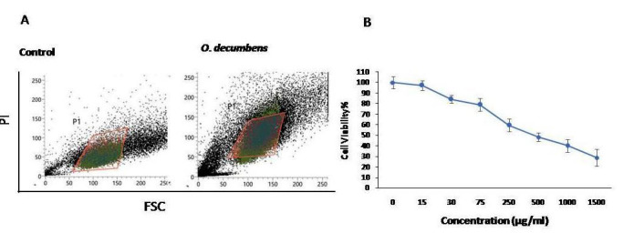
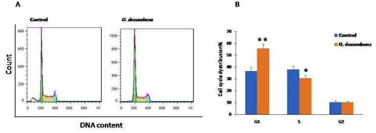
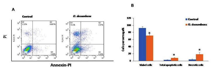
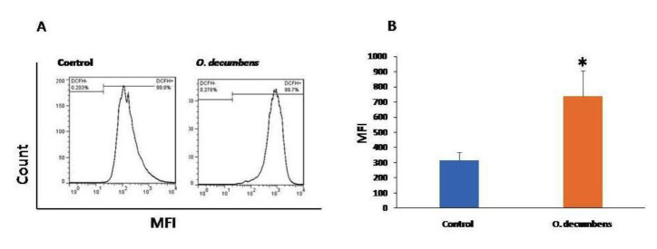
Globally, skin cancer is a main public health challenge whose incidence is continuously increasing. Given the limitations of conventional t herapies, new research and novel therapies may be promising for reducing skin cancer morbidity and mortality. Phytochemicals are attractive resources for new therapy design in cancer research due to their cost-effectiveness and lower side effects. In the present study, the anti-cancer activity of Oliveria decumbens (O. decumbens) extract was investigated on the human skin cancer A431 cell line A431. The aqueous extract of the O. decumbens plant was prepared using the traditional method. Then IC50 was determined using 3-(4,5-dimethylthiazol-2-yl)-2,5-diphenyl-2H-tetrazolium bromide (MTT) assay under different concentrations of O. decumbens. Cell apoptosis was investigated by Annexin V-FITC/Propidium Iodide (PI) and flow cytometry. Cell cycle was investigated by PI staining and flow cytometry. Reactive oxygen species (ROS) production was analyzed by DCFH-DA (2', 7' -dichlorofluorescein-diacetate) staining and flowcytometry.IC50 for cell viability was determined 475g/ml. Cell cycle analyses showed G1 arrest in treated cells compared to control cell. Results also confirmed significant increase of apoptotic cells (8.2%1, P<0.05) under IC50 concentration of the extract in comparison to the control group (2.50.99%). A significant increase in ROS level was observed in O.ecumbens treated cells compared to control cells (738 170 vs 31655 in the control group, P<0.05.).Overall, the present results indicate that O. decumbens extract can inhibit skin cancer cell proliferation via inhibition of cell cycle and apoptosis. It seems that ROS production plays a critical role in the anticancer effect of O. decumbens extract. Therefore, its potential option for future treatment of skin cancer should be considered.
Keywords: A431cell line; Aqueous extract; Oliveriadecumbens; ROS; Skin cancer.
Conflict of interest statement
The authors declare that they have no conflict of interests.
Figures
Similar articles
-
In-vitro and in-vivo anti-breast cancer activity of OEO (Oliveria decumbens vent essential oil) through promoting the apoptosis and immunomodulatory effects.J Ethnopharmacol. 2020 Feb 10;248:112313. doi: 10.1016/j.jep.2019.112313. Epub 2019 Oct 23. J Ethnopharmacol. 2020. PMID: 31655147
-
Pterospermum acerifolium (L.) wild bark extract induces anticarcinogenic effect in human cancer cells through mitochondrial-mediated ROS generation.Mol Biol Rep. 2018 Dec;45(6):2283-2294. doi: 10.1007/s11033-018-4390-6. Epub 2018 Sep 28. Mol Biol Rep. 2018. PMID: 30267191
-
Viscum articulatum Burm. f. aqueous extract exerts antiproliferative effect and induces cell cycle arrest and apoptosis in leukemia cells.J Ethnopharmacol. 2018 Jun 12;219:91-102. doi: 10.1016/j.jep.2018.03.005. Epub 2018 Mar 16. J Ethnopharmacol. 2018. PMID: 29555410
-
Ethyl acetate fraction of Garcina epunctata induces apoptosis in human promyelocytic cells (HL-60) through the ROS generation and G0/G1 cell cycle arrest: a bioassay-guided approach.Environ Toxicol Pharmacol. 2013 Nov;36(3):865-74. doi: 10.1016/j.etap.2013.07.015. Epub 2013 Aug 6. Environ Toxicol Pharmacol. 2013. PMID: 23981377
-
Anticancer activity of caffeic acid n‑butyl ester against A431 skin carcinoma cell line occurs via induction of apoptosis and inhibition of the mTOR/PI3K/AKT signaling pathway.Mol Med Rep. 2018 Apr;17(4):5652-5657. doi: 10.3892/mmr.2018.8599. Epub 2018 Feb 13. Mol Med Rep. 2018. Retraction in: Mol Med Rep. 2021 May;23(5):372. doi: 10.3892/mmr.2021.12011. PMID: 29436638 Free PMC article. Retracted.
References
MeSH terms
Substances
LinkOut - more resources
Full Text Sources
Medical
Research Materials
Miscellaneous
