

Paraneoplastic leukocytoclastic vasculitis mimicking ulcus cruris as rare initial manifestation of smoldering myeloma IgG kappa
- PMID: 40257478
- PMCID: PMC12141142
- DOI: 10.1007/s00277-025-06366-2
Paraneoplastic leukocytoclastic vasculitis mimicking ulcus cruris as rare initial manifestation of smoldering myeloma IgG kappa
Abstract
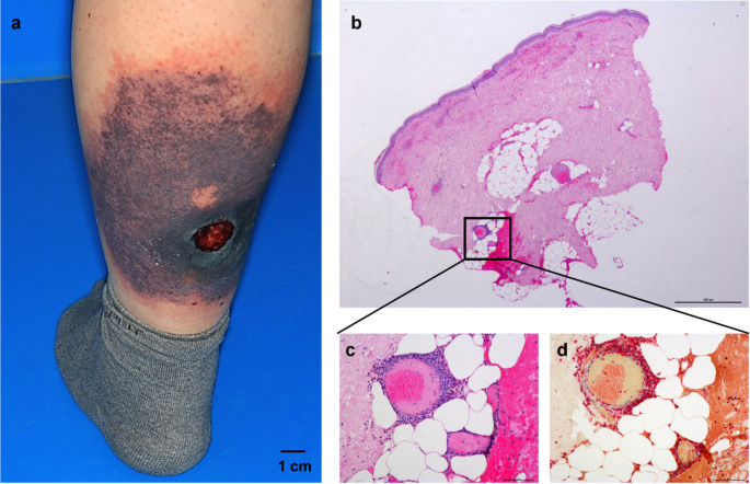
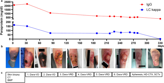
Leukocytoclastic vasculitis (LCV) has been reported as a rare paraneoplastic phenomenon associated with several hematologic disorders, including indolent lymphomas such as Waldenström macroglobulinemia. However, there are very few cases of LCV in the context of plasma cell disorders. We present the case of a 58-year-old female who developed a rapidly progressive, ulceronecrotic skin lesion on her left lower leg due to leukocytoclastic vasculitis. The lesion was initially suspected to be an ulcerative chronic wound (ulcus cruris) but represented an atypical manifestation of leukocytoclastic vasculitis as primary and only clinical sign of smoldering multiple myeloma IgG kappa. After standard induction chemoimmunotherapy with daratumumab, bortezomib, lenalidomide, and dexamethasone, the patient proceeded to high-dose chemotherapy with melphalan, followed by autologous stem cell transplantation for consolidation. Despite a bacterial skin superinfection, myeloma treatment was successfully completed without any major complications. The skin lesion healed concurrently with the reduction in paraprotein levels, and there was no need for plastic surgical intervention. LCV mimicking ulcus cruris can represent a rare and atypical initial manifestation of plasma cell neoplasia. In this case report, systemic myeloma treatment proved to be effective for inducing complete remission of advanced ulceronecrotic skin damage. This case extends the spectrum of reported monocloncal gammopathies of cutaneous significance.
Keywords: Leukocytoclastic vasculitis; Monoclonal gammopathy of cutaneous significance; Paraneoplastic phenomenon; Skin.
© 2025. The Author(s).
Conflict of interest statement
Declarations. Ethical approval: Informed consent was obtained from the patient for the publication of this case report including any potentially identifiable images or data shown in this article. Data statement: The raw data supporting the conclusion of this article will be made available by the authors, without undue reservation. Competing interests: The authors declare no competing interests.
Figures


References
-
- Carlson JA, Chen K-R (2006) Cutaneous vasculitis update: small vessel neutrophilic vasculitis syndromes. Am J Dermatopathol 28:486–506. 10.1097/01.dad.0000246646.45651.a2 - PubMed
-
- Abate MS, Battle LR, Emerson AN, Gardner JM, Shalin SC (2019) Dermatologic urgencies and emergencies: what every pathologist should know. Arch Pathol Lab Med 143:919–942. 10.5858/arpa.2018-0239-RA - PubMed
-
- Zax RH (1990) Cutaneous leukocytoclastic vasculitis: serial histopathologic evaluation demonstrates the dynamic nature of the infiltrate. Arch Dermatol 126:69. 10.1001/archderm.1990.01670250075012 - PubMed
-
- Wick MR, Patterson JW (2019) Cutaneous paraneoplastic syndromes. Semin Diagn Pathol 36:211–228. 10.1053/j.semdp.2019.01.001 - PubMed
-
- Bayer-Garner IB, Smoller BR (2003) Leukocytoclastic (Small Vessel) vasculitis in multiple myeloma: leukocytoclastic vasculitis. Clin Exp Dermatol 28:521–524. 10.1046/j.1365-2230.2003.01324.x - PubMed
Publication types
MeSH terms
Substances
LinkOut - more resources
Full Text Sources
Medical
