. 2025 Apr 21;20(1):44.
doi: 10.1007/s11481-025-10199-1.

Cranberry Extract Ameliorates Diabetic Cognitive Impairment in Rats Via LncRNA GAS-5 Downregulation and Pyroptosis Pathway Inhibition

Affiliations

Cranberry Extract Ameliorates Diabetic Cognitive Impairment in Rats Via LncRNA GAS-5 Downregulation and Pyroptosis Pathway Inhibition

Mariam Ali Abo-Saif et al. J Neuroimmune Pharmacol. .

Abstract

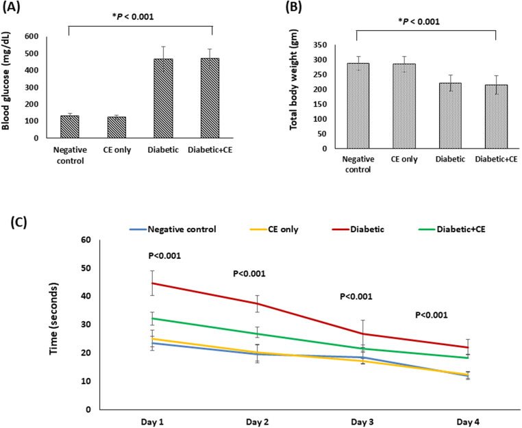
The pathophysiology of diabetes-induced brain injury involves pyroptosis, an inflammatory programmed cell death. This study aimed to investigate the potential protective effect of cranberry extract (CE) against diabetes-induced brain injury. Type 1 diabetes was induced by intraperitoneal injection of streptozotocin in rats. Brain tissue samples were investigated for biochemical determination of the reduced glutathione (GSH), superoxide dismutase (SOD), and malondialdehyde (MDA), and the quantitative RT-PCR for the gene expression of glial cell-derived neurotrophic factor (GDNF), lncRNA GAS-5, and pyroptosis markers. ELISA was used to determine the caspase-1 level and immunohistochemical staining for assessing IL-1β. Prophylactic dosing of the CE in diabetic rats improved cognitive behavior and significantly suppressed MDA concentration, pyroptosis genes expression (gasdermin D and caspase 1), and lncRNA GAS-5. In addition, CE significantly elevated GSH concentration, SOD activity, and gene expression of GDNF and markedly reduced IL-1β positive stained cells score in the brain. Phytochemical characterization of the CE by FT-IR and UPLC-PDA-MS/MS revealed cyanidin arabinoside, procyanidins, quercetin, and isorhamnetin as key components. CE protects against diabetes-induced cognitive dysfunction in rats by targeting redox-related signaling pathways and inducing an anti-inflammatory effect. LncRNA GAS-5 downregulation and pyroptosis pathway inhibition may contribute to its beneficial effects, suggesting its therapeutic potential.

Keywords: Caspase 1; Cranberry extract; Diabetes-induced cognitive impairment; GDNF; Gasdermin D; Growth-arrest-specific (GAS)-5; LncRNA GAS-5; Pyroptosis.


Conflict of interest statement

Declarations. Ethical Approval: The animal study protocol was approved by the Research Ethical Committee of the Faculty of Pharmacy, Tanta University (protocol code TP/RE/11/22P-0060). Consent To Participate: not applicable. Institutional Review Board Statement: The animal study protocol was approved by the Research Ethical Committee of the Faculty of Pharmacy, Tanta University (protocol code TP/RE/11/22P-0060). Competing Interests: The authors declare no competing interests.

Figures

Fig. 1
Fig. 1
IR spectrum of the cranberry extract
Fig. 2
Fig. 2
UPLC-ESI-MS total ion chromatogram of the cranberry extract in negative ion mode. Compounds identified are labelled in red (1–10)
Fig. 3
Fig. 3
Effect of the cranberry extract (CE) prophylactic treatment on (A) Random blood glucose level, (B) Total body weight, and (C) The mean escape latencies of the studied rat groups. Data are revealed as mean ± SD, n = 6. Negative control (NC): non-diabetic rats taking the vehicle, CE only: non-diabetic rats treated with the CE 250 mg/kg/day orally for eight weeks, positive control (PC): diabetic rats taking the vehicle, and diabetic + CE: diabetic rats treated with the CE 250 mg/kg/day orally for eight weeks. Type 1 diabetes was induced in the rats by intraperitoneal injection of a single dose of 60 mg/kg streptozotocin
Fig. 4
Fig. 4
Effect of the cranberry extract (CE) prophylactic treatment on (A) Malondialdehyde (MDA) level, (B) Glutathione (GSH) level, and (C) Superoxide dismutase (SOD) activity, (D) Caspase 1 level in the brain of rats in the studied groups. Data are revealed as mean ± SD, n = 6. Negative control (NC): non-diabetic rats taking the vehicle, CE only: non-diabetic rats treated with the CE 250 mg/kg/day orally for eight weeks, positive control (PC): diabetic rats taking the vehicle, and diabetic treated: diabetic rats treated with the CE 250 mg/kg/day orally for eight weeks. Type 1 diabetes was induced in the rats by intraperitoneal injection of a single dose of 60 mg/kg streptozotocin
Fig. 5
Fig. 5
Fold gene expression analysis in the brain of the studied rat groups (A) Glial cell-derived neurotrophic factor (GDNF), (B) Long non-coding RNA growth-arrest-specific-5 (LncRNA GAS-5), (C) Gasdermin D, and (D) Caspase 1. Data are revealed as mean ± SD, n = 3. Negative control (NC): non-diabetic rats taking the vehicle, CE only: non-diabetic rats treated with the cranberry extract 250 mg/kg/day orally for eight weeks, positive control (PC): diabetic rats taking the vehicle, and diabetic + CE: diabetic rats treated with the cranberry extract 250 mg/kg/day orally for eight weeks. Type 1 diabetes was induced in the rats by intraperitoneal injection of a single dose of 60 mg/kg streptozotocin
Fig. 6
Fig. 6
Microscopic pictures of H&E-stained cerebral cortex of the studied rat’s groups. (A and B): Cerebral cortical sections from the negative control group and cranberry extract-only treated group, respectively, showing normal neurons (blue arrows), glial cells (blue arrowheads) and neuropil. (C -E): Cerebral cortical sections from the positive control group (diabetic rats) showed marked shrinkage and degeneration of many neurons (black arrows), many apoptotic glial cells (black arrowheads), prominent neurophagia (yellow arrows) and vacuolation in neuropil (red arrows). (F-H): Cerebral cortical section from diabetic rats treated with cranberry extract showing decreased numbers of affected neurons (black arrows) and glial cells (black arrowheads), mild neurophagia (yellow arrows) and mild vacuolation in neuropil (red arrows). magnification X: 400 bar 50 μm
Fig. 7
Fig. 7
Microscopic pictures of H&E-stained hippocampal sections showing: A, B) Normal neurons (black arrows) in the pyramidal layer in the control group and normal rats treated with cranberry. C) Marked shrinkage & degeneration of many neurons (black arrows), few apoptotic neurons (blue arrows) with many vacuolations (black arrowheads) in the diabetic group. D) Few degenerated neurons (black arrow) and few apoptotic neurons (blue arrows) in the diabetic group pre-treated with cranberry. Magnification: x400, scale bar 50 μm
Fig. 8
Fig. 8
Microscopic pictures of immunostained brain sections against IL-1β in the studied rat groups. (A and B): Brain sections from rats in the negative control group showed negative staining in neurons and glial cells in the examined cerebral cortex and hippocampal pyramidal layer, respectively. (C and D): Brain sections from non-diabetic rats treated with cranberry extract showed negative staining in neurons and glial cells in the examined cerebral cortex and hippocampal pyramidal layer, respectively. (E and F): Brain sections from diabetic rats showing marked positive staining in glial cells (arrows) in the cerebral cortex and neurons (arrows) of the hippocampal pyramidal layer, respectively. (G and H): Brain sections from diabetic rats treated with cranberry extract showed weak positive staining in a few glial cells and a few cerebral cortical sections (arrows) and negative staining in neurons of the hippocampal pyramidal layer, respectively. Magnification: x400, scale bar 50 μm

Similar articles

References

    1. Afzal M, Redha A, AlHasan R (2019) Anthocyanins potentially contribute to defense against Alzheimer’s disease. Mol Basel Switz 24:4255 - PMC - PubMed
    1. Ahmadiantehrani S, Barak S, Ron D (2014) GDNF is a novel ethanol-responsive gene in the VTA: implications for the development and persistence of excessive drinking. Addict Biol 19:623 - PMC - PubMed
    1. Anitha M et al (2006) GDNF rescues hyperglycemia-induced diabetic enteric neuropathy through activation of the PI3K/Akt pathway. J Clin Invest 116:344–356 - PMC - PubMed
    1. Assar DH et al (2021a) Wound healing potential of licorice extract in rat model: antioxidants, histopathological, immunohistochemical and gene expression evidences. Biomed Pharmacother Biomedecine Pharmacother 143:112151 - PubMed
    1. Assar DH et al (2021b) Ameliorative effects of Aspergillus Awamori against the initiation of hepatocarcinogenesis induced by diethylnitrosamine in a rat model: regulation of Cyp19 and p53 gene expression. Antioxid Basel Switz 10:922 - PMC - PubMed

MeSH terms

LinkOut - more resources