. 2025 Apr 1;66(4):60.
doi: 10.1167/iovs.66.4.60.

Detailed Clinical, Ophthalmic, and Genetic Characterization of MYO7A-Associated Usher Syndrome

Affiliations

Detailed Clinical, Ophthalmic, and Genetic Characterization of MYO7A-Associated Usher Syndrome

Juan C Romo-Aguas et al. Invest Ophthalmol Vis Sci. .

Abstract

Purpose: To analyze the clinical spectrum and natural history of MYO7A-associated Usher syndrome type I (USH1).

Methods: Patients with molecularly confirmed MYO7A-associated USH1 in a single tertiary referral center. Data was extracted from physical and electronic case notes, including imaging and electrophysiology. Genetic results were reviewed, and the detected variants were assessed. Main outcome measures were clinical findings, qualitative and quantitative analysis of retinal imaging, and electrophysiology.

Results: Eighty patients were identified and evaluated longitudinally. The mean age (±SD) of onset of symptoms was 12.0 ± 5.8 years of age, and a mean follow-up time of 16.2 years. BCVA was 0.4 ± 0.5 LogMAR at baseline, and 0.7 ± 0.8 LogMAR at the last visit for both eyes. The change in BCVA over time was 0.02 LogMAR per year. A hyperautofluorescent (hyperAF) ring was present in 51% of the patients. The mean ellipsoid zone width (EZW) at baseline was 2568.2 ± 1528.9 µm OD and 2527.9 ± 1609.3 µm OS, which decreased to 2012.3 ± 1705.1 µm OD and 1806.3 ± 1647.1 µm OS at last visit. Electrophysiology revealed rod and cone dysfunction with relative or complete sparing of macular function. There were statistically significant changes in BCVA, EZW, and hyperAF ring between baseline and follow-up. Genetic analysis identified 83 variants in MYO7A, including 18 novel variants.

Conclusions: Longitudinal analysis shows that the majority of patients retain central visual function and structure until the fifth decade of life, which informs advice on prognosis and the window for therapeutic intervention.


Conflict of interest statement

Disclosure: J.C. Romo-Aguas, None; T.A.C. de Guimarães, None; A. Kalitzeos, None; N. Aychoua, None; C. Tsika, None; A.G. Robson, None; Y. Fujinami-Yokokawa, None; K. Fujinami, None; O.A. Mahroo, None; A.R. Webster, None; M. Michaelides, MeiraGTx (C), Janssen Pharmaceuticals (C), Saliogen (C), AAVantgarde (C)

Figures

Figure 1.
Figure 1.
Bland-Altman Plot. The graph shows the distribution of EZW in µm and hyperAF area in mm2 of both eyes at baseline and follow-up. The red lines represent the mean bias, and the green lines represent the upper and lower 95% confidence interval.
Figure 2.
Figure 2.
Baseline-follow up values indicating change over time. Plotted from observations of (A) BCVA (n = 74), (B) EZW) (n = 35, and (C) hyperAF ring area (n = 34). Each datapoint pair represents the right eye of a subject across their follow-up time. Datapoints with hollow circles represent patients with NDN genotype, and datapoints with filled circles represent patients with DN genotype. (C) The distinct group of five datapoint pairs extending beyond 80 mm2 hyper-AF ring area belong to patients within the mild phenotype.
Figure 3.
Figure 3.
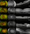
Multimodal imaging of five patients from different age groups. Imaging findings in patients with MYO7A-associated Usher syndrome type 2. Here we show a composition of pseudocolor wide-field fundus photography, fundus autofluorescence, and optical coherence tomography from five unrelated patients of different groups of age. Patients MYO7A 018 (A), MYO7A 002 (B), MYO7A 003 (C), MYO7A 024 (D), and MYO7A 056 (E), respectively.
Figure 4.
Figure 4.
Multimodal imaging of four patients with a milder phenotype. Pseudocolor wide-field fundus photography, fundus autofluorescence and optical coherence tomography, of two patients with mild generalized retinitis pigmentosa and two patients with sector retinitis pigmentosa, (A, B) MYO7A 066 [age 21], MYO7A 046 (C, D) [age 39], MYO7A 066 (E, F) [age 45], and MYO7A 040 (G, H) [age 56].
Figure 5.
Figure 5.
Kaplan-Meier survival analysis showing the percentage of patients with a range of baseline best-corrected visual acuities. (A) Percentage of patients with BCVA ≤0.3 LogMAR (6/12) in at least one eye (50% at 53 years of age) and (B) the percentage of patients with legal blindness (≥1 LogMAR, 50% at 62 years of age).
Figure 6.
Figure 6.
Full-field ERG and PERG P50 findings. Full field and pattern ERG findings in 19 subjects that were tested according to ISCEV standard methods. (A) The amplitudes of the DA 0.01 ERG, DA 10 ERG a- wave, LA 30 Hz ERG and LA 3 ERG b-wave are plotted as a percentage of the age-matched 5th percentile of the (“normal”) reference range, with values arranged in ascending order of DA10 ERG a-wave amplitude for clarity. (B) The LA 30 Hz peak times are plotted as a difference from the age-matched 95th percentile of the reference range (x-axis indicates 0 ms delay). (C) The ISCEV Standard PERG P50 amplitudes of the same patients, expressed as a percentage of the lower limit of the (“normal”) reference range. Subjects 5 and 9 had double null MYO7A variants. All electrophysiological recordings showed a high degree of interocular symmetry and are shown for right eyes only.
Figure 7.
Figure 7.
A schematic diagram of the genetic and protein structures of myosin-VIIa. The positions of detected MYO7A variants in our cohort. IQ1-5, 1-5 isoleucine-glutamine motifs; FERM, radixin-moesin; MyTH4, myosin tail homology 4; SAH, stable single a-helix; SH3, SRC homology 3.

References

    1. Yan D, Liu XZ.. Genetics and pathological mechanisms of Usher syndrome. J Hum Genet. 2010; 55: 327–335. - PMC - PubMed
    1. Kimberling WJ, Hildebrand MS, Shearer AE, et al. .. Frequency of Usher syndrome in two pediatric populations: Implications for genetic screening of deaf and hard of hearing children. Genet Med. 2010; 12: 512–516. - PMC - PubMed
    1. Millán JM, Aller E, Jaijo T, Blanco-Kelly F, Gimenez-Pardo A, Ayuso C.. An update on the genetics of Usher syndrome. J Ophthalmol. 2011; 2011: 1–8. - PMC - PubMed
    1. Boughman JA, Vernon M, Shaver KA.. Usher syndrome: Definition and estimate of prevalence from two high-risk populations. J Chronic Dis. 1983; 36: 595–603. - PubMed
    1. Ullah F, Zeeshan Ali M, Ahmad S, et al. .. Current updates on genetic spectrum of usher syndrome. Nucleosides Nucleotides Nucleic Acids. 2025; 44: 337–360. - PubMed

MeSH terms