

Exploring the mechanism of bone marrow mesenchymal stromal cell exosomes in respiratory syncytial virus infection based on miRNA sequencing
- PMID: 40258894
- PMCID: PMC12012132
- DOI: 10.1038/s41598-025-98160-3
Exploring the mechanism of bone marrow mesenchymal stromal cell exosomes in respiratory syncytial virus infection based on miRNA sequencing
Abstract
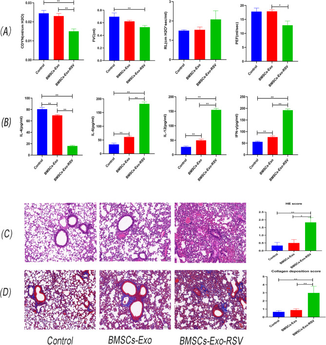
The role of Bone marrow mesenchymal stem cells (BMSCs) and their exosomes in regulating the host response to viral infections has garnered significant attention, yet research on their specific mechanisms in response to respiratory syncytial virus (RSV) infection remains limited. This study analyzes changes in cytokine levels and exosomal miRNA expression profiles in BMSCs supernatants following RSV infection. The findings reveal that RSV infection leads to a significant decrease in IL-4 levels in BMSCs supernatants, alongside notable increases in IL-6, IL-12, and IFN-γ levels. Additionally, expressions of RSV F protein, G protein, and N gene were detected in the exosomes. Further in vivo experiments demonstrated that exosomes from RSV-treated BMSCs significantly enhanced the inflammatory response in RSV-infected mice, indicated by elevated serum inflammatory cytokines, lung dysfunction, airway inflammation, and increased mucus secretion. In contrast, exosomes from untreated BMSCs showed minimal effects on airway inflammation and damage in infected mice. miRNA sequencing analysis of the exosomes identified differential miRNAs enriched in multiple key signaling pathways, suggesting that RSV infection alters the functional characteristics of BMSCs exosomes, shifting their role from anti-inflammatory and repair mechanisms to a pro-inflammatory function. This transformation may be mediated by changes in the miRNA expression profile.
Keywords: Exosomes; High-throughput sequencing; Mesenchymal stem cells; Respiratory syncytial virus infection; miRNA.
© 2025. The Author(s).
Conflict of interest statement
Declarations. Competing interests: The authors declare no competing interests.
Figures


Similar articles
-
Development and Comparative Study of a Mouse Model of Airway Inflammation and Remodeling Induced by Exosomes Derived from Bone Marrow Mesenchymal Stem Cells.Bull Exp Biol Med. 2024 Aug;177(4):544-551. doi: 10.1007/s10517-024-06221-w. Epub 2024 Sep 16. Bull Exp Biol Med. 2024. PMID: 39279005
-
Respiratory syncytial virus infection in human bone marrow stromal cells.Am J Respir Cell Mol Biol. 2011 Aug;45(2):277-86. doi: 10.1165/rcmb.2010-0121OC. Epub 2010 Oct 22. Am J Respir Cell Mol Biol. 2011. PMID: 20971883 Free PMC article.
-
Effects of WuHuTang on the function and autophagy of dendritic cells treated with exosomes induced by RSV.J Ethnopharmacol. 2024 Oct 5;332:118397. doi: 10.1016/j.jep.2024.118397. Epub 2024 May 26. J Ethnopharmacol. 2024. PMID: 38806137
-
A chimeric A2 strain of respiratory syncytial virus (RSV) with the fusion protein of RSV strain line 19 exhibits enhanced viral load, mucus, and airway dysfunction.J Virol. 2009 May;83(9):4185-94. doi: 10.1128/JVI.01853-08. Epub 2009 Feb 11. J Virol. 2009. PMID: 19211758 Free PMC article.
-
Respiratory syncytial virus (RSV) evades the human adaptive immune system by skewing the Th1/Th2 cytokine balance toward increased levels of Th2 cytokines and IgE, markers of allergy--a review.Virus Genes. 2006 Oct;33(2):235-52. doi: 10.1007/s11262-006-0064-x. Virus Genes. 2006. PMID: 16972040 Review.
Cited by
-
Applications of Osteoimmunomodulation Models in Evaluating Osteogenic Biomaterials.J Funct Biomater. 2025 Jun 11;16(6):217. doi: 10.3390/jfb16060217. J Funct Biomater. 2025. PMID: 40558903 Free PMC article. Review.
References
-
- Billard, M. N. & Bont, L. J. The link between respiratory syncytial virus infection during infancy and asthma during childhood. Lancet401(10389), 1632–1633 (2023). - PubMed
-
- Hartert, T. V., Wu, P. & Brunwasser, S. M. Respiratory syncytial virus and asthma: Untying the Gordian knot. Lancet Respir. Med.9(10), 1092–1094 (2021). - PubMed
-
- Backman, K., Ollikainen, H., Piippo-Savolainen, E., Nuolivirta, K. & Korppi, M. Asthma and lung function in adulthood after a viral wheezing episode in early childhood. Clin. Exp Allergy48(2), 138–146 (2018). - PubMed
MeSH terms
Substances
Grants and funding
LinkOut - more resources
Full Text Sources
Medical
