. 2025 Apr 21;15(1):13759.
doi: 10.1038/s41598-025-96075-7.

Enhanced peri-implantitis management through purple-LED irradiation coupled with silver ion application and calcium phosphate gene transfection carrier coating

Affiliations

Enhanced peri-implantitis management through purple-LED irradiation coupled with silver ion application and calcium phosphate gene transfection carrier coating

Taito Iwabuchi et al. Sci Rep. .

Abstract

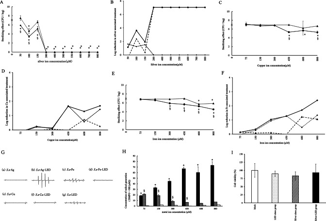
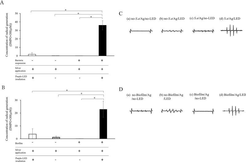
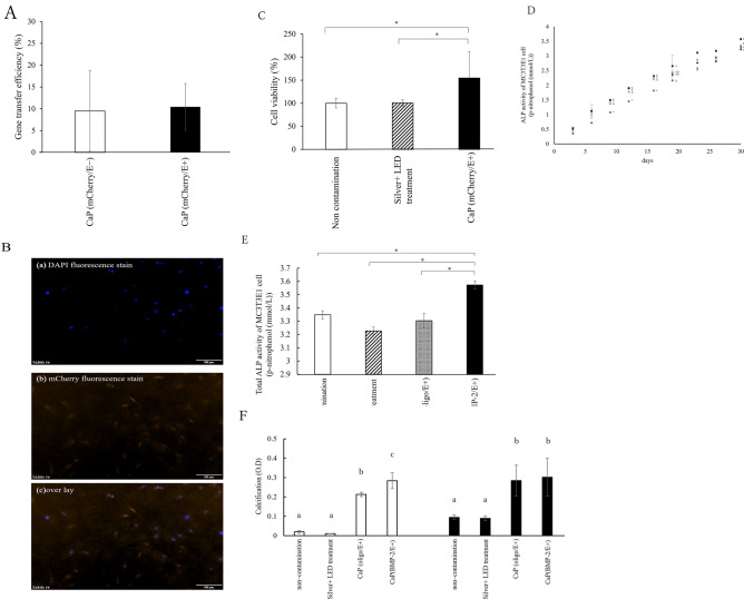
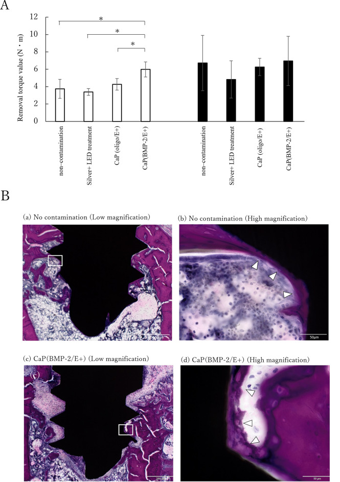
The aim of this study was to investigate the bactericidal effect and recovery of biocompatibility of contaminated titanium surfaces using a combination treatment involving silver, copper, or iron ion application along with 400 nm purple-LED light irradiation. Additionally, the study sought to develop a functional calcium phosphate (CaP) coating treatment on titanium surfaces following disinfection, to promote re-osseointegration. A purple-LED emitting light at 400 nm was utilized to irradiate Staphylococcus aureus suspensions and biofilms in the presence of various concentrations of silver, copper, and iron solutions for 1 min. The bactericidal effect and electron spin resonance (ESR) spectrum were subsequently evaluated. Additionally, the hydrophilicity of the titanium surface and cell viability of MC3T3-E1 cells after combination treatment with silver ion was evaluated. Furthermore, a titanium surface coating with CaP gene transfection carrier containing plasmid DNA was developed using an electric current. The activity of hard tissue formation was then evaluated both in vitro and in vivo post-treatment. The bactericidal effect of the combination treatment with silver ions was attributed to the generation of hydroxyl radicals, whereas the effects from iron and copper treatments were not radical-mediated. The silver treatment significantly restored the hydrophilicity and cell affinity of the titanium surface. Moreover, CaP coating applied via an electric current (30 µA for 5 min) enhanced hard tissue formation activity on the titanium surface in both in vitro and in vivo settings. The combination treatment utilizing silver ions and purple-LED irradiation significantly enhanced bactericidal effects by generating high levels of hydroxyl radicals. Additionally, coating the titanium surface with functionalized CaP promoted early osseointegration, suggesting a promising strategy for improving implant outcomes.

Keywords: Calcium phosphate; Gene transfection; Hydroxyl radicals; Purple LED; Silver; Titanium surface.


Conflict of interest statement

Declarations. Competing interests: The authors declare no competing interests. Ethics statement: All methods are reported in accordance with ARRIVE guidelines ( https://arriveguidelines.org ).

Figures

Fig. 1
Fig. 1
(A) Bactericidal effect of Ag-purple LED treatment on suspended bacteria. Symbol ●, ▲, and × demonstrate Silver + LED treatment, Silver − No LED treatment, and Control (water) − No LED groups, respectively. *Significant differences compared to Control (water)-No LED group (p < 0.05). §Significant differences between Silver + LED treatment and Silver-No LED treatment groups (p < 0.05). (B) Log reduction (●) between Silver + LED treatment group and Control (water)-No LED groups, (▲) Silver-No LED treatment group and Control (water)-No LED groups, (◆) Silver + LED treatment group and Silver-No LED treatment group. (C) Bactericidal effect of Cu-purple LED treatment on suspended bacteria Symbols ●, ▲, and × demonstrate Copper + LED treatment, Copper-No LED treatment, and Control (water)-No LED groups, respectively. *Significant differences compared to Control (water)-No LED group (p < 0.05). §Significant differences between Copper + LED treatment and Copper-No LED treatment groups (p < 0.05). (D) Log reduction (●) between Copper + LED treatment group and Control (water)-No LED groups, (▲) Copper-No LED treatment group and Control (water)-No LED groups, (◆) Copper + LED treatment group and Copper-No LED treatment group. (E) Bactericidal effect of Fe-purple LED treatment on suspended bacteria Symbols ●, ▲, and × demonstrate Iron + LED treatment, Iron-No LED treatment, and Iron-No LED treatment groups, respectively. *Significant differences compared to Control (water)-No LED group (p < 0.05). §Significant differences between Iron + LED treatment and Iron-No LED treatment groups (p < 0.05). (F) Log reduction (●) between Iron + LED treatment group and Control (water)-No LED groups, (▲) Iron-No LED treatment group and Control (water)-No LED groups, (◆) Iron + LED treatment group and Iron-No LED treatment group. (G) Representative electron spin resistance (ESR) spectra of the radicals generated by (a) S. a-Ag, (b) S. a-Ag-LED, (c) S. a-Fe, (d) S. a-Fe-LED, (e) S. a-Cu, (f) S. a-Cu-LED, and (g) S. a-LED. (H) Radical generation at metal ion concentrations Symbols ■, ▩, and □ demonstrate Silver + LED treatment, Copper + LED treatment, and Iron + LED treatment groups, respectively. *Significant differences between Silver + LED treatment and Copper + LED treatment groups (p < 0.05). §Significant differences between Iron + LED treatment and Copper + LED treatment groups (p < 0.05). (I) Comparison of cell viability of the combination treatment (Silver + LED), silver ion application (Silver alone), or purple LED irradiation (LED alone). No significant differences were noted among all groups.
Fig. 2
Fig. 2
(A) Generated radicals in silver ion solution with and without bacterial suspension and purple LED irradiation. *Significant differences (p < 0.05). (B) Radicals generated in silver ion solution with and without biofilms on titanium disks, under purple LED irradiation. *Significant difference (p < 0.05). + indicates application or irradiation. (C) Representative electron spin resistance (ESR) spectra of the radicals generated by the following treatments: (a) bacterial suspension(−) silver application(+) Purple-LED irradiation(−), (b) bacterial suspension(−) silver application(+) Purple-LED irradiation(+), (c) bacterial suspension(+) silver application(+) Purple-LED irradiation(−), and (d) bacterial suspension(+) silver application(+) Purple-LED irradiation(+). (D) Representative ESR spectra of the radical generated by the following treatments: (a) Titanium with biofilm(−) silver application(+) Purple-LED irradiation(−), (b) Titanium with biofilm(−) silver application(+) Purple-LED irradiation(+), (c) Titanium with biofilm(+) silver application(+) Purple-LED irradiation(−), and (d) Titanium with biofilm(+) silver application(+) Purple-LED irradiation(+).
Fig. 3
Fig. 3
(A) Scanning electron microscopy (SEM) images of titanium surfaces in a) non-contamination group, b) biofilm, c) Silver-No LED treatment group, and d) Silver-LED treatment group. Symbols ⇒ and △ shows bacteria and granule deposits, respectively. (B) Bactericidal effects of silver ion on biofilm on titanium. Symbols ●, ▲, and × demonstrate Silver + LED treatment, Silver-No LED treatment, and Control (water)-No LED groups, respectively. *Significant differences between Silver + LED treatment and Control (water)-No LED groups (p < 0.05). §Significant differences between Silver + LED treatment and Silver-No LED treatment groups (p < 0.05). (C) Log reduction (●) between Silver + LED treatment group and Control (water)-No LED groups, (▲) Silver-No LED treatment group and Control (water)-No LED groups, (◆) Silver + LED treatment group and Silver-No LED treatment group. (D) Evaluation of contact angle in non-contamination, bacteria-attached group, Silver-No LED treatment group, and Silver-No LED treatment group. * Significant difference (P < 0.05). (E) An example of contact angle of titanium disk in a) Non contamination group: 17.4°, b) Biofilm group: 40.1°, c) Silver-No LED treatment group: 22.4°, and d) Control (water)-No LED group: 21.9°.
Fig. 4
Fig. 4
Comparison of cell viability under conditions of silver application and/or LED treatment. *Significant difference (p < 0.05).
Fig. 5
Fig. 5
(A) Scanning electron microscopy (SEM) images of a) CaP(oligo), b) titanium surface c) CaP(oligo) coating on titanium surface. (B) CaP adhesion rate on titanium surface based on different current values. (C) CaP adhesion rate on titanium surface based on different energization times at 30 μA. (D) CaP adhesion rate on titanium surface based on different coating conditions. (E) Comparison of CaP adhesion rates under different titanium surface conditions with and without Ag-purple LED treatment. *Significant difference (p < 0.05); CaP calcium phosphate nanoparticles.
Fig. 6
Fig. 6
(A) Gene transfection efficiency of CaP loaded with plasmid DNA encoding mCherry with CaP (mCherry/E+) and without CaP (mCherry/E−), electric current set to 30 μA. (B) Example images of a) DAPI fluorescence stain, b) mCherry fluorescence stain, and c) overlay of the gene transfection efficiency of CaP on the titanium surface after Silver + LED treatment. (C) Viability of MC3T3E1 cells on treated titanium surface after 3 days of application with CaP (mCherry) with electronic current set at 30 μA. *Significant difference (p < 0.05). (D) ALP activity of MC3T3E1 cells after the application of CaP (Oligo) or CaP(BMP-2) with electronic current at 30 μA. Symbols ×, ●, ◆, and ■□ represent non-contamination, Silver + LED treatment, CaP(oligo/E+), and CaP(BMP-2/E+), respectively. Significant differences (p < 0.05) between groups are denoted by different superscript letters (the same letter indicates no significant difference). (E) Total ALP activity of MC3T3E1 cells after 30 days the application of CaP (Oligo) or CaP (BMP-2) with electronic current at 30 μA. *Significant difference (p < 0.05). (F) Cell calcification on the titanium surface treated with Silver + LED treatment followed by the application of CaP (Oligo) or CaP (BMP-2) with electronic current set at 30 μA in MC3T3E1 cells at 14 (□) or 28 days (■□). Significant differences (p < 0.05) between groups are denoted by different superscript letters (the same letter is not significantly different).
Fig. 7
Fig. 7
(A) Removal torque test in rat tibia 14 days (□) and 28 days (■□) post-implantation. *denotes significant differences (p < 0.05). (B) Histological images of the tissue surrounding implanted titanium screws 28 days after implantation in the rat tibia. Histological assessment of the non-contamination titanium screw (a,b) and titanium screws in the CaP(BMP-2/E +) group (c,d) after staining with hematoxylin and eosin (H–E). Scale bars: 500 μm (a, c) and 50 μm (b, d). H&E-stained histology around the titanium screws 28 days post-implantation in rat femurs. △ shows direct bone attachment on the implant screw surface.

Similar articles

References

    1. Duong, H. Y. et al. Oral health-related quality of life of patients rehabilitated with fixed and removable implant-supported dental prostheses. Periodontol 200088(1), 201–237 (2022). - PMC - PubMed
    1. Zhu, Y. et al. Prediction models for the complication incidence and survival rate of dental implants—A systematic review and critical appraisal. Int. J. Implant. Dent.11(1), 5 (2025). - PMC - PubMed
    1. Wada, M. et al. Prevalence and risk indicators for peri-implant diseases: A literature review. Jpn. Dent. Sci. Rev.57, 78–84 (2021). - PMC - PubMed
    1. Hasuike, et al. Evidence and future challenges for diagnosis, risk factors, and treatment of peri-implantitis. J. Japan. Assoc. Periodontol.65, 81–92 (2023).
    1. Almoharib, H. Erbium-doped yttrium aluminium garnet (Er:YAG) lasers in the treatment of peri-implantitis. Cureus.17(1), e78279 (2025). - PMC - PubMed

LinkOut - more resources