. 2025 Apr 21;15(1):13678.
doi: 10.1038/s41598-025-97164-3.

The primary somatosensory sensory cortex-basolateral amygdala pathway contributes to comorbid depression in spared nerve injury-induced neuropathic pain

Affiliations

The primary somatosensory sensory cortex-basolateral amygdala pathway contributes to comorbid depression in spared nerve injury-induced neuropathic pain

Guo Chen et al. Sci Rep. .

Abstract

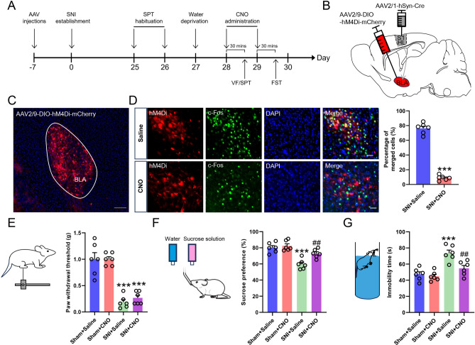
Comorbid depression in chronic pain is a prevalent health problem, yet the underlying neural mechanisms remain largely unexplored. This study identified a dedicated neural circuit connecting the hind limb region of the primary somatosensory cortex (S1HL) to the basolateral amygdala (BLA) that mediated neuropathic pain-induced depression. We demonstrated that depressive-like behaviors in the chronic phase of a mouse neuropathic pain model were associated with heightened activity in the S1HL and BLA. Using viral tracing and RNAscope in situ hybridization, we characterized the circuit architecture of S1HL glutamatergic projections to BLA cholecystokinin (CCK) neurons (S1HLGlu → BLACCK). In vivo fiber photometry calcium imaging revealed that both the S1HL BLA-projecting afferents and the BLA S1HL-innervating neurons exhibited hyperactivity in neuropathic pain-induced depressive states. Chemogenetic inhibition of the S1HL → BLA circuit could block neuropathic pain-induced depressive-like behaviors. In addition, specific knockdown of CCK expression in BLA S1HL-innervating neurons alleviated these depressive-like behaviors. Our findings demonstrated that the cortical-amygdala circuit S1HLGlu → BLACCK drove the transition from chronic pain to depression, thus suggesting a potential neural circuit basis for treating chronic pain-related depressive disorders.

Keywords: Basolateral amygdala; Cholecystokinin; Chronic pain; Depression; Primary somatosensory cortex.


Conflict of interest statement

Declarations. Competing interests: The authors declare no competing interests.

Figures

Fig. 1
Fig. 1
SNI induced pain sensitization and depression-like behavior. (A) Schedule for the SNI procedure and subsequent behavioral experiments. (B) Ipsilateral and contralateral paw withdrawal thresholds of mice in Sham, SNI-2d, SNI-7d, and SNI-28d groups. (C) Sucrose preference of mice in Sham, SNI-2d, SNI-7d, and SNI-28d groups. (D) Immobility time of mice in Sham, SNI-2d, SNI-7d, and SNI-28d groups. Data were presented as mean ± SEM, ***P < 0.001 versus Sham group; ##P < 0.01, ###P < 0.001 versus SNI-2d group; &P < 0.05 versus SNI-7d group, n = 6. SNI: spared sciatic nerve injury, VF: Von Frey test, SPT: sucrose preference test, FST: force swimming test, SNI-2d: 2 days after SNI, SNI-7d: 7 days after SNI, SNI-28d: 28 days after SNI.
Fig. 2
Fig. 2
SNI indued the activation of S1HL and BLA. (AC) Immunofluorescence staining showed the expression of c-Fos in the contralateral S1HL and BLA of sham and SNI mice (Scale bar = 200 μm/50 μm), data were expressed as mean ± SEM, ***P < 0.001 versus Sham group; ###P < 0.001 versus SNI-2d group; &&&P < 0.001 versus SNI-7d group, n = 6.
Fig. 3
Fig. 3
The establishment of S1HLGlu → BLACCK circuit. (A1) Schematic of retrograde tracing strategies used to validate the distribution of BLA-projecting neurons of S1; (A2) Representative image of AAV2/Retro-EGFP injection site in BLA; (A3-4) Representative image of AAV2/Retro-EGFP marked the S1 BLA-projecting soma. (B) RNAscope in situ hybridization showed the colocalization of S1HL BLA-projecting neurons (green) with Slc17a7 mRNA (red). (C1) Schematic of anterograde tracing strategies used to validate the distribution of S1HL-innervating neurons of BLA; (C2) Representative image of AAV2/9-DIO-EGFP injection site in BLA; (C3) Representative image of AAV2/9-DIO-EGFP marked the BLA S1HL-innervating neurons. (D, E) RNAscope in situ hybridization showed the colocalization of BLA S1HL-innervating neurons (green) with CCK mRNA and Slc17a7 (red). (F) Immunofluorescence staining showed the expression of c-Fos in the BLA-projecting neurons of contralateral S1HL of sham and SNI mice, data were expressed as mean ± SEM, ***P < 0.001 versus Sham group; ###P < 0.001 versus SNI-2d group; &&P < 0.01 versus SNI-7d group, n = 6. (Scale bar = 200 μm/50 μm).
Fig. 4
Fig. 4
Hyperexcitation of S1HL → BLA circuit in mice with neuropathic pain-induced depression. (A) Schematic diagram of fiber photometry used to examine the activity of S1HL BLA-projecting afferents. (BC) Representative image showed the expression of GCaMP7f. in S1HL and the optical fiber configuration. (DF) Example trace and heatmap of afferent Ca2+ signals under Von Frey stimulus in Sham, SNI-7d, and SNI-28d groups. (G) Changes of peak ΔF/F (%) under Von Frey stimulus in Sham, SNI-7d, and SNI-28d groups. (H) Schematic diagram of fiber photometry used to examine the activity of BLA S1HL-innervating neurons. (I) Representative image showed the expression of GCaMP7f in BLA and the optical fiber configuration. (J-L) Example trace and heatmap of neuronal Ca2+ signals under forced swimming stress in Sham, SNI-7d, and SNI-28d groups. (M) Changes of peak ΔF/F (%) under forced swimming stress in Sham, SNI-7d, and SNI-28d groups. Data were presented as mean ± SEM, *P < 0.05, ***P < 0.001 versus pre-stimulus or out water phase of same group; #P < 0.05, ###P < 0.001 versus Sham group; &P < 0.05 &&&P < 0.001 versus SNI-7d group, n = 5. (Scale bar = 100 μm).
Fig. 5
Fig. 5
Chemogenetic inhibition of the S1HL BLA-projecting neurons alleviated the depressive-like behaviors of SNI mice. (A) Schedule of experiments. (B) Schematic diagram of chemogenetic inhibition of S1HL BLA-projecting neurons with intra-BLA injection of AAV2/retro-hSyn-Cre and intra-S1HL injection of AAV2/9-DIO-hM4Di-mCherry. (C) Representative image showed the expression of AAV2/9-DIO-hM4Di-mCherry in S1HL. (D) Immunofluorescence staining showed the expression of c-Fos in the hM4Di-expressing neurons of S1HL after CNO or saline i.p. administration. (E) Ipsilateral paw withdrawal thresholds of sham and SNI mice with or without chemogenetic inhibition of S1HL BLA-projecting neurons. (F) Sucrose preference in SPT of sham and SNI mice with or without chemogenetic inhibition of S1HL BLA-projecting neurons. (G) Immobility time in FST of sham and SNI mice with or without chemogenetic inhibition of S1HL BLA-projecting neurons. Data are mean ± SEM, **P < 0.01 ***P < 0.001 Sham + Saline group versus SNI + Saline group or Sham + CNO group versus SNI + CNO group; #P < 0.01 SNI + Saline group versus SNI + CNO group, n = 6. VF: Von Frey test, SPT: sucrose preference test, FST: force swimming test. VF: Von Frey test, SPT: sucrose preference test, FST: force swimming test. (Scale bar = 200 μm/50 μm).
Fig. 6
Fig. 6
Chemogenetic inhibition of the BLA S1HL-innervating neurons ameliorated the depressive-like behaviors of SNI mice. (A) Schedule of experiments. (B) Schematic diagram of chemogenetic inhibition of BLA S1HL-innervating neurons with intra-S1HL injection of AAV2/1-hSyn-Cre and intra-BLA injection of AAV2/9-DIO-hM4Di-mCherry. (C) Representative image showed the expression of AAV2/9-DIO-hM4Di-mCherry in BLA. (D) Immunofluorescence staining showed the expression of c-Fos in the hM4Di-expressing neurons of BLA after CNO or saline i.p. administration. (E) Ipsilateral paw withdrawal thresholds of sham and SNI mice with or without chemogenetic inhibition of BLA S1HL-innervating neurons. (F) Sucrose preference in SPT of sham and SNI mice with or without chemogenetic inhibition of BLA S1HL-innervating neurons. (G) Immobility time in FST of sham and SNI mice with or without chemogenetic inhibition of BLA S1HL-innervating neurons. Data are mean ± SEM, ***P < 0.001 Sham + Saline group versus SNI + Saline group or Sham + CNO group versus SNI + CNO group; ##P < 0.01 SNI + Saline group versus SNI + CNO group, n = 6. VF: Von Frey test, SPT: sucrose preference test, FST: force swimming test. (Scale bar = 200 μm/50 μm).
Fig. 7
Fig. 7
CCK was responsible for the BLA S1HL-innervating neurons-mediated depressive-like behaviors after SNI. (A) Schedule of experiments. (B) Schematic diagram of specific ablation of CCK mRNA expression in BLA S1HL-innervating neurons with intra-S1HL injection of AAV2/1-hSyn-Cre and intra-BLA injection of AAV2/9-DIO-CCK-KD-mCherry or AAV2/9-DIO-CCK-NC-mCherry. (C) Representative image showed the expression of AAV2/9-DIO-CCK-KD-mCherry in BLA. (D) RNAscope staining and immunofluorescence co-staining showed the expression of CCK mRNA (green) in the S1HL-innervated BLA neurons (red) after viral injection, data are mean ± SEM, ***P < 0.01 CCK-NC group versus CCK-KD group, n = 6. (E) Ipsilateral paw withdrawal thresholds of sham and SNI mice with the intra-BLA injection of AAV2/9-DIO-CCK-KD-mCherry or AAV2/9-DIO-CCK-NC-mCherry. (F) Sucrose preference in SPT of sham and SNI mice with the intra-BLA injection of AAV2/9-DIO-CCK-KD-mCherry or AAV2/9-DIO-CCK-NC-mCherry. (G) Immobility time in FST of sham and SNI mice with the intra-BLA injection of AAV2/9-DIO-CCK-KD-mCherry or AAV2/9-DIO-CCK-NC-mCherry. Data are expressed as mean ± SEM, ***P < 0.01 Sham + CCK-NC group versus SNI + CCK-NC group or Sham + CCK-KD group versus SNI + CCK-KD group; #P < 0.05 SNI + CCK-NC group versus SNI + CCK-KD group, n = 6. VF: Von Frey test, SPT: sucrose preference test, FST: force swimming test. (Scale bar = 200 μm/50 μm).

Similar articles

References

    1. Cohen, S. P., Vase, L. & Hooten, W. M. Chronic pain: an update on burden, best practices, and new advances. Lancet397, 2082–2097. 10.1016/s0140-6736(21)00393-7 (2021). - PubMed
    1. Malhi, G. S. & Mann, J. J. Depression. Lancet392, 2299–2312. 10.1016/s0140-6736(18)31948-2 (2018). - PubMed
    1. Meda, R. T. et al. Chronic pain-induced depression: A review of prevalence and management. Cureus14, e28416. 10.7759/cureus.28416 (2022). - PMC - PubMed
    1. Zis, P. et al. Depression and chronic pain in the elderly: Links and management challenges. Clin. Interv. Aging12, 709–720. 10.2147/cia.s113576 (2017). - PMC - PubMed
    1. Emptage, N. P., Sturm, R. & Robinson, R. L. Depression and comorbid pain as predictors of disability, employment, insurance status, and health care costs. Psychiatr Serv56, 468–474. 10.1176/appi.ps.56.4.468 (2005). - PubMed

Substances

LinkOut - more resources