. 2025 May;27(5):790-800.
doi: 10.1038/s41556-025-01655-4. Epub 2025 Apr 21.

Mitochondrial NADPH fuels mitochondrial fatty acid synthesis and lipoylation to power oxidative metabolism

Affiliations

Mitochondrial NADPH fuels mitochondrial fatty acid synthesis and lipoylation to power oxidative metabolism

Dohun Kim et al. Nat Cell Biol. 2025 May.

Erratum in

Abstract

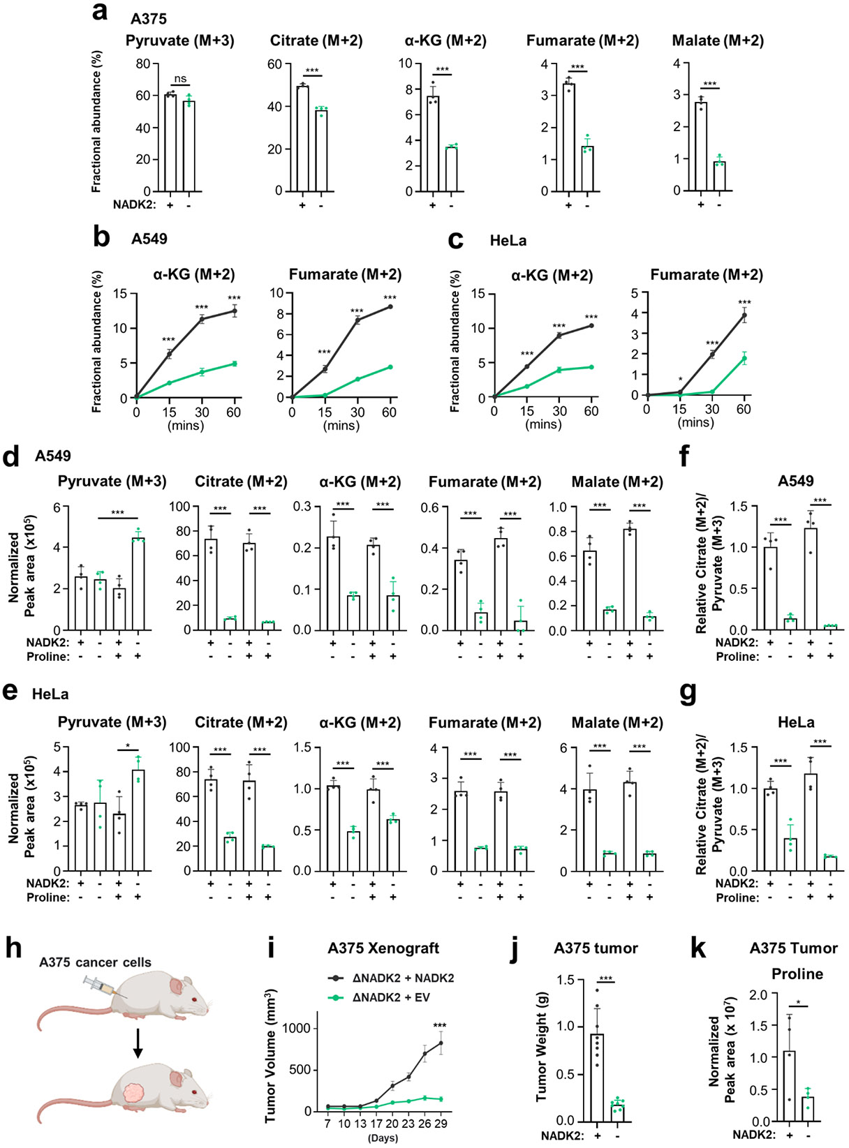
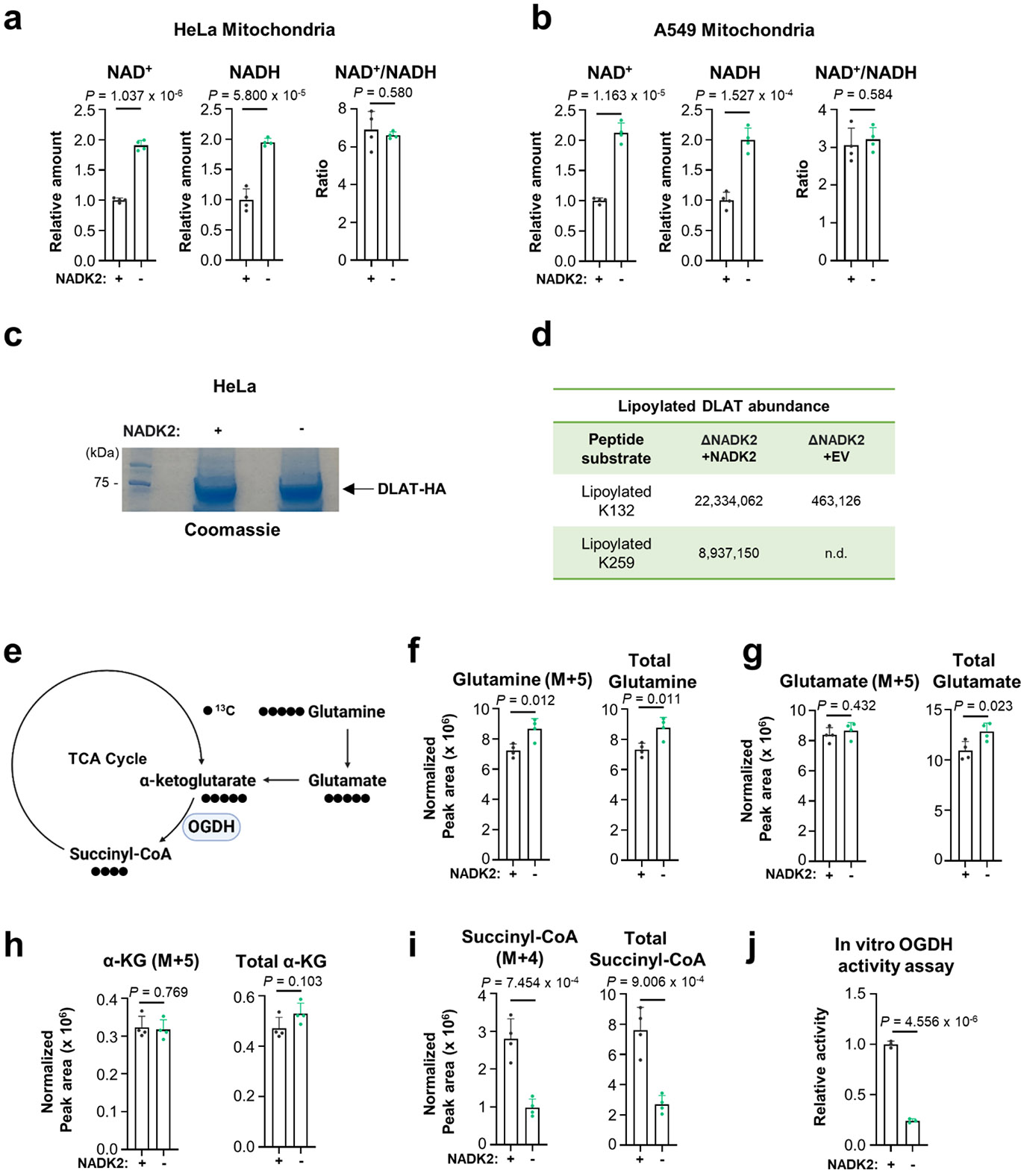
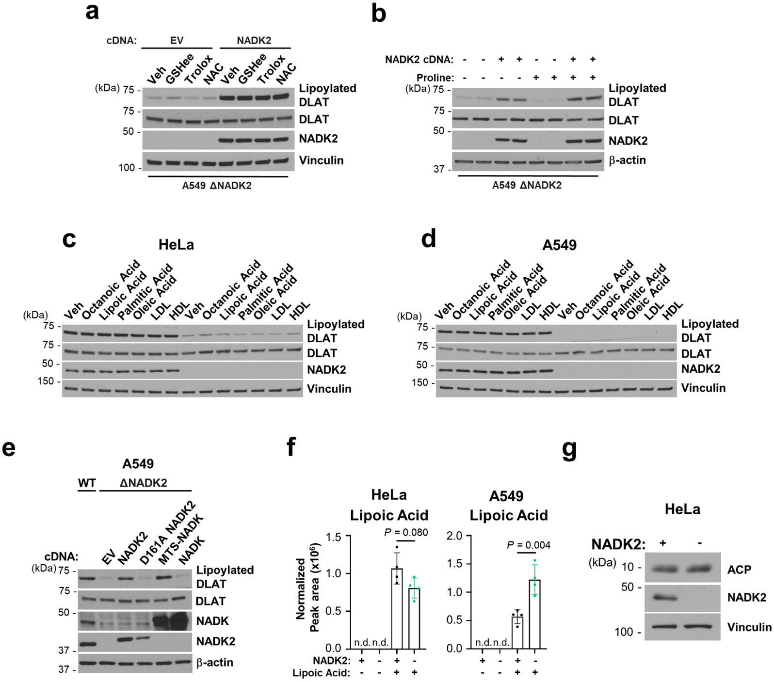
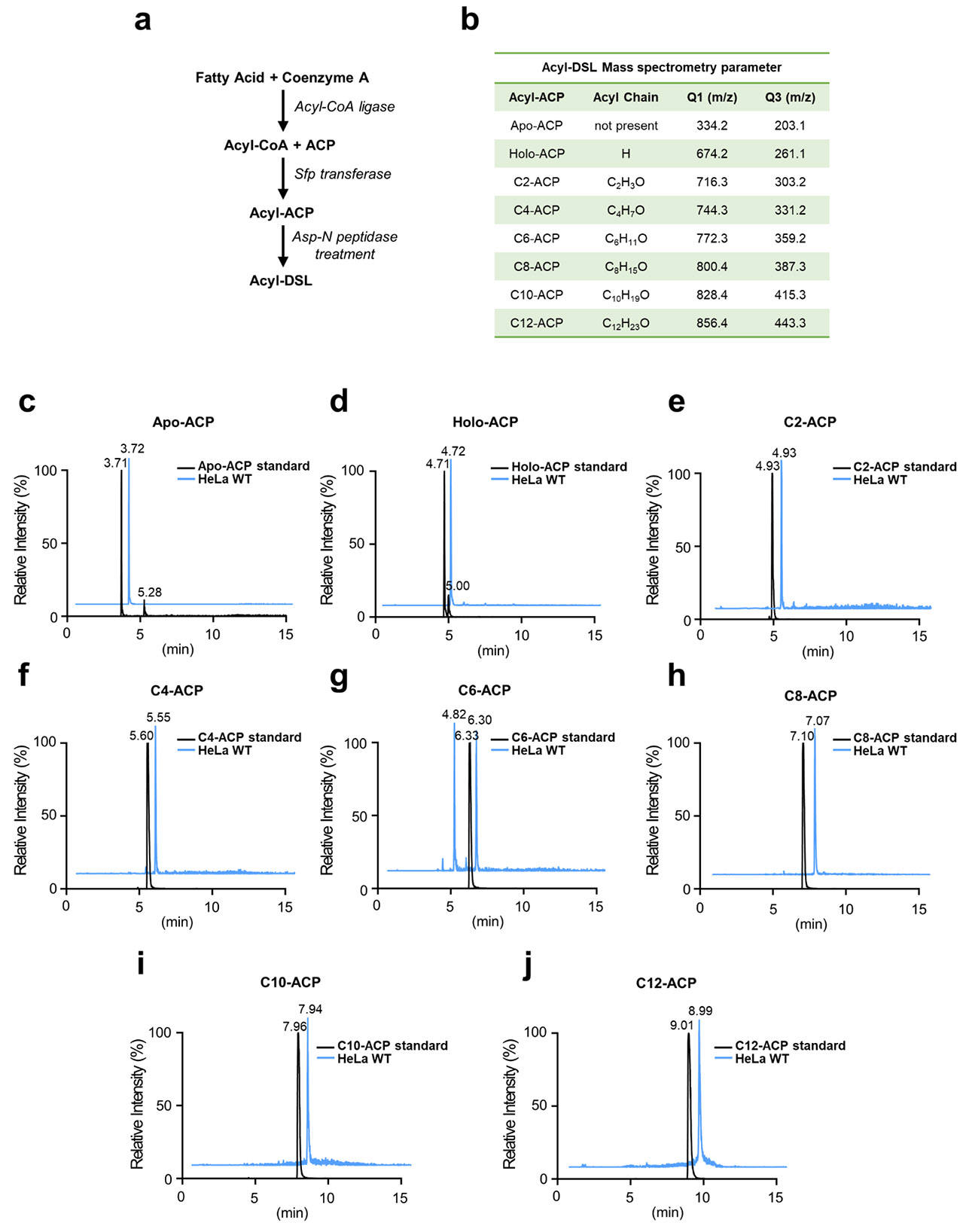
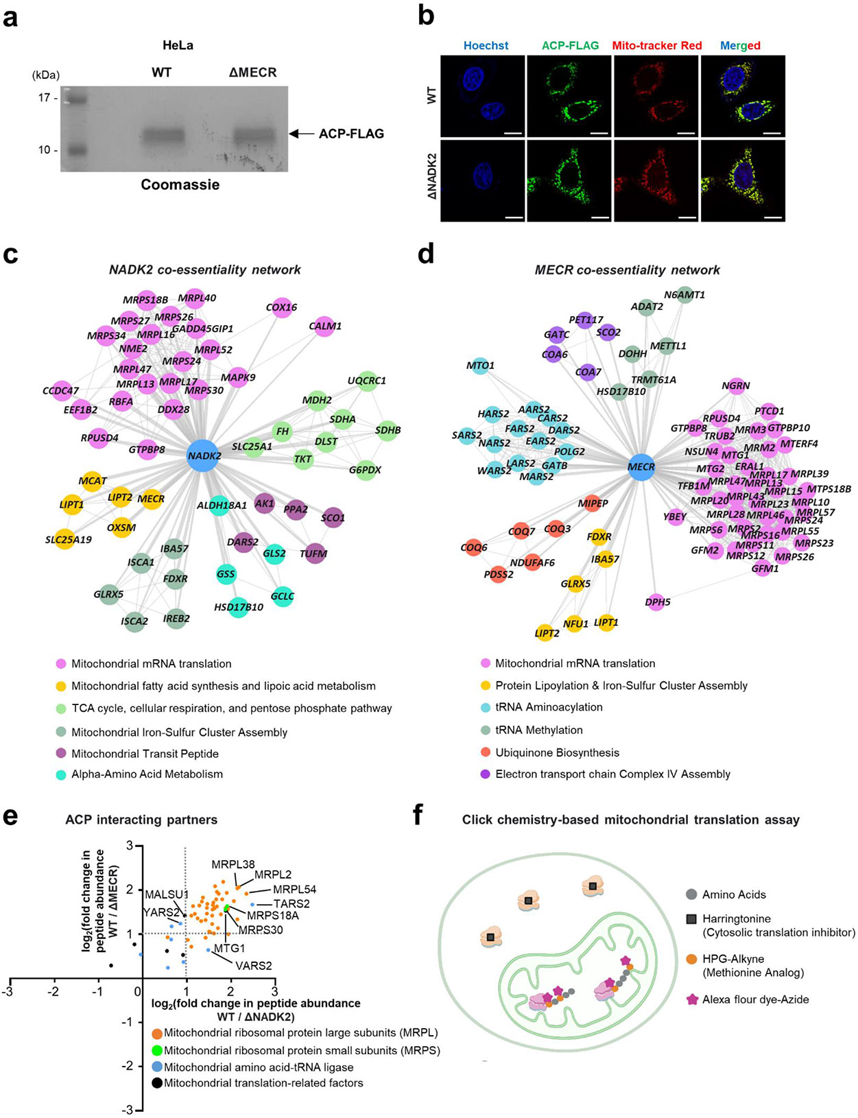
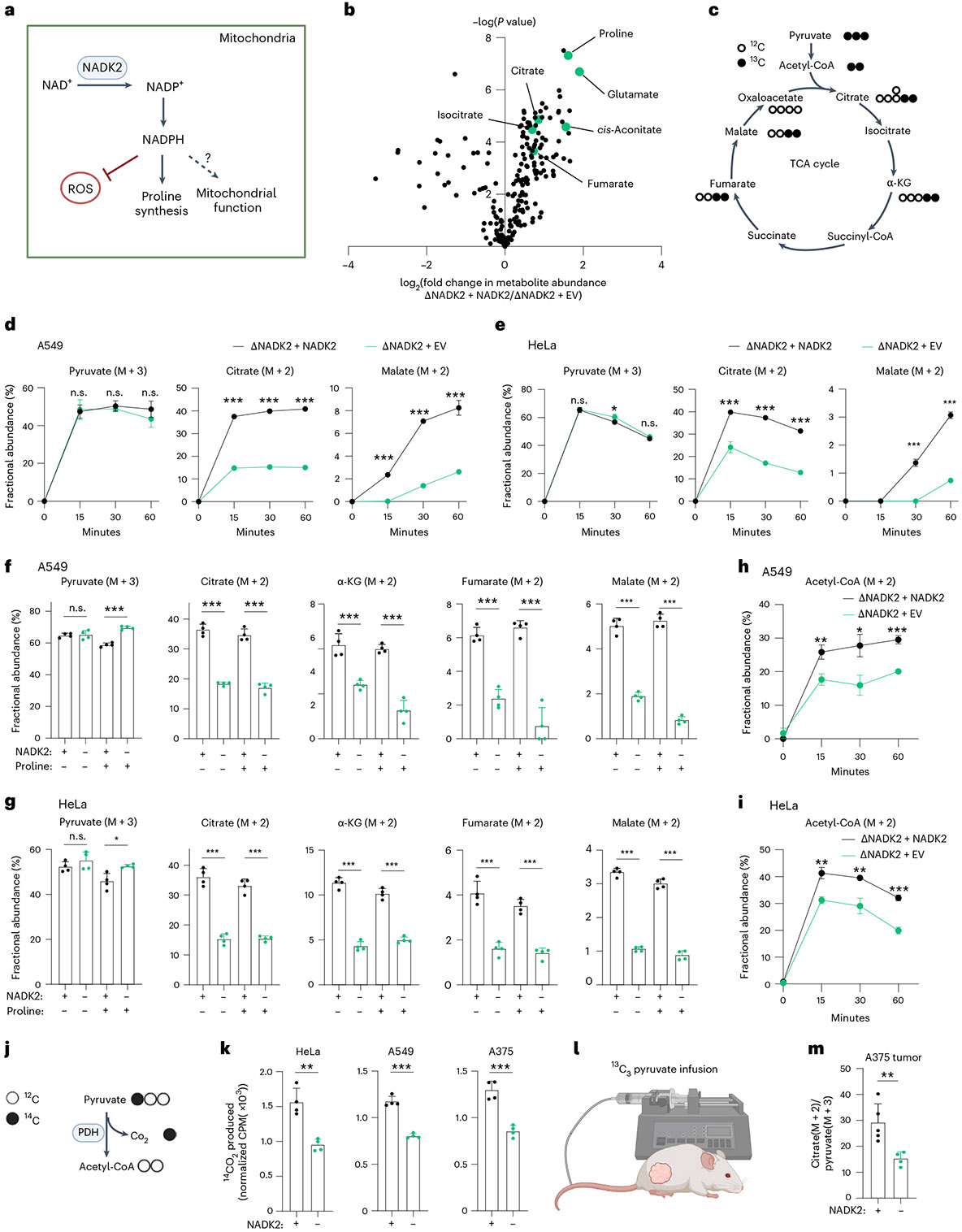
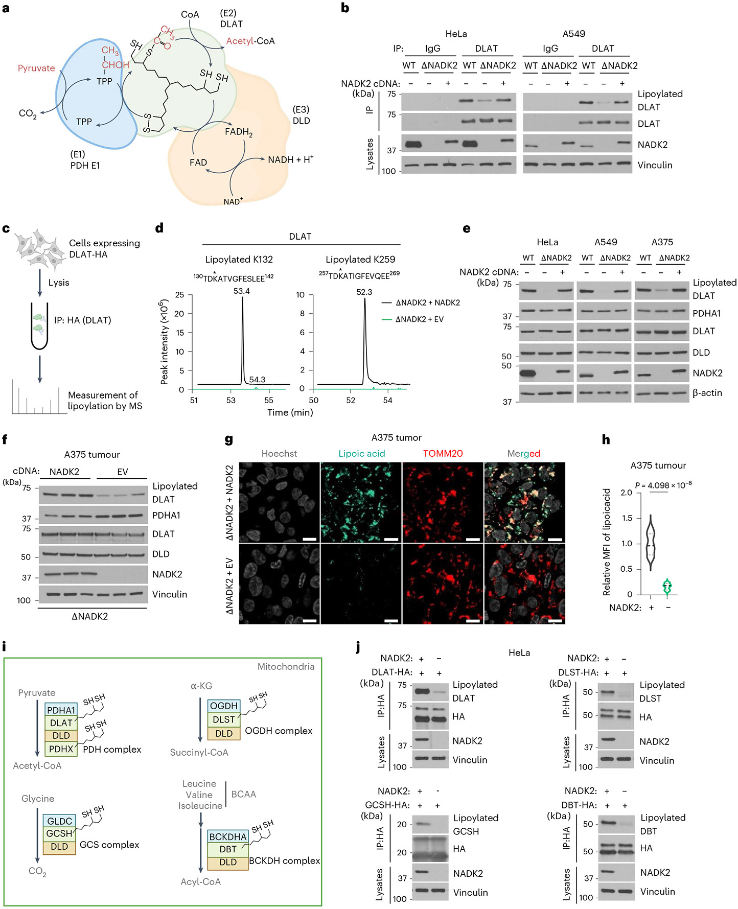
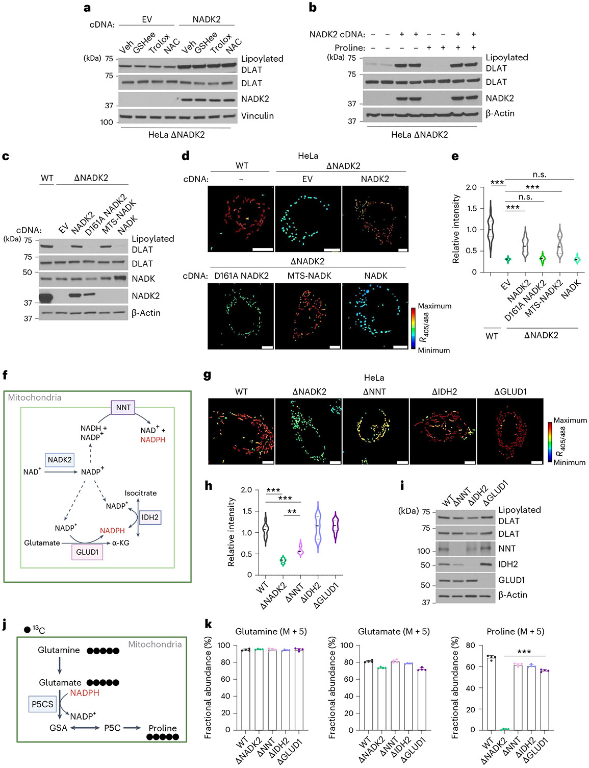
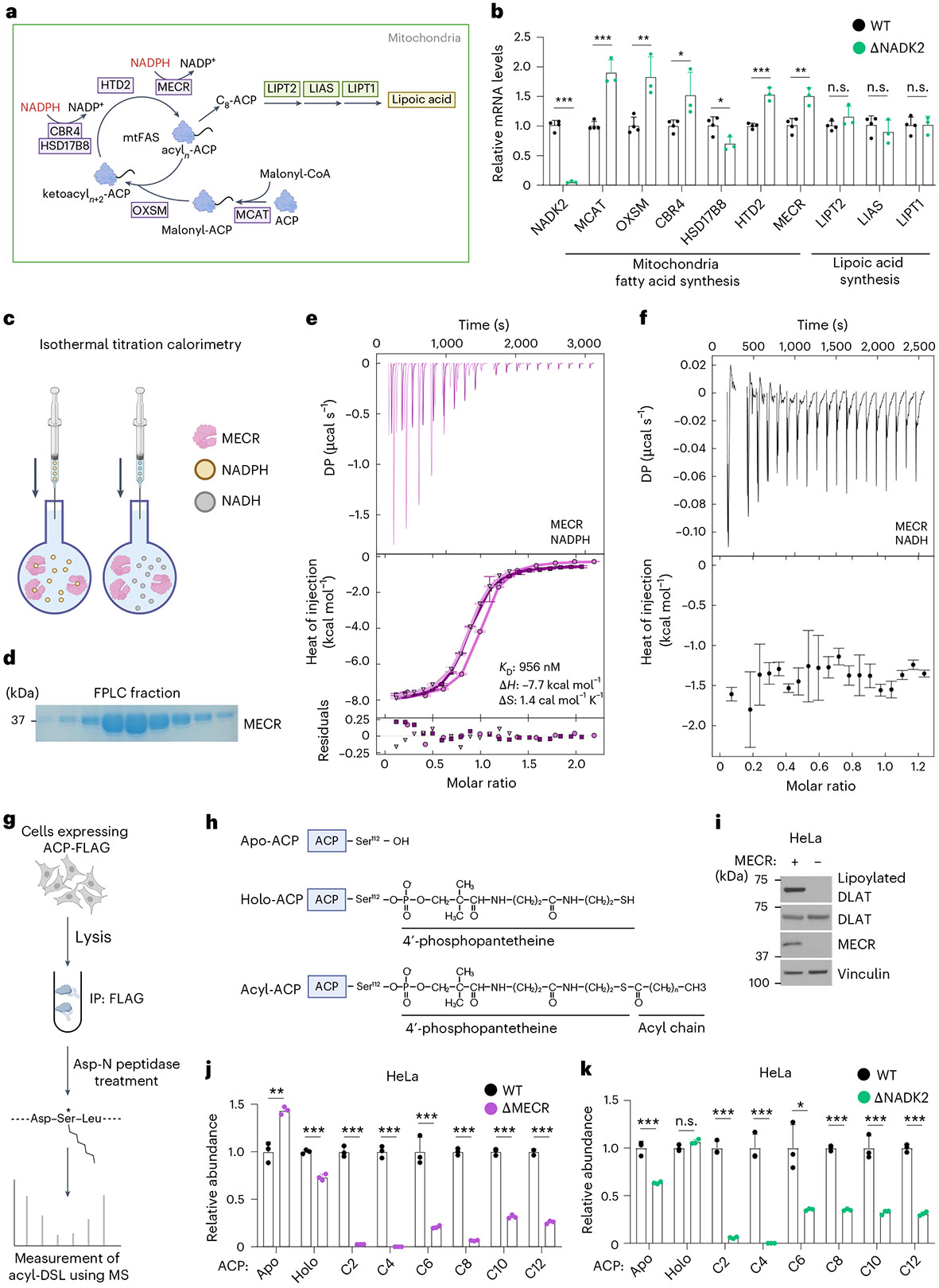
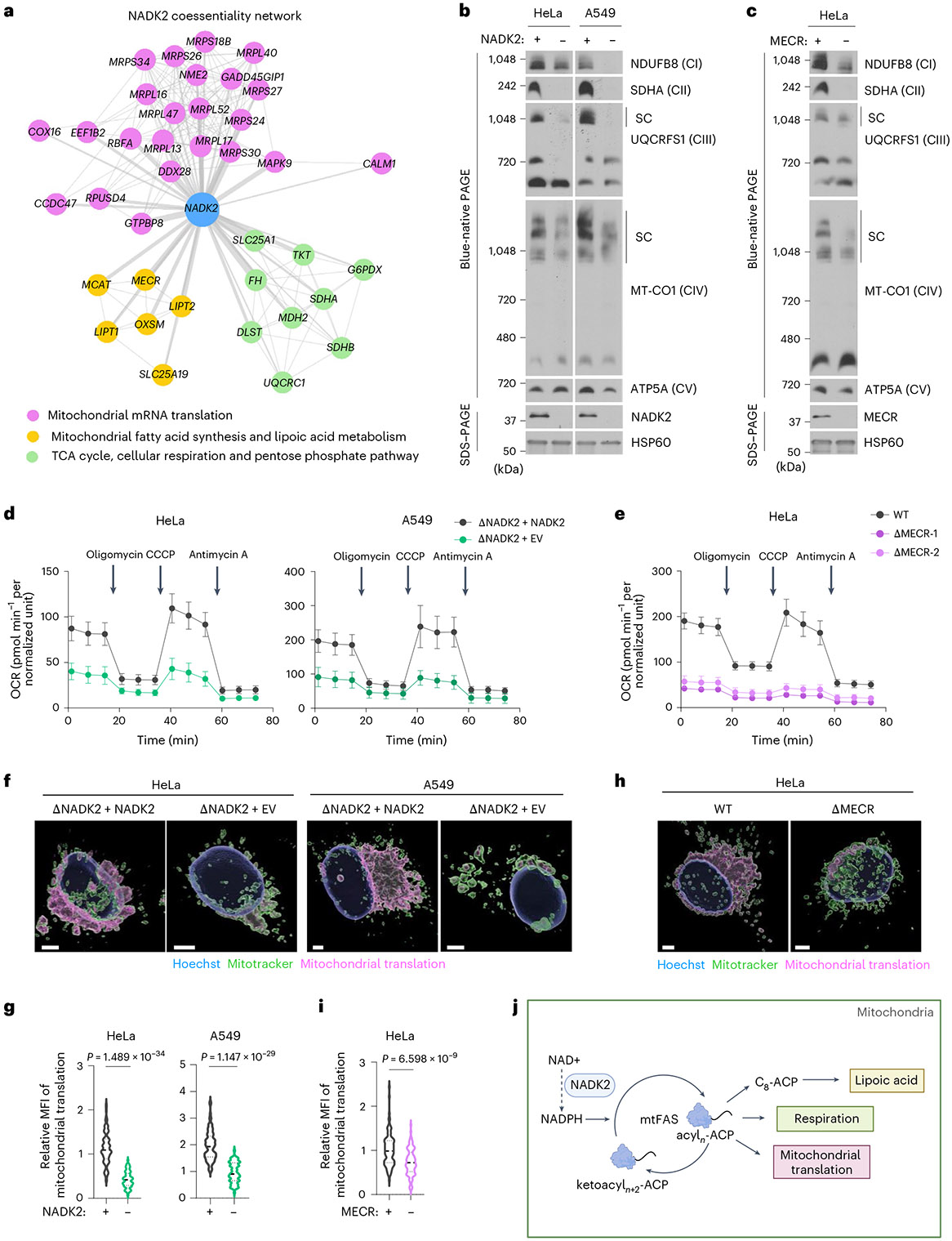
Nicotinamide adenine dinucleotide phosphate (NADPH) is a vital electron donor essential for macromolecular biosynthesis and protection against oxidative stress. Although NADPH is compartmentalized within the cytosol and mitochondria, the specific functions of mitochondrial NADPH remain largely unexplored. Here we demonstrate that NAD+ kinase 2 (NADK2), the principal enzyme responsible for mitochondrial NADPH production, is critical for maintaining protein lipoylation, a conserved lipid modification necessary for the optimal activity of multiple mitochondrial enzyme complexes, including the pyruvate dehydrogenase complex. The mitochondrial fatty acid synthesis (mtFAS) pathway utilizes NADPH for generating protein-bound acyl groups, including lipoic acid. By developing a mass-spectrometry-based method to assess mammalian mtFAS, we reveal that NADK2 is crucial for mtFAS activity. NADK2 deficiency impairs mtFAS-associated processes, leading to reduced cellular respiration and mitochondrial translation. Our findings support a model in which mitochondrial NADPH fuels the mtFAS pathway, thereby sustaining protein lipoylation and mitochondrial oxidative metabolism.


Conflict of interest statement

Competing interests: The authors declare no competing interests.

Figures

Extended Data figure 1.
Extended Data figure 1.. NADK2 supports pyruvate oxidation and tumor growth
(a) Fractional abundance (%) of the indicated TCA cycle metabolites from isogenic ΔNADK2 A375 cells expressing either empty vector or NADK2 and labeled with [13C3]-pyruvate for 30 minutes in 0.2 mM proline. (n = 4 biological replicates) (b) As in (a), but the experiments were performed in isogenic ΔNADK2 A549 cells expressing either empty vector or NADK2 and labeled with [13C3]-pyruvate in the presence of 0.2 mM proline for 15, 30, or 60 minutes. (n = 4 biological replicates). Related to Fig. 1d. (c) As in (b), but the experiments were performed in isogenic ΔNADK2 HeLa cells expressing either empty vector or NADK2. (n = 4 biological replicates). Related to Fig. 1e. (d) As in (b), but the cells were labeled with [13C3]-pyruvate for 1 hour. The peak area was normalized to protein abundance. (n = 4 biological replicates). (e) As in (d), but the experiments were performed in isogenic ΔNADK2 HeLa cells expressing either empty vector or NADK2. (n = 4 biological replicates). (f) Relative citrate (M+2)/ pyruvate (M+2) ratio from (d). (n = 4 biological replicates). (g) Relative citrate (M+2)/ pyruvate (M+2) ratio from (e). (n = 4 biological replicates). (h) Schematic of A375 tumor xenograft. Related to Fig. 1l,m. (i) Tumor growth from isogenic ΔNADK2 A375 cells expressing either empty vector or NADK2. (n = 8 biologically independent animals). Related to Fig. 1l,m. (j) The size of tumors derived from isogenic ΔNADK2 A375 cells expressing either empty vector or NADK2 at the end of the experiment. (n = 8 biologically independent tumors). Related to Fig. 1l,m. (k) Normalized proline abundance from tumors derived from isogenic ΔNADK2 A375 cells expressing either empty vector or NADK2. (n = 4 biologically independent tumors). Data are presented as the mean ± standard deviation (a-g, j, and k) or mean ± s.e.m. (i). *P < 0.05, **P < 0.01, and ***P < 0.001 were calculated using a two-tailed Student’s t-test (a-c, and i-k) or one-way ANOVA with Tukey’s post hoc test (d-g).
Extended Data figure 2.
Extended Data figure 2.. Impact of NADK2 on redox ratios and the TCA cycle
(a) The relative levels of NAD+ and NADH and NAD+/NADH ratio in mitochondria isolated from isogenic ΔNADK2 HeLa cells expressing either empty vector or NADK2. The cells were cultured in the presence of 0.2 mM proline. (n = 4 biologically independent replicates) (b) The relative levels of NAD+ and NADH and NAD+/NADH ratio in mitochondria isolated from isogenic ΔNADK2 A549 cells expressing either empty vector or NADK2. The cells were cultured in the presence of 0.2 mM proline. (n = 4 biologically independent replicates) (c) Coomassie staining of immunoprecipitated DLAT-HA from isogenic ΔNADK2 HeLa cells expressing either empty vector or NADK2 and transfected with DLAT-HA. The cells were cultured in the presence of 0.2 mM proline. The bands were cut out and subjected to mass spectrometry for detecting lipoylated peptides. (d) The abundance of lipoylated DLAT as described in (c). (e) Schematic of [13C5]-glutamine tracing. (f) The normalized peak area (labeled and total pools) of glutamine in isogenic ΔNADK2 HeLa cells expressing either empty vector or NADK2 labeled with [13C5]-glutamine for 1 hour. The peak areas were normalized to the protein amount assessed with a BCA assay. (n = 4 biological replicates). (g) As in (f), but the normalized peak area (labeled and total pools) of glutamate are shown. (n = 4 biological replicates) (h) As in (f), but the normalized peak area (labeled and total pools) of alpha-ketoglutarate are shown. (n = 4 biological replicates) (i) As in (f), but the normalized peak area (labeled and total pools) of succinyl-CoA are shown. (n = 4 biological replicates) (j) The relative OGDH activity from mitochondrial extracts of isogenic ΔNADK2 HeLa cells expressing either empty vector or NADK2, which were grown in DMEM containing 10% FBS and 0.2 mM proline. (n = 3 biological replicates). Data are presented as the mean ± standard deviation (a,b,f-j). *P < 0.05, **P < 0.01, and ***P < 0.001 were calculated using a two-tailed Student’s t-test (a,b,f-j).
Extended Data figure 3.
Extended Data figure 3.. Impact of ROS, proline, and lipids on protein lipoylation
(a) Immunoblots showing DLAT lipoylation, DLAT, NADK2, and vinculin levels from isogenic ΔNADK2 A549 cells expressing either empty vector or NADK2. Cells were grown in the presence of 0.2 mM proline and treated with the indicated antioxidants for 48 hours. GSHee; glutathione ethyl ester (5 mM), NAC; N-acetyl cysteine (1 mM), and Trolox (5 μM). (b) Immunoblotting was done as in (a), but cells were grown in the presence or absence of 0.2 mM proline. (c) Immunoblotting was done as in (a), but cells were grown in the presence of 0.2 mM proline and treated with octanoic acid (50 μM), lipoic acid (50 μM), palmitic acid (100 μM), oleic acid (100 μM), low-density lipoprotein (LDL, 50 μg/ml), or high-density lipoprotein (HDL, 50 μg/ml) for 48 hours. (d) Immunoblotting was done as in (a) but in ΔNADK2 A549 cells expressing either empty vector or NADK2. (e) Immunoblotting was done as in (a), but from WT or isogenic ΔNADK2 A549 cells expressing empty vector, NADK2, D161A NADK2, MTS-NADK, or cytosolic NADK. D161A NADK2: kinase-dead NADK2; MTS: Mitochondrial Targeting Sequence. (f) The abundance of intracellular lipoic acid from isogenic ΔNADK2 HeLa or A549 cells expressing either empty vector or NADK2 treated with either vehicle or lipoic acid (50μM) for 48 hours assessed via LC/MS. The peak areas were normalized to the protein amount. (n = 4 biologically independent replicates). Data are presented as the mean ± standard deviation. **P < 0.01 was calculated using a two-tailed Student’s t-test. (g) Immunoblots showing ACP, NADK2, and vinculin levels from WT or ΔNADK2 HeLa cells.
Extended Data figure 4.
Extended Data figure 4.. The synthesis and detection of acyl-ACP standards via LC/MS.
(a) Schematic illustrating workflow for synthesizing acyl-DSL standard for acyl-ACP measurement. (b) The mass spectrometry parameters of various acyl-DSLs. (c-j) The chromatogram of synthesized standards (black) and biological HeLa WT (blue) for Apo-ACP (c), Holo-ACP (d), C2-ACP (e), C4-ACP (f), C6-ACP (g), C8-ACP (h), C10-ACP (i), C12-ACP (j) run on LC/MS.
Extended Data figure 5.
Extended Data figure 5.. Impact of NADK2 and mtFAS on mitochondrial translation
(a) Coomassie staining of purified ACP-FLAG from WT and ΔMECR HeLa cells, which was then subjected to Asp-N peptidase treatment for assessment of acyl-ACP species via LC/MS. (b) Representative immunofluorescence images for ACP-FLAG (green) detected with anti-FLAG antibody MitoTracker Red (red), and nuclei (blue) stained with Hoechst for WT and ΔNADK2 HeLa cells cultured in the presence of 0.2 mM proline. Scale bars, 10 μm. (c) The NADK2 coessentiality network showing the top 6 enrichment gene clusters. (d) The MECR coessentiality network showing the top 6 enrichment gene clusters. (e) Volcano plot illustrating the log2 fold change of mitoribosome-related proteins identified in ACP immunoprecipitates from wild-type (WT), ΔNADK2, and ΔMECR HeLa cells. Proteins belonging to the mitochondrial ribosomal protein large subunit (MRPL) or small subunit (MRPS), as well as other significant interactors, are indicated and color-coded. (f) Schematic illustrating the workflow of click chemistry-based mitochondrial translation assay.
Fig. 1 |
Fig. 1 |. NADK2 loss impairs pyruvate oxidation.
a, A schematic illustrating the synthesis and roles of mitochondrial NADP(H). b, A volcano plot showing the log2 fold change of indicated metabolites from steady-state metabolomics of isogenic ΔNADK2 HeLa cells expressing an empty vector (EV) or NADK2, grown with 0.2 mM proline (n = 4 for ΔNADK2 + EV; n = 3 for ΔNADK2 + NADK2 biologically independent replicates). c, A schematic of [13C3]-pyruvate tracing. d, The fractional abundance (%) of the indicated TCA cycle metabolites from isogenic ΔNADK2 A549 cells expressing either EV or NADK2 and labelled with [13C3]-pyruvate in the presence of 0.2 mM proline for 15, 30 or 60 min (n = 4 biologically independent replicates). n.s., not significant. e, As in d, but the experiments were performed in isogenic ΔNADK2 HeLa cells expressing either EV or NADK2 (n = 4 biologically independent replicates). f, As in d, but the cells were labelled with [13C3]-pyruvate for 1 h (n = 4 biologically independent replicates). g, As in e, but the cells were labelled with [13C3]-pyruvate for 1 h (n = 4 biologically independent replicates). h, The fractional abundance (%) of acetyl-CoA (M + 2) from isogenic ΔNADK2 A549 cells expressing either EV or NADK2 and labelled with [13C3]-pyruvate in the presence of 0.2 mM proline for 15, 30 or 60 min. (n = 3 biologically independent replicates). i, As in h, but the experiments were performed in isogenic ΔNADK2 HeLa cells expressing either EV or NADK2 (n = 3 biologically independent replicates). j, A schematic of [1-14C]-pyruvate oxidation and 14CO2 release. k, Normalized counts per minute (CPM) of captured 14CO2 during a 2-h period from isogenic ΔNADK2 HeLa, A549 or A375 cells expressing either EV or NADK2 incubated with [1-14C]-pyruvate (n = 4 biologically independent replicates). l, A schematic of [13C3]-pyruvate intravenous infusion. m, Ratio of citrate (M + 2)/pyruvate (M + 3) from the tumours after intravenous infusion with [13C3]-pyruvate (n = 4 for ΔNADK2 + EV; n = 5 for ΔNADK2 + NADK2 biologically independent replicates). The data are presented as the mean ± standard deviation for di, k and m. *P < 0.05, **P < 0.01 and ***P < 0.001 were calculated using a two-tailed Student’s t-test for b, d, e, h, i, k and m or one-way ANOVA with Tukey’s post hoc test for f and g. Created with BioRender.com (a, j and l).
Fig. 2 |
Fig. 2 |. NADK2 is required for protein lipoylation.
a, A schematic illustrating the PDH complex components (PDHA1, DLAT and DLD) and the reaction intermediates. TPP, thiamine pyrophosphate. b, The immunoblots of indicated proteins from immunoprecipitates (IP) of endogenous DLAT or control (IgG) in wild-type (WT) or ΔNADK2 HeLa or A549 cells expressing either empty vector or NADK2. The cells were grown in 0.2 mM proline. c, A schematic of the workflow for assessing lipoylated peptides via mass spectrometry (MS). d, The chromatograms of lipoylated DLAT peptides from isogenic ΔNADK2 HeLa cells expressing either empty vector (EV) or NADK2 and transfected with DLAT-HA. e, Immunoblots showing DLAT lipoylation and indicated proteins from HeLa, A549 or A375 WT or NADK2-deficient cells expressing EV vector or NADK2 grown in the presence of 0.2 mM proline. Actin, loading control. f, As in e, but immunoblotting was done on tumours derived from isogenic ΔNADK2 A375 cells expressing either an EV or NADK2. g, Representative immunofluorescence images of protein lipoylation (cyan), TOMM20 (red) and nuclei (Hoechst, grey) in tumour slides from isogenic ΔNADK2 A375 cells expressing EV or NADK2. Scale bars, 10 μm. h, A relative MFI of the lipoylation signal from g. The P value was calculated using a two-tailed Student’s t-test (n = 8 different tumour slides). i, A schematic showing the five lipoylated protein subunits within distinct mitochondrial complexes. GCS, glycine cleavage system; BCKDH, branched-chain alpha-keto acid dehydrogenase. j, The immunoblots of the indicated proteins from purified HA-tagged DLAT, DLST, GCSH and DBT expressed in isogenic ΔNADK2 HeLa cells expressing either an EV or NADK2. Created with BioRender.com (a, c and i).
Fig. 3 |
Fig. 3 |. Mitochondrial NADPH, but not ROS, or proline, is required for protein lipoylation.
a, The immunoblots showing DLAT lipoylation and the indicated proteins from isogenic ΔNADK2 HeLa cells expressing an empty vector (EV) or NADK2. The cells were grown in the presence of 0.2 mM proline and treated with the indicated antioxidants for 48 h. Veh, vehicle (water); GSHee, glutathione ethyl ester (5 mM); NAC, N-acetyl cysteine (1 mM) and Trolox (5 μM). b, Immunoblotting done as in a but with cells grown in the presence or absence of 0.2 mM proline. c, Immunoblotting done as in a but from wild-type (WT) HeLa cells or isogenic ΔNADK2 HeLa cells expressing empty vector, NADK2, D161A NADK2, MTS-NADK or cytosolic NADK. D161A NADK2, kinase-dead NADK2. d, Representative images of the mitochondrial NADPH biosensor (MTS-iNap1) expressed in WT or isogenic ΔNADK2 HeLa cells expressing an EV, NADK2, kinase-dead D161A NADK2, MTS-NADK or cytosolic NADK. The colours of the images present the R405/R488 ratio. Scale bars, 5 μm. e, A quantification of MTS-iNap1 from d (n = 18 different cells). f, A schematic indicating mitochondrial NADP(H)-utilizing enzymes. n.s., not significant. g, Representative images of the mitochondrial NADPH biosensor as in d but from WT or ΔNADK2, ΔNNT, ΔIDH2 and ΔGLUD1 HeLa cells. Scale bars, 5 μm. h, A quantification of mitochondrial NADPH from images in g (n = 9 different cells). i, The levels of lipoylated DLAT and indicated proteins from HeLa WT or NNT, IDH2 and GLUD1 knockouts. j, A schematic of [13C5]-glutamine tracing into proline. k, The fractional abundance (%) of the glutamine (M + 5), glutamate (M + 5) and proline (M + 5) from HeLa WT or ΔNADK2, ΔNNT, ΔIDH2 and ΔGLUD1 labelled with [13C5]-glutamine for 3 h. The data are presented as the mean ± standard deviation (n = 4 biologically independent replicates). *P < 0.05, **P < 0.01 and ***P < 0.001 were calculated with one-way ANOVA test and Tukey’s post hoc test for e, h and k. Created with BioRender.com (f and j).
Fig. 4 |
Fig. 4 |. Mitochondrial NADPH sustains mtFAS.
a, A schematic indicating the mtFAS (pink) and the lipoic acid synthesis (green) pathways. b, The relative mRNA levels of NADK2 and mtFAS or lipoic acid synthesis enzymes from HeLa wild-type (WT) and ΔNADK2 cells grown in the presence of 0.2 mM proline (n = 4 for WT and n = 3 for ΔNADK2 biologically independent replicates). n.s., not significant. c, A schematic of an ITC measuring MECR binding affinity for NADPH and NADH. d, A Coomassie stain of purified MECR. FPLC, fast protein liquid chromatography. e, Top: The thermograms from three independent ITC experiments of MECR-NADPH. The corrected heat (μcal s−1) is plotted against time. Bottom: The corresponding binding isotherms for MECR-NADPH interactions. The integrated heat changes (kcal mol−1) are plotted against the molar ratio of NADPH:MECR. The dissociation constant (Kd = 1/Ka) is indicated in the graph. ΔH, enthalpy change; ΔS, entropy change. f, Top: a thermogram of one ITC experiment of MECR-NADH. Bottom: a corresponding binding isotherm for MECR-NADH interaction. No binding curve could be fitted. DP, differential power. g, A schematic illustrating the workflow for acyl-ACP measurement by LC–MS. Asterisks indicate acylation on the serine residue. h, The chemical structure of Apo-ACP, Holo-ACP and acyl-ACP. MS, mass spectrometry. i, Immunoblots showing DLAT lipoylation and the indicated proteins from WT or ΔMECR HeLa cells. j, The relative levels of the indicated ACP or acyl-ACP species in WT and ΔMECR HeLa cells transfected with ACP-FLAG. ACP-FLAG was purified and subjected to Asp-N peptidase to release the acyl-chains, which were then measured via LC–MS (n = 3 biological independent replicates). k, As in j, but the measurements were performed in WT and ΔNADK2 HeLa cells (n = 3 biological replicates). The data are presented as the mean ± standard deviation for b, e, f, j and k. *P < 0.05, **P < 0.01 and ***P < 0.001, were calculated using a two-tailed Student’s t-test for b, j and k. Created with BioRender.com (a, c, g and h).
Fig. 5 |
Fig. 5 |. NADK2 is crucial for cellular respiration and mitochondrial translation.
a, The NADK2 coessentiality network showing the top three enrichment gene clusters, including mitochondrial translation, mtFAS and lipoic acid metabolism and the TCA cycle and cellular respiration. b, An assembly of ETC complexes (CI, CII, CIII, CIV and CV) assessed by blue-native PAGE of isogenic ΔNADK2 HeLa or A549 cells expressing either empty vector (EV) or NADK2 from cells grown in the presence of 0.2 mM proline. SC; super complex. c, As in b, but the experiments were performed in wild-type (WT) and ΔMECR HeLa cells. d, The OCR of isogenic ΔNADK2 HeLa and A549 cells expressing either EV or NADK2 grown in the presence of 0.2 mM proline (n = 12 for ΔNADK2 + NADK2 HeLa, n = 11 for ΔNADK2 + EV HeLa and n = 15 for A549 biologically independent replicates). e, The OCR from WT and ΔMECR HeLa cells. Two clones of ΔMECR were analysed (n = 8 biologically independent replicates). f, Representative images of mitochondrial translation of ΔNADK2 HeLa or A549 cells expressing either EV or NADK2 grown in the presence of 0.2 mM proline. The mitochondrial translation was assessed via HPG click-chemistry (pink), with the mitochondria and nuclei stained with MitoTracker (green) and Hoechst (blue), respectively. Scale bars, 5 μm. g, A relative MFI of mitochondrial mRNA translation from the experiment shown in f (n = 110 for ΔNADK2 + NADK2 HeLa, n = 72 for ΔNADK2 + EV HeLa, n = 93 for ΔNADK2 + NADK2 A549 and n = 79 for ΔNADK2 + EV A549, where nrepresents distinct cells from biologically independent experiments). h, The mitochondrial translation was measured as in f from WT and ΔMECR HeLa cells. Scale bars, 5 μm. i, A relative MFI of mitochondrial mRNA translation from h (n = 110 for WT cells, n = 108 for ΔMECR cells, where nshows distinct cells from biologically independent experiments). j, A model depicting NADK2 and mitochondrial NADPH regulating mtFAS, lipoic acid synthesis, cellular respiration and mitochondrial mRNA translation. The data are presented as the mean ± s.e.m. for d and e. The Pvalues were calculated using a two-tailed Student’s t-test for g and i. Created with BioRender.com (j).

Similar articles

References

    1. Pollak N, Dolle C & Ziegler M The power to reduce: pyridine nucleotides—small molecules with a multitude of functions. Biochem. J 402, 205–218 (2007). - PMC - PubMed
    1. Chandel NS NADPH—the forgotten reducing equivalent. Cold Spring Harb. Perspect. Biol 13, a040550 (2021). - PMC - PubMed
    1. Ohashi K, Kawai S & Murata K Identification and characterization of a human mitochondrial NAD kinase. Nat. Commun 3, 1248 (2012). - PMC - PubMed
    1. Bartman CR, Faubert B, Rabinowitz JD & DeBerardinis RJ Metabolic pathway analysis using stable isotopes in patients with cancer. Nat. Rev. Cancer 23, 863–878 (2023). - PMC - PubMed
    1. Praharaj PP et al. Cryo-EM structure and regulation of human NAD kinase. Sci. Adv 11, eads2664 (2025). - PMC - PubMed

LinkOut - more resources