. 2025 Apr;29(8):e70566.
doi: 10.1111/jcmm.70566.

rhCC16 Suppresses Cellular Senescence and Ameliorates COPD-Like Symptoms by Activating the AMPK/Sirt1-PGC-1-α-TFAM Pathway to Promote Mitochondrial Function

Affiliations

rhCC16 Suppresses Cellular Senescence and Ameliorates COPD-Like Symptoms by Activating the AMPK/Sirt1-PGC-1-α-TFAM Pathway to Promote Mitochondrial Function

Ying-Jie Ren et al. J Cell Mol Med. 2025 Apr.

Abstract

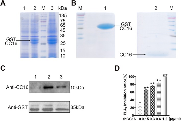
Chronic obstructive pulmonary disease (COPD) is a widespread lung disease marked by alveolar wall damage, leading to inflammation and fibrosis. Key risk factors include age, smoking, sex, and education, with smoking being the most crucial. These factors are globally consistent and linked with aging. Club cell secretory protein 16 (CC16), primarily secreted by non-ciliated bronchial epithelial cells, is crucial for pulmonary health, offering anti-inflammatory and antioxidant benefits. CC16 levels are notably reduced in COPD, suggesting its enhancement as a potential treatment. In this study, cellular senescence of BEAS-2B cells was stimulated using cigarette smoke extract (CSE) and the function of recombinant human CC16 protein (rhCC16) in cellular senescence was assessed by detecting the levels of β-galactosidase, p16, p21, ROS and the underlined mechanism was revealed by measuring mitochondrial biogenesis and metabolism. Additionally, COPD mice were prepared, and rhCC16's role on the cellular senescence of lung tissues was examined. Our findings showed that rhCC16 ameliorated cellular senescence in BEAS-2B cells and lung tissues of COPD mice accompanied by lower levels of β-galactosidase, p16, p21 and ROS. Mechanically, rhCC16 mitigated senescence via triggering PGC-1α expression through the AMPK/SIRT1 pathway and fostering mitochondrial biogenesis and metabolism to reduce the levels of ROS. Furthermore, the results also indicated that rhCC16 exerted its effect via both integrin α4β1 and clathrin-mediated endocytosis. Collectively, rhCC16 suppresses cellular senescence and ameliorates COPD-like symptoms by activating the AMPK/Sirt1-PGC-1-α-TFAM pathway to foster mitochondrial function.

Keywords: AMPK; COPD; cellular senescence; mitochondrial function; rhCC16.


Conflict of interest statement

The authors declare no conflicts of interest.

Figures

FIGURE 1
FIGURE 1
Preparation of rhCC16. (A) Induction of GST‐hCC16 in the host Escherichia coli strain using IPTG. Lane 1: Uninduced protein; Lane 2: Induced total protein; Lane 3: The protein in the supernatant. (B) Lane 1: GST‐rhCC16 bound to the solid‐phase carrier after the removal of contaminants. Lane 2: Purified rhCC16 obtained after enzymatic digestion of GST. (C) Verification of the obtained GST‐rhCC16 using Western blot. (D) Evaluation of the bioactivity of rhCC16 using acid–base titration. The data were presented as the mean ± SEM. *p < 0.05 and **p < 0.01, compared to the control group.
FIGURE 2
FIGURE 2
Treatment with rhCC16 ameliorated cellular senescence in mouse lung tissue cells and COPD‐like pathological changes in mice with COPD. (A) The levels of CC16 protein in the lung tissues of mice at different ages were measured using ELISA. (B) Representative immunohistochemical images of CC16 and GLB1 in the lung tissues of mice; scale bar: 100 μm. (C) The statistical graph of the data obtained from the immunohistochemistry analysis presented in (B) were expressed as positive cell area fractions. (D) Results of lung function tests conducted with mice at different ages. (E) Representative images of P16, P21, and GLB1 obtained by immunohistochemical analysis in the control, COPD, and rhCC16 groups; scale bar: 200 μm. (F) Representative images of H&E‐stained mouse lung tissue samples from the control, COPD, and rhCC16 groups. The mice in the COPD group and rhCC16 group were exposed to smoke for 24 weeks, 5 days a week, for 2 h each day. In the 21st week, the mice in the rhCC16 group were treated with intranasal administration 2 h before smoke exposure, and the treatment lasted for 1 month. After 24 weeks, fresh lung tissues were collected, embedded, and paraffin sections were prepared for HE staining. The data were presented as the mean ± SEM. *p < 0.05 and **p < 0.01, ***p < 0.001, compared to the control group; # p < 0.05, ## p < 0.01, compared to the COPD group.
FIGURE 3
FIGURE 3
rhCC16 inhibited the senescence induced by CSE. (A) The protein levels of P16 and P21 in CSE‐stimulated BEAS‐2B cells were determined using Western blot. (B) The mRNA expression of P16 and P21 in CSE‐stimulated BEAS‐2B cells was determined through RT–qPCR. (C) Representative images of the senescence‐associated β‐galactosidase staining experiment conducted with CSE‐stimulated BEAS‐2B cells; scale bar: 20 μm. (D) The protein levels of P16 and P21 in CSE‐stimulated or rhCC16‐treated BEAS‐2B cells were determined using Western blot. (E) The mRNA expression of P16 and P21 in CSE‐stimulated or rhCC16‐treated BEAS‐2B cells was determined through RT–qPCR. (F) Representative images of senescence‐associated β‐galactosidase staining in CSE‐stimulated or rhCC16‐treated BEAS‐2B cells; scale bar: 20 μm. The data were presented as the mean ± SEM. *p < 0.05, **p < 0.01, and ***p < 0.001, compared to the control group; # p < 0.05, ## p < 0.01, compared to the 5% CSE group.
FIGURE 4
FIGURE 4
rhCC16 restrained cellular senescence by reducing ROS production induced by CSE. (A) Representative images of the detection of ROS levels using a DCFH probe (green) and nuclei stained with DAPI (blue); scale bar: 100 μm. (B) Representative images of the detection of mitochondrial ROS levels using the MitoSOX Red probe (red) and nuclei stained with DAPI (blue); scale bar: 100 μm. (C) The fluorescence intensity was calculated using a multifunctional microplate reader at an excitation wavelength of 488 nm and an emission wavelength of 525 nm. (D) The fluorescence intensity was calculated using a multifunctional microplate reader at an excitation wavelength of 510 nm and an emission wavelength of 580 nm. (E) Flow cytometry was used to observe the changes in the intracellular total ROS levels. (F) Flow cytometry was used to observe the changes in the mitochondrial ROS levels. (G) Western blot was used to determine the protein expression levels of SOD1, SOD2, CAT, and GPX‐4. (H) The mRNA expression levels of SOD1, SOD2, CAT, and GPX‐4 were determined through RT–qPCR. (I) The mRNA expression levels of IL‐6 and IL‐8 were determined through RT–qPCR. The data were presented as the mean ± SEM. *p < 0.05 and **p < 0.01, ***p < 0.001 compared to the control group; # p < 0.05, ## p < 0.01, compared to the 5% CSE group.
FIGURE 5
FIGURE 5
rhCC16 inhibited ROS accumulation by regulating mitochondrial function. (A) The levels of the oxidative phosphorylation complex I, complex II, and complex III proteins were determined using Western blot. (B) The intracellular ATP levels were determined using an ATP assay kit. (C) The ultrastructure of intracellular mitochondria in CSE‐stimulated or rhCC16‐treated BEAS‐2B cells, as observed under a transmission electron microscope; yellow arrows represent mitochondria with normal morphology; red arrows represent mitochondria with abnormal morphology and functional damage; blue arrows represent mitochondria with restored morphology and function. Scale bar: 1 μm. (D) The levels of NAD+ and NADH in CSE‐stimulated or rhCC16‐treated BEAS‐2B cells, based on which the NAD+/NADH ratio was calculated. (E) The levels of p‐AMPK, AMPK, SIRT1, PGC‐1α, and TFAM proteins in CSE‐stimulated or rhCC16‐treated BEAS‐2B cells were determined using Western blot. (F) The levels of p‐AMPK, AMPK, SIRT1, PGC‐1α, and TFAM proteins in CSE‐stimulated or rhCC16‐treated BEAS‐2B cells treated with Ex527 (10 μM) or dorsomorphin (1 μM) were determined using Western blot. (G) The intracellular concentrations of NAD+ and NADH in CSE‐stimulated or rhCC16‐treated BEAS‐2B cells treated with dorsomorphin (1 μM), based on which the NAD+/NADH ratio was calculated. The data were presented as the mean ± SEM. *p < 0.05 and **p < 0.01, compared to the control group; # p < 0.05, ## p < 0.01 compared to the 5% CSE group.
FIGURE 6
FIGURE 6
rhCC16 improved mitochondrial function via the AMPK/Sirt1/PGC‐1α/TFAM pathway both in vitro and in vivo. (A) Representative images of the oxidative phosphorylation complex I, complex II, and complex III proteins obtained via immunohistochemistry analysis of mouse lung tissue samples from the control, COPD, and rhCC16 treatment groups; scale bar: 200 μm. (B) Representative images of p‐AMPK, SIRT1, PGC‐1α, and TFAM obtained from immunohistochemical analysis of mouse lung tissue samples from the control, COPD, and rhCC16 treatment groups; scale bar: 200 μm. (C) Statistical bar chart plot for Figure A and Figure B. (D) Representative TEM images of mouse lung tissue samples from the control, COPD, and rhCC16 treatment groups; yellow arrows represent mitochondria with normal morphology; red arrows represent mitochondria with abnormal morphology and functional damage; blue arrows represent mitochondria with restored morphology and function. Scale bar: 500 nm. (E) The levels of p16 and p21 proteins in CSE‐stimulated or rhCC16‐treated BEAS‐2B cells treated with BIO‐1211 (10 μM) or M‐β‐CD (5 μM) were determined using Western blot. The data were presented as the mean ± SEM. *p < 0.05, **p < 0.01, and ***p < 0.001, compared to the control group; # p < 0.05, ## p < 0.01, and ### p < 0.001, compared to the 5% CSE group; & p < 0.05, && p < 0.01, compared to the 5% CSE + rhCC16 group.

Similar articles

References

    1. Fang L., Gao P., Bao H., et al., “Chronic Obstructive Pulmonary Disease in China: A Nationwide Prevalence Study,” Lancet Respiratory Medicine 6 (2018): 421–430. - PMC - PubMed
    1. Zhang X., Lei Z., Wu Y., et al., “Prevalence and Risk Factors for COPD in an Urbanizing Rural Area in Western China: A Cross‐Sectional Study,” International Journal of Chronic Obstructive Pulmonary Disease 18 (2023): 459–468, 10.2147/COPD.S400213. - DOI - PMC - PubMed
    1. Mannino D. M. and Buist A. S., “Global Burden of COPD: Risk Factors, Prevalence, and Future Trends,” Lancet 370 (2007): 765–773. - PubMed
    1. Rutten E. P. A., Gopal P., Wouters E. F. M., et al., “Various Mechanistic Pathways Representing the Aging Process Are Altered in COPD,” Chest 149 (2016): 53–61. - PubMed
    1. Liao Y., Huang C., Wang J., and Fan X., “Association of Surfactant‐Associated Protein D Gene Polymorphisms With the Risk of COPD: A Meta‐Analysis,” Clinics (São Paulo, Brazil) 74 (2019): e855. - PMC - PubMed

MeSH terms