. 2025 Apr 21;31(1):143.
doi: 10.1186/s10020-025-01177-7.

Distinctive features of blood- and ascitic fluid-derived extracellular vesicles in ovarian cancer patients

Affiliations

Distinctive features of blood- and ascitic fluid-derived extracellular vesicles in ovarian cancer patients

Francesca Gorini et al. Mol Med. .

Abstract

Background: Ovarian cancer (OC) is a highly aggressive malignancy characterized by early dissemination of cancer cells from the surface of the ovary to the peritoneum. To gain a deeper understanding of the mechanisms associated with this intraperitoneal spread, we aimed to characterize the role of extracellular vesicles (EVs) in metastatic colonization in OC.

Methods: To this purpose, a total of 150 samples of ascitic fluids, blood serum, tumor and normal tissues from 60 OC patients, were extensively analyzed to characterize the EVs released in blood and ascitic fluids of OC patients, in terms of size, expression of superficial epitopes and abundance of miRNAs biocargo.

Results: A statistically significant difference in the size of EVs derived from ascitic fluid and serum was identified. Analysis of surface protein expression highlighted twenty epitopes with a significant difference between the two biological matrices, of which 18 were over- and two were under-expressed in ascitic fluid. With regard to miRNA levels, Principal Component Analysis (PCA) assessed four distinct clusters representing tumor tissue, normal tissue, ascitic fluid, and serum. A prominent difference in circulating miRNAs was observed in serum and ascitic fluid highlighting 98 miRNAs significantly deregulated (P-adj < 0.05) between the two bodily fluids. Deregulated miRNAs and epitopes underline an enrichment in ascites in components contributing to the metastatic spread.

Conclusion: The results highlight a clear difference between the two biological fluids, suggesting that tumor selectively releases specific EVs populations in serum or ascites. In this context, it seems that ascites-derived EVs play a major role in modulating EMT and metastatic cascade, which is a key feature of OC.

Keywords: Extracellular vesicles; Metastasis; Metastatic spread; Ovarian cancer; miRNA.


Conflict of interest statement

Declarations. Ethics approval and consent to participate: Patients’ samples were collected after providing informed consent and the study was approved by IRB (788/2021/Sper/AOUBo). The study was performed in accordance with the Declaration of Helsinki. Consent for publication: Not applicable. Competing interests: The authors declare no competing interests.

Figures

Fig. 1
Fig. 1
A Violin plots representing the vesicle size and concentration in serum and ascitic fluid samples, analyzed by NTA. Median and quartiles are represented by the bold line and dotted lines, respectively. **** = P < 0.0001; * = P < 0.05; ns not significant. B Representative images of EVs retrieved from blood serum (on the right) and ascitic fluid (AF, on the left) samples, analyzed by transmission electron microscopy (TEM)
Fig. 2
Fig. 2
A Heatmap showing the expression of significant epitopes in serum (S) and ascitic fluid (AF). B Different expression of specific epitopes in EVs derived from ascitic fluid and serum. **** = P < 0.0001; *** = P < 0.001; ** = P < 0.01: * = P < 0.05
Fig. 3
Fig. 3
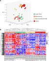
A PCA showing miRNAs levels in ascitic fluid, serum, tumor and non-tumor (normal) tissue samples. B Unsupervised Hierarchical clustering of miRNAs profile in blood serum, ascitic fluid, normal and tumor tissue of OC patients. Red and blue represent higher or lower levels of miRNAs (median centered), respectively. AF: ascitic fluid
Fig. 4
Fig. 4
Venn diagram summarizing the results obtained from the different comparisons. AF: ascitic fluid, TU: tumor
Fig. 5
Fig. 5
Relative abundance of the selected miRNAs. Median and interquartile ranges are presented. **** = P < 0.0001
Fig. 6
Fig. 6
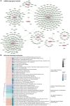
A Potential epigenetic networks between miRNAs and target genes. miRNAs are presented as central nodes colored pink, while their predicted target genes are shown in green. Some miRNAs have shared common targets. B Functional enrichment of Gene Ontology (GO) terms based on the significantly enriched miRNA target genes. The terms are divided into biological process (BP), molecular function (MF) and cellular compartment (CC). Size of dots indicates the number of genes involved in the specific pathways while their color indicates the adjusted P-value.
Fig. 7
Fig. 7
Expression level of ZEB1, PTEN, CDH1 (E-Cadherin) and CDH2 (N-Cadherin) mRNA after miRNAs mimics (miR-429 mimic, miR-21 mimic and miR-200 mimic) transfection in SKOV-3, and OVCAR-3; *** = P < 0.001; ** = P < 0.01: * = P < 0.05
Fig. 8
Fig. 8
A Progression free survival (PFS) and overall survival (OS) of patients with high and low CD326/EpCAM expression on EVs released in ascitic fluid (AF). B PFS and OS of patients with high and low miR-181b-5p in ascites-derived EVs

Similar articles

References

    1. Bachmann C. New achievements from molecular biology and treatment options for refractory/relapsed ovarian cancer-a systematic review. Cancers. 2023;15:5356. - PMC - PubMed
    1. Bandini E, Rossi T, Scarpi E, et al. Early detection and investigation of extracellular vesicles biomarkers in breast cancer. Front Mol Biosci. 2021;8:732900. - PMC - PubMed
    1. Becker A, Thakur BK, Weiss JM, Kim HS, Peinado H, Lyden D. Extracellular vesicles in cancer: cell-to-cell mediators of metastasis. Cancer Cell. 2016;30:836–48. - PMC - PubMed
    1. Brabletz S, Brabletz T. The ZEB/miR-200 feedback loop-a motor of cellular plasticity in development and cancer? EMBO Rep. 2010;11:670–7. - PMC - PubMed
    1. Cavallari I, Ciccarese F, Sharova E, et al. The miR-200 family of microRNAs: fine tuners of epithelial-mesenchymal transition and circulating cancer biomarkers. Cancers. 2021;13:5874. - PMC - PubMed

LinkOut - more resources