Review
. 2025 Apr 21;25(1):107.
doi: 10.1186/s12902-025-01919-y.

Scaffold-free endocrine tissue engineering: role of islet organization and implications in type 1 diabetes

Affiliations
Review

Scaffold-free endocrine tissue engineering: role of islet organization and implications in type 1 diabetes

Tugba Bal. BMC Endocr Disord. .

Abstract

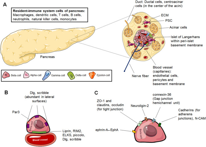
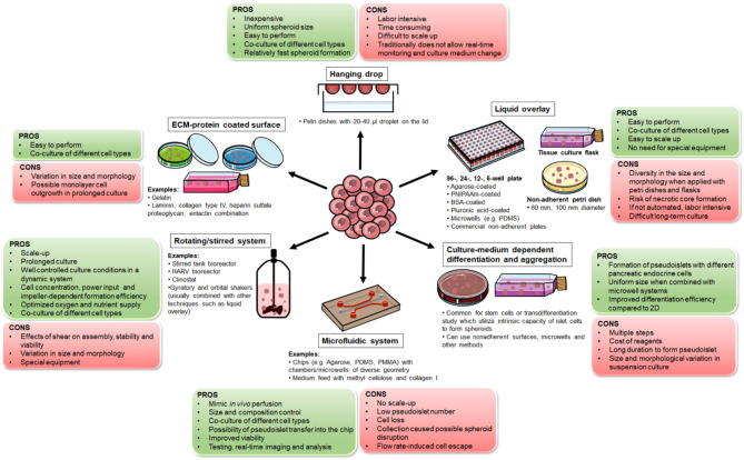
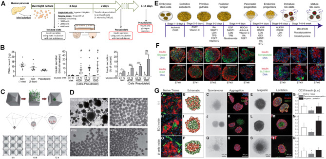
Type 1 diabetes (T1D) is a chronic hyperglycemia disorder emerging from beta-cell (insulin secreting cells of the pancreas) targeted autoimmunity. As the blood glucose levels significantly increase and the insulin secretion is gradually lost, the entire body suffers from the complications. Although various advances in the insulin analogs, blood glucose monitoring and insulin application practices have been achieved in the last few decades, a cure for the disease is not obtained. Alternatively, pancreas/islet transplantation is an attractive therapeutic approach based on the patient prognosis, yet this treatment is also limited mainly by donor shortage, life-long use of immunosuppressive drugs and risk of disease transmission. In research and clinics, such drawbacks are addressed by the endocrine tissue engineering of the pancreas. One arm of this engineering is scaffold-free models which often utilize highly developed cell-cell junctions, soluble factors and 3D arrangement of islets with the cellular heterogeneity to prepare the transplant formulations. In this review, taking T1D as a model autoimmune disease, techniques to produce so-called pseudoislets and their applications are studied in detail with the aim of understanding the role of mimicry and pointing out the promising efforts which can be translated from benchside to bedside to achieve exogenous insulin-free patient treatment. Likewise, these developments in the pseudoislet formation are tools for the research to elucidate underlying mechanisms in pancreas (patho)biology, as platforms to screen drugs and to introduce immunoisolation barrier-based hybrid strategies.

Keywords: Beta-cell models; Beta-cell replacement therapy; Cell-cell interactions; Diabetes; Drug screening; Pancreas (patho)biology; Pseudoislet.


Conflict of interest statement

Declarations. Ethical approval and consent to participate: Not applicable. Consent for publication: Not applicable. Competing interests: The authors declare no competing interests.

Figures

Fig. 1
Fig. 1
Organization in pancreas. (A) Pancreas, its compartments and microenvironment. Pancreas has two compartments: Endocrine and exocrine pancreas. Endocrine pancreas contains islets wrapped by peri-islet basement membrane. In islets, 5 different endocrine cells are present: Alpha-cells, beta-cells, delta-cells, epsilon-cells and gamma-cells. Exocrine pancreas is formed by acinar cells and ducts that contain ductal cells and centroacinar cells. In addition, resident-immune system cells and PSCs, as well as nerve fibers are found in the pancreas tissue. The pancreas also accommodates blood vessels. In figure as example, capillaries are shown. In general, capillaries are made of endothelial cells facing the blood and they are covered by pericytes and basement membrane. The acellular part of the pancreas is ECM that is rich in proteins, glycosaminoglycans and proteoglycans. (B) Polarity determinants for beta-cells. Beta-cells express Dlg and scribble in different regions, but their expression is enriched in lateral surfaces. Par3 is abundant in the apical domain away from vasculature. The capillary interface/vascular face is marked with the presynaptic scaffold proteins of liprin, RIM2, ELKS and piccolo. (C) Proteins on beta-cells used to form junctions for cell-cell interactions. ZO-1, claudins and occludin containing tight junctions, connexin-36-based gap junctions provide intercellular communication between beta-cells. Adherens junctions established between beta-cell and beta-cell and, between beta-cell and alpha-cell contain several cadherin family members and N-CAM. Apart from these junctions, EphA receptor tyrosine kinases and their ephrin-A ligands are located on beta-cells and alpha-cells for cellular communication and insulin secretion. Transcellular neuroligin-2 is an important protein of beta-cells to control insulin secretion. Dlg: Discs large protein, ECM: Extracellular matrix, N-CAM: Neural cell adhesion molecule, Par3: Partitioning defective 3 homologue, PSC: Pancreatic stellate cell, RIM2: Rab3-interacting protein. In labeling of endocrine cells for (A), the shapes and sizes of the cell representations are not relevant to the actual morphology and size of the cells. Figure 1 contains images from Servier Medical Art, which were modified in color and size where necessary. Servier Medical Art (smart.servier.com) is licensed under a Creative Commons Attribution 4.0 International License (CC BY 4.0) (https://creativecommons.org/licenses/by/4.0/)
Fig. 2
Fig. 2
The most common pseudoislet assembly techniques. Although less widely applied, techniques such as magnetic levitation, assembly through layer-by-layer coated single cells, encapsulation and bioprinted molds are also reported (not shown in the figure). Figure 2 contains images from Servier Medical Art, which were modified in color, shape and size where necessary. Servier Medical Art (smart.servier.com) is licensed under a Creative Commons Attribution 4.0 International License (CC BY 4.0) (https://creativecommons.org/licenses/by/4.0/)
Fig. 3
Fig. 3
Examples of several methods to form pseudoislets and contributions to artificial islet research. (A) Design of human pseudoislets by hanging drop technique with different initial cell numbers. (B) (Left) Comparison of DNA content of native islets and human pseudoislets (cultured for 2 days and initial cell number: 2000 cells). (Middle) Fold stimulation of native islets and human pseudoislets in GSIS. (Right) Insulin secretion (% of insulin content) of native islets and human pseudoislets in 2.8 mM and 12 mM glucose. (C) Human pseudoislets formed via centrifugation of a suspension of single cells in square-pyramidal microwells for 0–72 h. (D) Controlled, consistent pseudoislet size in square-pyramidal wells with initial cell numbers of 1000, 750 and 500 human islet cells per well. Scale bars: 200 μm. (E) Differentiation protocol for SC-islets from human embryonic stem cell line, H1. (F) Immunostaining of SC-islets obtained via protocol in (E) for insulin, glucagon, somatostatin and SLC18A1, and DNA staining at stage 7 culture. Scale bars: 100 μm. (G) (Left) Brightfield and immunofluorescence images. (Right) Quantification of CD31+ cells of native human islets, pseudoislets (EndoC-βH3 cells and HUVECs) by spontaneous aggregation and magnetic levitation. Scale bars: 50 μm for native tissue and 200 μm for pseudoislets. GSIS: Glucose stimulated insulin secretion, HUVEC: Human umbilical vein endothelial cells. (A) and (B) are reprinted from Lorza-Gil et al. [274] published by Springer Nature and are licensed under Creative Commons Attribution 4.0 International License (http://creativecommons.org/licenses/by/4.0/). Labels (A, B, C, D) of original article are removed. (C) and (D) are reprinted from Yu et al. [272] published Springer Nature and are licensed under Creative Commons Attribution 4.0 International License (http://creativecommons.org/licenses/by/4.0/). Labels (a, b, c, d) of original article are removed. (E) and (F) are reprinted from Balboa et al. [311] published by Springer Nature and are licensed under Creative Commons Attribution 4.0 International License (http://creativecommons.org/licenses/by/4.0/). Labels (a, b) of original article are removed. (G) is reprinted from Urbanczyk et al. [286] published by Mary Ann Liebert, Inc. and is licensed under Creative Commons CC BY (http://creativecommons.org/licenses/by/4.0/)

Similar articles

References

    1. Röder PV, Wu B, Liu Y, Han W. Pancreatic regulation of glucose homeostasis. Exp Mol Med. 2016;48(3):e219. - PMC - PubMed
    1. Karpińska M, Czauderna M. Pancreas—its functions, disorders, and physiological impact on the mammals’ organism. Front Physiol. 2022;13. - PMC - PubMed
    1. Pociot F, Lernmark Å. Genetic risk factors for type 1 diabetes. Lancet. 2016;387(10035):2331–9. - PubMed
    1. Hörber S, Achenbach P, Schleicher E, Peter A. Harmonization of immunoassays for biomarkers in diabetes mellitus. Biotechnol Adv. 2020;39:107359. - PubMed
    1. Undlien DE, Kockum I, Rønningen KS, Lowe R, Saanjeevi CB, Graham J, et al. HLA associations in type 1 diabetes among patients not carrying high-risk DR3-DQ2 or DR4-DQ8 haplotypes. Tissue Antigens. 1999;54(6):543–51. - PubMed

MeSH terms