

Prenatal diagnosis of pulmonary atresia with intact ventricular septum: a single-center study in China
- PMID: 40259328
- PMCID: PMC12013191
- DOI: 10.1186/s12947-025-00348-0
Prenatal diagnosis of pulmonary atresia with intact ventricular septum: a single-center study in China
Abstract
Objectives: To evaluate the efficacy of prenatal ultrasound in diagnosing pulmonary atresia with intact ventricular septum (PA/IVS).
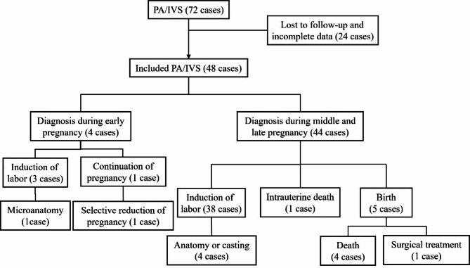
Methods: This retrospective study analyzed 48 cases of PA/IVS at the Fujian Maternity and Child Health Hospital between January 2013 and December 2023. Prenatal ultrasound was used to characterize and classify the features of PA/IVS. Pregnancy outcomes were followed up, and the results were compared with post-termination pathological anatomical findings or postnatal imaging. This study aims to enhance the understanding of PA/IVS and improve the accuracy of its prenatal diagnosis.
Results: Among the 48 PA/IVS cases, four were diagnosed during early pregnancy and 44 during mid-to-late pregnancy. In the mid-to-late pregnancy group, there were 29 cases of type I (TV-Z scores ranging from - 1.77 to 5.22), 10 cases of type II (TV-Z scores ranging from - 3.50 to -2.06), and five cases of type III (TV-Z scores ranging from - 4.29 to -7.41). The cohort included 41 singleton pregnancies and seven twin pregnancies. Ventriculo-coronary artery communication (VCAC) was observed in 19 cases. Additional abnormalities included Ebstein's anomaly (EA) in three cases, restricted opening of the foramen ovale in one case, increased inner diameter of the foramen ovale in one case, reversal or deepening of the a-wave of the ductus venosus in six cases, and umbilical vein pulsation in one case. Genetic testing (amniocentesis, NIPT, or SNP-array) was performed in 19 cases, with one case revealing a genomic copy number deletion in the q22.3 region of chromosome 21. Pregnancy outcomes included 41 terminations (five with pathologic dissection or vascular casting), five live births, one selective reduction, and one intrauterine death.
Conclusion: Fetal echocardiography is an effective tool for diagnosing PA/IVS. While PA/IVS can be diagnosed in early gestation, it remains diagnostical challenging. Given the progressive nature of PA/IVS in utero, sequential ultrasound examinations during the second and third trimesters are essential for monitoring disease progression and hemodynamic changes. Additionally, a comprehensive evaluation for associated intracardiac and extracardiac anomalies should be systematically conducted throughout the pregnancy.
Keywords: Fetus; Pregnancy outcome; Prenatal ultrasound; Pulmonary atresia with intact ventricular septum.
© 2025. The Author(s).
Conflict of interest statement
Declarations. Ethics approval and consent to participate: The study was approved by the Ethics Committee of Fujian Maternity and Child Health Hospital (2024KY309). The patients/participants provided their written informed consent to participate in this study. Written informed consent was obtained from the individual(s) for the publication of any potentially identifiable images or data included in this article. Competing interests: The authors declare no competing interests.
Figures
References
-
- Cheung EW, Richmond ME, Turner ME, Bacha EA, Torres AJ. Pulmonary Atresia/intact ventricular septum: influence of coronary anatomy on single-ventricle outcome. 2014; 98 (4): 1371–7. - PubMed
-
- Todros T, Paladini D, Chiappa E, Russo MG, Gaglioti P, Pacileo G, Cau MA, Martinelli P. Pulmonary stenosis and Atresia with intact ventricular septum during prenatal life. Ultrasound Obstet Gynecol. 2003;21(3):228–33. - PubMed
-
- Gardiner HM, Belmar C, Tulzer G, Barlow A, Pasquini L, Carvalho JS, Daubeney PEF, Rigby ML, Gordon F, Kulinskaya E. Morphologic and functional predictors of eventual circulation in the fetus with pulmonary Atresia or critical pulmonary stenosis with intact septum. J Am Coll Cardiol. 2008;51(13):1299–308. - PubMed
-
- Salvin JW, McElhinney DB, Colan SD, Gauvreau K, del Nido PJ, Jenkins KJ, Lock JE, Tworetzky W. Fetal tricuspid valve size and growth as predictors of outcome in pulmonary Atresia with intact ventricular septum. Pediatrics. 2006;118(2):e415–20. - PubMed
-
- Roman KS, Fouron JC, Nii M, Smallhorn JF, Chaturvedi R, Jaeggi ET. Determinants of outcome in fetal pulmonary valve stenosis or Atresia with intact ventricular septum. Am J Cardiol. 2007;99(5):699–703. - PubMed
MeSH terms
Supplementary concepts
Grants and funding
- 2023Y9377/Joint Funds for the innovation of science and Technology, Fujian province
- 2023Y9377/Joint Funds for the innovation of science and Technology, Fujian province
- 2023Y9377/Joint Funds for the innovation of science and Technology, Fujian province
- 2023Y9377/Joint Funds for the innovation of science and Technology, Fujian province
- 2023Y9377/Joint Funds for the innovation of science and Technology, Fujian province
- 2023Y9377/Joint Funds for the innovation of science and Technology, Fujian province
- 2023Y9377/Joint Funds for the innovation of science and Technology, Fujian province
- 2023QNA057/provincial health technology project
- 2023QNA057/provincial health technology project
- 2023QNA057/provincial health technology project
- 2023QNA057/provincial health technology project
- 2023QNA057/provincial health technology project
- 2023QNA057/provincial health technology project
- 2023QNA057/provincial health technology project
LinkOut - more resources
Full Text Sources
Medical
