

MRI patterns of thigh muscle involvement in immune-mediated necrotizing myopathy and dermatomyositis
- PMID: 40259367
- PMCID: PMC12010673
- DOI: 10.1186/s41927-025-00500-3
MRI patterns of thigh muscle involvement in immune-mediated necrotizing myopathy and dermatomyositis
Abstract
Background: Immune-mediated necrotizing myopathy (IMNM) and dermatomyositis (DM) are characterized by weakness, hyperCKemia, associated autoantibodies, and varying extramuscular manifestations. Muscle MRI, currently subordinate to histopathology and serology in idiopathic inflammatory myopathy (IIM) classification, has an evolving role. Our study aims to define thigh muscle MRI involvement in IMNM and DM by direct comparison.
Methods: This single-center, retrospective, cross-sectional study included 25 participants, who met IIM classification criteria (14 IMNM, 11 DM) and had available thigh MRI. Clinical and paraclinical data were available and reviewed. 11 muscles were graded for edema on MRI using a semi-quantitative scale (0: normal, 1: <30% of muscle involvement, 2: 31-75%, 3: > 75%). For 3 participants with no significant muscle edema, muscle fatty infiltration was scored according to the same scale. Using linear mixed-effects models, muscle scores were compared between the two groups and a secondary analysis was performed of only edema scores, excluding the 3 participants with fatty infiltration scores.
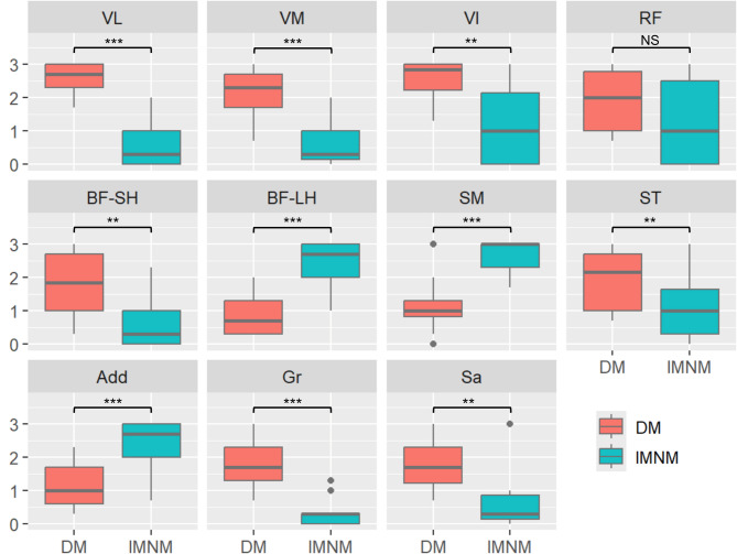
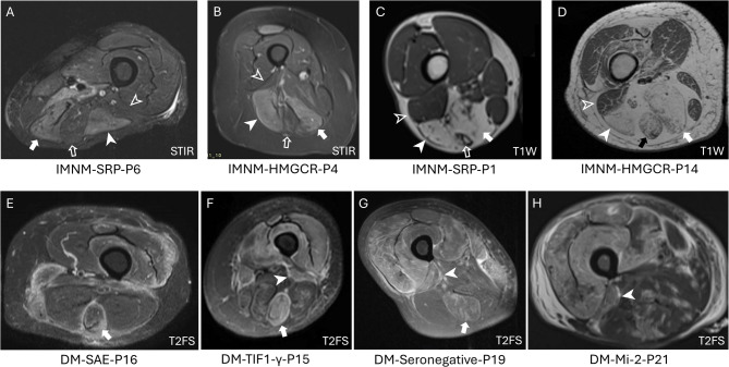
Results: The most affected muscles in IMNM were the semimembranosus (3.0 [2.7-3.0] {median [IQR]}), biceps femoris-long head (BF-LH) (2.7 [2.0-3.0]), and adductors (2.5 [2.0-3.0]). In DM, the most affected muscles were the vastus lateralis (2.7 [2.3-3.0]), vastus intermedius (2.9 [2.2-3.0]), vastus medialis (2.3 [1.7-2.7]), semitendinosus (2.2 [1.0-2.7]), rectus femoris (RF) (2.0 [1.0-2.8]), biceps femoris-short head (BF-SH) (1.9 [1.0-2.7]), gracilis, and sartorius. Intergroup statistical difference of scores was significant (p < 0.01) for 10/11 thigh muscles excluding the RF (p = 0.19), supporting an inverse relationship of muscle involvement for DM and IMNM. The secondary comparative analysis of only muscle edema scores was significant (p < 0.05) for the same 10/11 muscles with a consistent direction for all comparisons.
Conclusion: DM and IMNM affect disparate thigh muscles on MRI. DM preferentially affects the anterior thigh, semitendinosus and BF-SH in the posterior thigh, and gracilis in the medial thigh, whereas IMNM preferentially affects the posterior thigh (semimembranosus and BF-LH) and adductors in the medial thigh.
Keywords: Classification; Dermatomyositis; Immune-mediated necrotizing myopathy; Inflammatory myopathy; MRI.
© 2025. The Author(s).
Conflict of interest statement
Declarations. Ethics approval and consent to participate: This study was conducted in accordance with the Declaration of Helsinki and met criteria for waiving informed consent as outlined by the Common Rule (45 CFR 46.116(f)). Ethical approval was waived by the Oregon Health & Science University Institutional Review Board in view of the retrospective nature of the study and given all the procedures being performed were part of the routine clinical care of the participants. Consent for publication: Not applicable. Competing interests: The authors declare no competing interests.
Figures
References
LinkOut - more resources
Full Text Sources
Miscellaneous
