. 2025 Apr 21;23(1):189.
doi: 10.1186/s12964-025-02204-0.

Mannose and PMI depletion overcomes radiation resistance in HPV-negative head and neck cancer

Affiliations

Mannose and PMI depletion overcomes radiation resistance in HPV-negative head and neck cancer

Tongchuan Wang et al. Cell Commun Signal. .

Abstract

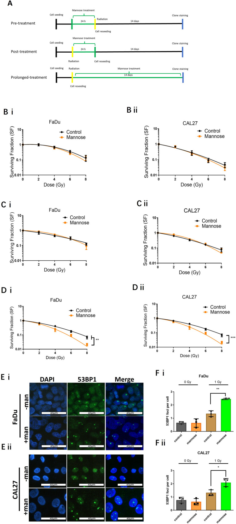
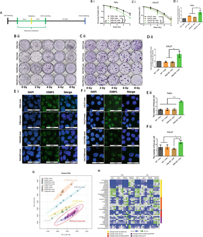
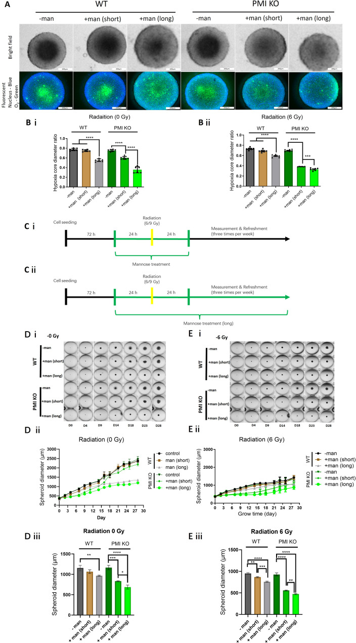
Radiotherapy is critical component of multidisciplinary cancer care, used as a primary and adjuvant treatment for patients with head and neck squamous cell carcinoma. This study investigates how mannose, a naturally occurring monosaccharide, combined with phosphomannose isomerase (PMI) depletion, enhances the sensitivity of HPV-negative head and neck tumour models to radiation. Isogenic PMI knockout models were generated by CRISPR/Cas9 gene editing, yielding a 20-fold increase in sensitivity to mannose in vitro, and causing significant tumour growth delay in vivo. This effect is driven by metabolic reprogramming, resulting in potent glycolytic suppression coupled with consistent depletion of ATP and glycolytic intermediates in PMI-depleted models. Functionally, these changes impede DNA damage repair following radiation, resulting in a significant increase in radiation sensitivity. Mannose and PMI ablation supressed both oxygen consumption rate and extracellular acidification, pushing cells towards a state of metabolic quiescence, effects contributing to increased radiation sensitivity under both normoxic and hypoxic conditions. In 3D-tumoursphere models, metabolic suppression by mannose and PMI depletion was shown to elevate intra-tumoursphere oxygen levels, contributing to significant in vitro oxygen-mediated radiosensitisation. These findings position PMI as a promising anti-tumour target, highlighting the potential of mannose as a metabolic radiosensitiser enhancing cancer treatment efficacy.

Keywords: Head and neck cancer; Mannose; Phosphomannose isomerase; Radiotherapy; Tumour metabolism.

Plain language summary

Radiotherapy is a vital treatment for head and neck cancer, however, many of these tumours poses areas that are poorly oxygenated, a feature that that causes resistance to radiotherapy. This study explores how mannose, a natural sugar, combined with reducing the activity of a protein called phosphomannose isomerase (PMI), makes HPV-negative head and neck tumours more sensitive to the effects of radiation. By dampening the ability of PMI to metabolise mannose, we report that the accumulation mannose metabolites disrupt tumour metabolism, depleting the tumour cell supply of energy (ATP), supressing the ability to repair DNA damage following radiation, and importantly helping to overcome resistance to radiation treatment caused by low-oxygen conditions. In 3D-tumour models, this combination resulted in increased tumoursphere oxygenation, further enhancing the effectiveness of radiation. These findings suggest PMI coupled with mannose as a promising target to improve cancer treatment outcomes.


Conflict of interest statement

Declarations. Competing interests: The authors declare no competing interests.

Figures

Fig. 1
Fig. 1
PMI depletion enhances the anti-proliferative effect of mannose. A i & ii) Growth curves of FaDu and CAL27 WT cells following exposure to mannose (20 mM). B i & ii) PMI protein expression in FaDu and CAL27 WT and KO/KD cells, quantification of relative PMI expression by densitometry analysis. C i & ii) Concentration dependent (0 mM – 500 mM) impact of mannose on cell viability in PMI WT and KO/KD FaDu and CAL27 cells, measured 48 h post-treatment using Alamar blue reagent. D i & ii) Trypan blue exclusion assay demonstrating increased sensitivity to mannose (20 mM) following PMI KO over 48 h. E i) Assessment of the direct anti-tumour impact of mannose in WT and KO FaDu xenograft tumours (n = 8 mice for each group). Animals bearing established tumours (100 mm3) were administered 200 µmol mannose (200 µl of 1 M mannose) via oral gavage, daily for seven days. Additionally, drinking water was supplemented with 10% mannose (0.5 M). Tumour volume was measured and recorded three times weekly, plotting mean tumour volume +/- SEM. E ii) Mean percentage weight change compared relative to experimental starting weight +/-SEM. Data presented (panels A-D) are mean ± SD of three independent biological replicates. *p ≤ 0.05 - Two-way ANOVA -Bonferroni’s multiple comparisons test and unpaired Student t-test
Fig. 2
Fig. 2
Metabolic shifts in glycolysis and oxidative metabolism induced by mannose following PMI depletion. Differential phenotypic profiles of WT and PMI KO/KD A i) FaDu and A ii) CAL27 cells 300 min post-mannose exposure assessing alterations to extracellular acidification (ECAR) and oxygen consumption (OCR) obtained through Seahorse analysis. LC-MS metabolite analysis using uniformly labelled 13C6-Glc in WT and PMI KO FaDu cells treated with or without mannose. B) Intracellular ATP levels per cell in WT and KO/KD models of (i) FaDu and (ii) CAL27 cells in the presence or absence of mannose treatment (24 h). C-I) Quantitative analysis of the relative abundance of key intracellular metabolites, including C) mannose, D) pyruvate, E) lactate, F) fumarate, G) a-ketoglutarate, H) glutamate and I) alanine (n = 4 or 5 per group). Data presented (panels A & B) are mean ± SD of three independent biological replicates. *p ≤ 0.05 - One-way ANOVA – Dunnett’s multiple comparisons test for A) and Tukey’s multiple comparisons test for B)
Fig. 3
Fig. 3
Radiation response of WT HNSCC tumour models to radiation. A) The effect of mannose treatment schedules on clonogenic survival assays. Treatment conditions include: pre-treatment - mannose (20 mM) 24 h prior to radiation; post-treatment - mannose (20 mM) 24 h after radiation treatment; or prolonged-treatment - mannose (20 mM) supplementation throughout the 14-day colony forming period. B) Pre-treatment clonogenic survival data for (i) FaDu and (ii) CAL27 WT cells +/- mannose. C) Post-treatment clonogenic survival data for (i) FaDu and (ii) CAL27 WT cells +/- mannose. D) Prolonged treatment (14 day) clonogenic survival data for (i) FaDu and (ii) CAL27 WT cells +/- mannose. E) Immunofluorescence images of 53BP1 foci, a surrogate indicator of DNA double strand break damage in WT (i) FaDu and (ii) CAL27 cells treated with mannose (20 mM) for 72 h prior to radiation (1 Gy), then fixed 24 h post-radiation allowing repair. F) Quantified residual DNA DSB damage 24 h post radiation (1 Gy) treatment in WT (i) FaDu and (ii) CAL27 cells. Foci were calculated by scoring a minimum of 50 cells per replicate, scored from three independent biological replicates (n = 3). Scale bars: 40 μm. Data presented (panels B-F) are mean ± SD of three independent biological replicates. p ≤ 0.05 - two-way ANOVA and unpaired Student t-test
Fig. 4
Fig. 4
Radiosensitization in PMI ablated HNSCC models driven by combined PMI ablation and mannose mediated metabolic alterations. A) Schematic mannose treatment schedule of colony forming assay. Clonogenic assays in WT and PMI KO/KD B i & ii) FaDu and C i & ii) CAL27 cells, +/- mannose (20 mM) pre-treatment (48 h), followed by radiation (2–8 Gy). D) Relative ROS levels detected using 2’,7’-dichlorodihydrofluorescein diacetate (DCFH-DA) in WT and PMI KO/KD (i) FaDu and (ii) CAL27 cells +/- mannose (20 mM) pre-treatment (24 h) prior to radiation (4 Gy). ROS levels were measured 10 min post-radiation treatment. E) 53BP1 immunofluorescence in WT and PMI KO (i) FaDu cells with (ii) quantified differences calculated by scoring a minimum of 50 cells per independent replicate. Scale bars: 40 μm. F) 53BP1 immunofluorescence in WT and PMI KD (i) CAL27 cells with (ii) quantified differences calculated by scoring a minimum of 50 cells per independent replicate. Unlabelled LC-MS analysis of the metabolic profiles of WT and PMI KO FaDu cells treated with mannose, +/- radiation (4 Gy). Scale bars: 40 μm. G) Principal Component Analysis (PCA) depicting distinctive clustering of the metabolome in mannose exposed cells. H) A heatmap generated from thirty-nine dominant metabolites for each treatment group (n = 5 for each group). Data presented (panels B-F) are mean ± SD of three independent biological replicates. *p ≤ 0.05 - One-way ANOVA - Tukey’s multiple comparisons test B-D) and two-way ANOVA - Tukey’s multiple comparisons test A)
Fig. 5
Fig. 5
Overcoming hypoxia-Induced radioresistance through HIF-1α suppression by mannose and PMI Knockout. A) Schematic mannose treatment schedule of colony forming assay. Colony forming assays in WT and PMI KO/KD B i & ii) FaDu, and C i & ii) CAL27 cells +/- mannose (20 mM) for 24 h prior to radiation treatment (RT). Four hours before RT, cells were transferred to an InvivO2 hypoxic workstation set to 0.5% O2. D) Western blot and E) semi-quantitative analysis of relative HIF-1α levels in WT and PMI KO/KD cells. D i & E i) FaDu and D ii & E ii) CAL27 cells +/- mannose for 24 h, with a 4 h hypoxia incubation prior to sample collection. F) Relative levels of succinate in WT and PMI KO/KD i) FaDu and ii) CAL27 cells pretreated +/- mannose (20 mM) for 24 h, with a 4 h hypoxia incubation prior to sample collection. G) Relative ATP levels of WT and PMI KO/KD i) FaDu and ii) CAL27 cells pretreated +/- mannose (20 mM) for 24 h, with a 4 h hypoxia incubation prior to sample collection. Data presented (panels B, C, E-G) are mean ± SD of three independent biological replicates. *p ≤ 0.05 -One-way ANOVA - Tukey’s multiple comparisons test E-G) and two-way ANOVA - Tukey’s multiple comparisons test B&C)
Fig. 6
Fig. 6
Mannose sensitises PMI KO HNSCC tumourspheres to radiation. A) Confocal fluorescence (Image-iT™ Green Hypoxia Dye and Hoechst staining) and brightfield imaging of size matched (~ 600 μm) FaDu (WT/KO) tumourspheres pre-treated with mannose (20 mM). B) Diameter ratio of the necrotic/hypoxic core in (i) unirradiated or (ii) 6 Gy irradiated FaDu (WT/KO) tumourspheres treated +/- mannose (20 mM for 48 h for short-term exposure and sustained exposure to mannose for long-term treatment). C i & ii) Schematic diagram illustrating treatment scheduling for both WT and PMI KO FaDu tumoursphere models. D i) Representative images of unirradiated WT and PMI KO FaDu tumourspheres exposed to short-term (48 h) and long-term (sustained) mannose treatment. D ii) Dynamic growth data of unirradiated WT and PMI KO FaDu tumoursphere models. D iii) Differential tumoursphere diameter on Day 11 for WT and PMI KO tumourspheres treated with mannose for short-term (48 h) or long-term (sustained). E i) Representative images of irradiated (6 Gy) WT and PMI KO FaDu tumourspheres with short-term (48 h) and long-term (sustained) mannose treatment. E ii) Dynamic growth data of unirradiated WT and PMI KO FaDu tumoursphere models. E iii) Differential tumoursphere diameter on Day 11 for irradiated WT and PMI KO tumourspheres treated with mannose for short-term (48 h) or long-term (sustained). Data are presented as mean ± SD of three independent biological replicates. *p ≤ 0.05 (One-way ANOVA- Tukey’s multiple comparisons test (B, D &E)
Fig. 7
Fig. 7
Mechanistic overview. Tumour cells exhibit monosaccharide addiction, providing a means for preferential and competitive uptake of mannose via glucose transporters (GLUT1/2). This figure outlines how altered mannose metabolism through PMI depletion enhances tumour cell radiation sensitivity. Mannose potentiates glycolytic disruption, particularly in PMI-depleted cells, driving metabolic reprogramming characterised by ATP depletion, redox imbalance, and impaired DNA damage repair. Under hypoxia, mannose and PMI ablation reduced oxygen consumption rate and supressed TCA cycle activity, effects that led to destabilised HIF-1α under hypoxic conditions. These metabolic driven adaptions ultimately helped counteract hypoxia-mediated radiation resistance, though elevated intracellular oxygen. Together, these mechanisms act together to sensitise various in vitro tumour models to radiation, responses that require further in vivo validation, positioning PMI as a promising target to improve radiotherapy outcomes. These findings provide compelling evidence for the continued clinical exploration of mannose as a radiosensitiser, particularly in the context of PMI ablation

Similar articles

Cited by

References

    1. Anderson G, Ebadi M, Vo K, Novak J, Govindarajan A, Amini A. An updated review on head and neck Cancer treatment with radiation therapy. Cancers (Basel). 2021;13. - PMC - PubMed
    1. Zhou P, Yu Y-F, Lian C-L, Wang J, Zhuo R-G, Wu S-G. Survival outcomes and treatment decision by human papillomavirus status among patients with stage IVC head and neck squamous cell carcinoma. Front Oncol. 2021;11. - PMC - PubMed
    1. Johnson DE, Burtness B, Leemans CR, Lui VWY, Bauman JE, Grandis JR. Head and neck squamous cell carcinoma. Nat Rev Dis Primers. 2020;6:92. - PMC - PubMed
    1. Barsouk A, Aluru JS, Rawla P, Saginala K, Barsouk A, Epidemiology. Risk factors, and prevention of head and neck squamous cell carcinoma. Med Sci. 2023;11. - PMC - PubMed
    1. Rosenthal DI, Mohamed ASR, Garden AS, Morrison WH, El-Naggar AK, Kamal M, et al. Final report of a prospective randomized trial to evaluate the Dose-Response relationship for postoperative radiation therapy and pathologic risk groups in patients with head and neck cancer. Int J Radiat Oncol Biol Phys. 2017;98:1002–11. - PMC - PubMed

LinkOut - more resources