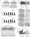
Calcineurin-mediated regulation of growth-associated protein 43 is essential for neurite and synapse formation and protects against α-synuclein-induced degeneration
- PMID: 40259946
- PMCID: PMC12009912
- DOI: 10.3389/fnagi.2025.1566465
Calcineurin-mediated regulation of growth-associated protein 43 is essential for neurite and synapse formation and protects against α-synuclein-induced degeneration
Abstract
Introduction: Elevated calcium (Ca2+) levels and hyperactivation of the Ca2+-dependent phosphatase calcineurin are key factors in α-synuclein (α-syn) pathobiology in Dementia with Lewy Bodies and Parkinson's Disease (PD). Calcineurin activity can be inhibited by FK506, an FDA-approved compound. Our previous work demonstrated that sub-saturating doses of FK506 provide neuroprotection against α-syn pathology in a rat model of α-syn neurodegeneration, an effect associated with the phosphorylation of growth-associated protein 43 (GAP-43).
Methods: To investigate the role of GAP-43 phosphorylation, we generated phosphomutants at the calcineurin-sensitive sites and expressed them in PC12 cells and primary rat cortical neuronal cultures to assess their effects on neurite morphology and synapse formation. Additionally, we performed immunoprecipitation mass spectrometry in HeLa cells to identify binding partners of these phosphorylation sites. Finally, we evaluated the ability of these phosphomutants to modulate α-syn toxicity.
Results: In this study, we demonstrate that calcineurin-regulated phosphorylation at S86 and T172 of GAP-43 is a crucial determinant of neurite branching and synapse formation. A phosphomimetic GAP-43 mutant at these sites enhances both processes and provides protection against α-syn-induced neurodegeneration. Conversely, the phosphoablative mutant prevents neurite branching and synapse formation while exhibiting increased interactions with ribosomal proteins.
Discussion: Our findings reveal a novel mechanism by which GAP-43 activity is regulated through phosphorylation at calcineurin-sensitive sites. These findings suggest that FK506's neuroprotective effects may be partially mediated through GAP-43 phosphorylation, providing a potential target for therapeutic intervention in synucleinopathies.
Keywords: FK506; GAP-43; calcineurin; neurite branching; neuroprotection; synapses; α-synuclein.
Copyright © 2025 Grebenik, Zaichick and Caraveo.
Conflict of interest statement
The authors declare that the research was conducted in the absence of any commercial or financial relationships that could be construed as a potential conflict of interest.
Figures


Similar articles
-
Harnessing IGF-1 and IL-2 as biomarkers for calcineurin activity to tailor optimal FK506 dosage in α-synucleinopathies.Front Mol Biosci. 2023 Nov 29;10:1292555. doi: 10.3389/fmolb.2023.1292555. eCollection 2023. Front Mol Biosci. 2023. PMID: 38094080 Free PMC article.
-
FKBP12 contributes to α-synuclein toxicity by regulating the calcineurin-dependent phosphoproteome.Proc Natl Acad Sci U S A. 2017 Dec 26;114(52):E11313-E11322. doi: 10.1073/pnas.1711926115. Epub 2017 Dec 11. Proc Natl Acad Sci U S A. 2017. PMID: 29229832 Free PMC article.
-
Alpha-synuclein overexpression increases phospho-protein phosphatase 2A levels via formation of calmodulin/Src complex.Neurochem Int. 2013 Sep;63(3):180-94. doi: 10.1016/j.neuint.2013.06.010. Epub 2013 Jun 22. Neurochem Int. 2013. PMID: 23796501
-
FK506 and the role of immunophilins in nerve regeneration.Mol Neurobiol. 1997 Dec;15(3):285-306. doi: 10.1007/BF02740664. Mol Neurobiol. 1997. PMID: 9457703 Review.
-
The contribution of alpha synuclein to neuronal survival and function - Implications for Parkinson's disease.J Neurochem. 2016 May;137(3):331-59. doi: 10.1111/jnc.13570. Epub 2016 Mar 23. J Neurochem. 2016. PMID: 26852372 Free PMC article. Review.
Cited by
-
Using network toxicology and molecular docking to identify core targets and pathways underlying tacrolimus-induced tremor in organ transplant recipients.Sci Rep. 2025 Jul 2;15(1):22817. doi: 10.1038/s41598-025-02381-5. Sci Rep. 2025. PMID: 40592862 Free PMC article.
References
-
- Alafuzoff I., Hartikainen P. (2017). Alpha-synucleinopathies. Handb. Clin. Neurol. 145 339–353. - PubMed
-
- Bosutti A., Dapas B., Grassi G., Bussani R., Zanconati F., Giudici F., et al. (2022). High eEF1A1 protein levels mark aggressive prostate cancers and the in vitro targeting of eEF1A1 reveals the eEF1A1-actin complex as a new potential target for therapy. Int. J. Mol. Sci. 23:4143. 10.3390/ijms23084143 - DOI - PMC - PubMed
LinkOut - more resources
Full Text Sources
Miscellaneous

