. 2025 Apr 7:13:1564827.
doi: 10.3389/fbioe.2025.1564827. eCollection 2025.

SHP099-containing multi-targeting hydrogel promotes rapid skin reconstruction through modulating a variety of cells

Affiliations

SHP099-containing multi-targeting hydrogel promotes rapid skin reconstruction through modulating a variety of cells

Zhixiao Liu et al. Front Bioeng Biotechnol. .

Abstract

Introduction: Adult wound scarring result in functional skin deficits. However, the development of effective measures to modulate the entire wound healing to encourage the skin function reconstruction is still a clinical challenge, as multiple cells are involved in wound healing hierarchically. Hydrogel scaffolds with long-lasting local release provide new insights into the clinical relevance of entire wound healing.

Methods: Herein, a multi-targeting hydrogel loaded with SHP099 (Gel-SHP) is designed to modulate multiple cells during wound repair.

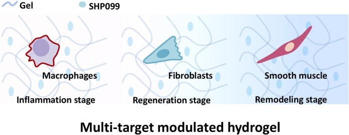
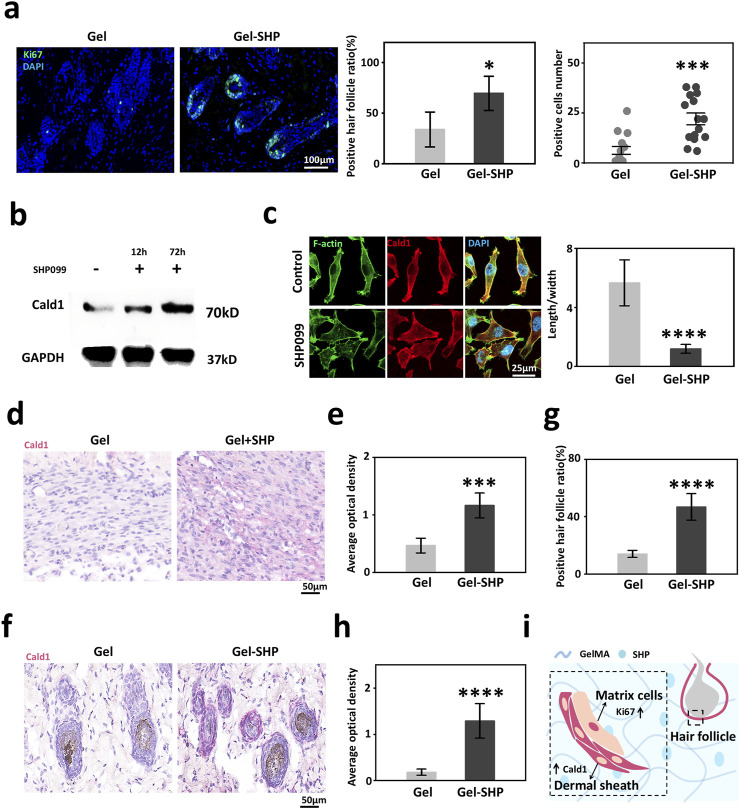
Results: Our results show that Gel-SHP promotes rapid reconstruction of wound skin by modulating macrophages in the inflammatory stage, fibroblasts in the regeneration stage and smooth muscle cells in the remodelling stage. Gel-SHP could increase M2 macrophage differentiation and remodel the dermal shell of hair follicles through in situ release. Moreover, Gel-SHP may modulate myofibroblasts to promote wound contraction through SHP099-scaffold synergistic interactions.

Discussion: Our results provide new insights into the design of functional hydrogels for tissue regeneration applications. Gel-SHP as a promising tool could provide new clues and new research paradigms for future studies and understanding of the wound healing process and dermal shell formation.

Keywords: SHP099; hair follicle dermal shell; macrophage; multi-targetinghydrogel; myofibroblast; wound healing.


Conflict of interest statement

The authors declare that the research was conducted in the absence of any commercial or financial relationships that could be construed as a potential conflict of interest. The author(s) declared that they were an editorial board member of Frontiers, at the time of submission. This had no impact on the peer review process and the final decision.

Figures

FIGURE 1
FIGURE 1
Gel loaded with SHP099 (SHP) to prepare multi-targeting hydrogel. The multi-targeting hydrogel is hypothesized to promote the reconstruction of wound skin structures by regulating macrophages, fibroblasts and smooth muscle cells at different stages of wound repair.
FIGURE 2
FIGURE 2
Gel-SHP promotes wound healing and skin remodeling. (a) Scanning electron microscopy (SEM) morphology characterization of Gel hydrogels (Gel) and Gel hydrogels loaded with the multi-targeting inhibitor SHP099 (Gel-SHP). (b) Cumulative release profile of SHP in Gel-SHP (n = 3). We hypothesized that Gel-SHP would regulate macrophages during the inflammatory stage and fibroblasts during the tissue regeneration stage. (c) Mouse wound model. (d, e) Gel-SHP promotes wound closure. (d) Photographs of wounds treated by Gel and Gel-SHP for day 0, 3 and 7 (1 mm per grid). (e) The wound closure rate between groups Gel and Gel-SHP was not significant on day 3 (n = 3, p = 0.15427), whereas the wound closure rate of Gel-SHP group was significantly higher than Gel group on day 7 (n = 3, p = 0.00453). (f) H&E-stained sections (left, day 7 of treatment) show the epidermal thickness (brown dash-dotted line) was significantly higher in group Gel-SHP than in group Gel (right, arrows, n = 3, p = 0.03237). (g) H&E-stained sections on day 14 treatment show (left) hair follicle number in Gel-SHP group was significantly higher than in Gel group (right, n = 3, p = p = 0.00158). *p < 0.05; **p < 0.01; ***p < 0.001; ****p < 0.0001.
FIGURE 3
FIGURE 3
Gel-SHP regulates macrophages during inflammatory stage and promotes tissue regeneration. (a) Q-PCR results of IL-6 expression in macrophages (RAW264.7; LPS-/SHP099- VS LPS+/SHP099-, n = 3, p = 1.71445E-7; LPS+/SHP099- VS LPS+/SHP099+, n = 3, p = 8.43562E-4). (b, c) Gel-SHP promotes M2 differentiation of macrophages in wounds. (b) Immunohistochemically (IHC) stained sections (day 3 of treatment; CD206, brown). (c) The percentage of macrophages positive for CD206 expression in Gel-SHP group was higher than in the Gel group (n = 5, p = 3.60348E-5). (d–f) Gel-SHP promotes collagen formation and fibroblast regeneration in the wound. (d) Masson stained sections (day 3 of treatment; collagen, blue; fibroblast, red) (e) The blue average optical density (AOD) representing collagen was significantly higher in Gel-SHP group than in Gel group (n = 5, p = 0.00135). (f) The blank area was significantly smaller in Gel-SHP group than in Gel group (n = 5, p = 0.00291). (g) Gel-SHP regulates the differentiation of wound macrophages from M1 to M2, while promoting fibroblast proliferation and facilitating collagen synthesis during the inflammatory stage. *p < 0.05; **p < 0.01; ***p < 0.001; ****p < 0.0001.
FIGURE 4
FIGURE 4
Gel-SHP regulates fibroblasts during regeneration stage and promotes angiogenesis. (a, b) Transcriptomic sequencing reveals Gel-SHP regulated genes and gene functions in the regeneration stage. (a) Volcano map of differentially expressed genes (day 7 of treatment; Gel-SHP vs Gel, n = 3). P < 0.05, log2 (Fold change) > 2 was defined as significant difference. (b) Gene Ontology (GO) enrichment analysis of significantly upregulated genes in biological progress (BP). Significant enrichment was defined as p value < 0.05 (Top 5 in BP). (c, d) Gel-SHP promotes fibroblast alpha-smooth muscle actin (alpha-SMA) expression in wounds. (c) IHC stained sections of alpha-SMA (day 7 of treatment; red). (d) The AOD of alpha-SMA in fibroblasts of Gel-SHP group is significantly higher than Gel group (n = 5, p = 0.00125). (e, f) Gel-SHP promotes angiogenesis of the wound. (e) H&E-stained sections (7 day of treatment). (f) Statistical results show that the number of blood vessels at the Gel-SHP group wound was significantly higher than that in Gel group (n = 5, p = 3.8429E-4). (g–i) Gel-SHP promotes alpha-SMA expression in neo-regenerated hair follicles. (g) IHC stained sections of alpha-SMA (day 7 of treatment; red). (h) The alpha-SMA positive hair follicle rate was significantly higher in Gel-SHP group than Gel group (n = 5, p = 0.00499). (i) The hair follicle alpha-SMA AOD in Gel-SHP group was significantly higher than that Gel group (n = 5, p = 2.88749E-6). (j, k) Gel-SHP did not significantly increase the number of hair follicles after day 7 of treatment. (j) H&E-stained sections (day 7 of treatment). (k) The difference in the number of hair follicles regenerated in the wound (Regeneration area: red box = 1 mm2) between Gel-SHP group and Gel group was not significant (n = 3, p = 0.34). (l) Gel-SHP modulates the differentiation of fibroblasts into myofibroblasts, promotes angiogenesis and promotes alpha-SMA expression during the regeneration stage. *p < 0.05; **p < 0.01; ***p < 0.001; ****p < 0.0001.
FIGURE 5
FIGURE 5
Gel-SHP promotes hair follicle regeneration via modulating Cald1 expression during the remodeling stage. (a) Gel-SHP promotes Ki67 expression in neonatal hair follicle matrix cells. Immunofluorescence (IF) stained sections (left; 7 day of treatment). The Ki67-positive follicles (follicles with Ki67-positive cells were defined as positive follicles) ratio (n = 5, p = 0.01081) and the number of positive cells in positive follicles (15 randomly selected hair follicles:3 follicles close to the wound per section, n = 15, p = 1.058E-4) were significantly higher in Gel-SHP group than in Gel group. (b) Western blot results of L929 showed that SHP099 promoted Cald1 expression in fibroblasts. (c) IF results (left, day 3 incubated) showed that SHP099 altered the morphology of fibroblasts and significantly decreased the length/width ratio of fibroblasts (n = 10, p = 1.17055E-9). (d, e) Gel-SHP promotes Cald1 expression in wound fibroblasts. (d) IHC stained sections of Cald1 (day7 of treatment; red). (e) Cald1 AOD in wound fibroblasts in Gel-SHP group was significantly higher than that in Gel group (n = 5, p = 2.62218E-4). (f–h) Gel-SHP promotes Cald1 expression in neo-regenerated hair follicles. (f) IHC stained sections of Cald1 (day 7 of treatment; red). (g) Cald1 positive neo-regenerated hair follicle percentage in Gel-SHP group was significantly higher than that in Gel group (n = 5, p = 6.04142E-5). (h) Cald1 AOD in neo-regenerated hair follicles in Gel-SHP group was significantly higher than that in Gel group (10 randomly selected hair follicles:2 follicles close to the wound per section, n = 10, p = 2.86295E-8). (i) Gel-SHP maintains the dermal shell of hair follicles to promote follicular regeneration by promoting Cald1 expression during the remodeling stage. *p < 0.05; **p < 0.01; ***p < 0.001; ****p < 0.0001.

Similar articles

References

    1. Aragona M., Dekoninck S., Rulands S., Lenglez S., Mascré G., Simons B. D., et al. (2017). Defining stem cell dynamics and migration during wound healing in mouse skin epidermis. Nat. Commun. 8, 14684. 10.1038/ncomms14684 - DOI - PMC - PubMed
    1. Burmeister B. T., Taglieri D. M., Wang L., Carnegie G. K. (2012). Src homology 2 domain-containing phosphatase 2 (Shp2) is a component of the A-kinase-anchoring protein (AKAP)-Lbc complex and is inhibited by protein kinase A (PKA) under pathological hypertrophic conditions in the heart. J. Biol. Chem. 287, 40535–40546. 10.1074/jbc.M112.385641 - DOI - PMC - PubMed
    1. Chambers S. E. J., Pathak V., Pedrini E., Soret L., Gendron N., Guerin C. L., et al. (2021). Current concepts on endothelial stem cells definition, location, and markers. Stem Cells Transl. Med. 10, S54–S61. 10.1002/sctm.21-0022 - DOI - PMC - PubMed
    1. Chen Y.-N. P., Lamarche M. J., Chan H. M., Fekkes P., Garcia-Fortanet J., Acker M. G., et al. (2016). Allosteric inhibition of SHP2 phosphatase inhibits cancers driven by receptor tyrosine kinases. Nature 535, 148–152. 10.1038/nature18621 - DOI - PubMed
    1. Cho D.-I., Kim M. R., Jeong H.-Y., Jeong H. C., Jeong M. H., Yoon S. H., et al. (2014). Mesenchymal stem cells reciprocally regulate the M1/M2 balance in mouse bone marrow-derived macrophages. Exp. & Mol. Med. 46, e70. 10.1038/emm.2013.135 - DOI - PMC - PubMed

LinkOut - more resources