. 2025 Mar 3:22:26.
doi: 10.25259/Cytojournal_212_2024. eCollection 2025.

Emerging regulators of gastric cancer angiogenesis: Synergistic effects of regulator of G protein signaling 4 and midkine

Affiliations

Emerging regulators of gastric cancer angiogenesis: Synergistic effects of regulator of G protein signaling 4 and midkine

Yanxin He et al. Cytojournal. .

Abstract

Objective: Globally, gastric cancer (GC) is among the most prevalent cancers. The development and spread of stomach cancer are significantly influenced by angiogenesis. However, the molecular mechanisms underlying this process remain unclear. This study aimed to investigate the role of the regulator of G protein signaling 4 (RGS4) in GC angiogenesis and its potential mechanisms.

Material and methods: Through in vitro and in vivo experiments, including tube formation assays and xenograft models in nude mice, we evaluated the effects of RGS4 on GC angiogenesis and metastasis. In addition, we employed techniques such as immunoprecipitation and immunofluorescence double staining to explore the interaction between RGS4 and midkine (MDK). Survival analysis was also performed to evaluate the association between the prognosis of patients with GC and the expression levels of RGS4 and MDK.

Results: Our findings revealed that RGS4 is a crucial factor in GC metastasis, significantly inducing angiogenesis. Further studies indicated that RGS4 directly interacts with MDK and upregulates its expression. By upregulating MDK, RGS4 stimulates the angiogenesis and metastasis of GC. Furthermore, a poor prognosis for patients with GC is directly linked to high expression of RGS4 and MDK.

Conclusion: This work is the first to clarify the molecular mechanism by which RGS4 upregulates MDK expression to increase GC angiogenesis. These findings not only enhance our understanding of the mechanisms underlying GC progression but also provide potential targets for developing new anti-angiogenic and antimetastatic therapies. RGS4 and MDK could serve as effective biomarkers for predicting the prognosis of patients with GC and offer new insights into personalized treatment approaches.

Keywords: Angiogenesis; Midkine; Regulator of G protein signaling 4.


Conflict of interest statement

The authors declare no conflict of interest.

Figures

Figure 1:
Figure 1:
RGS4 is a key factor in gastric cancer metastasis. (a and b) Validation of transfection efficiency in NCI–N87 cells after transfection with silencing plasmid. (c) Lung tissue was stained with H&E, objective: 200× and the metastases after silencing RGS4 were observed. Scale bar: 50 or 20 μm. (d) Statistical analysis of the number of lung metastatic foci (n = 6). (e and f) Validation of transfection efficiency in NCI– N87 cells after transfection with overexpression plasmid. (g) Lung tissue was stained with H&E, objective: 200× and the metastases after overexpression of RGS4 were observed. Scale bar: 50 or 20 μm. (h) Statistical analysis of the number of lung metastatic foci (n = 6). n = 3. P < 0.01, P < 0.001. RGS4: Regulator of G protein signaling 4, sh-control: Negative control to RGS4 shRNA, shRGS4: RGS4 shRNA, GAPDH: Glyceraldehyde-3-phosphate dehydrogenase, RGS4: Regulator of G protein signaling 4, H&E: Hematoxylin and eosin.
Figure 2:
Figure 2:
RGS4 induces angiogenesis in gastric cancer. (a and b) Relative expression of VEGF-A, VEGF-B, and VEGF-C in NCI–N87 cells overexpressing RGS4. (c) Relative level of VEGF-A, VEGF-B, and VEGF-C in the supernatant of NCI–N87 cells overexpressing RGS4. (d and e) Relative expression of VEGF-A, VEGF-B, and VEGF-C in RGS4 knockdown NCI–N87 cells. (f) Relative level of VEGF-A, VEGF-B, and VEGF-C in the supernatant of RGS4 knockdown NCI–N87 cells. (g and h) Migration and invasion assays, objective: 200×, of HUVECs treated with the supernatant of NCI–N87 cells overexpressing RGS4. Scale bar: 50 μm. (i and j) Migration and invasion assays, objective: 200×, of HUVECs treated with the supernatant of RGS4 knockdown NCI–N87 cells. Scale bar: 50 μm. (k and l) Tube formation assay of HUVECs treated with the supernatant of NCI–N87 cells overexpressing RGS4. Scale bar: 100 μm. (m and n) Tube formation assay of HUVECs treated with the supernatant of RGS4 knockdown NCI–N87 cells. Scale bar: 100 μm. n = 3. ns: No significant, P < 0.05, P < 0.01, P < 0.001. VEGF: Vascular endothelial growth factor, HUVECs: Human umbilical vein endothelial cells, RGS4: Regulator of G protein signaling 4.
Figure 3:
Figure 3:
Interaction between RGS4 and MDK. (a) Co-transfection of HEK293T cells with RGS4-Flag and MDK-GST plasmids, followed by immunoprecipitation of RGS4-Flag using anti-Flag antibody. (b) Co-transfection of HEK293T cells with RGS4-Flag and MDK-GST plasmids, followed by precipitation of MDK-GST using glutathione-Sepharose 4B beads. n = 3. GST: Glutathione-Stransferase, MDK: Midkine, RGS4: Regulator of G protein signaling 4.
Figure 4:
Figure 4:
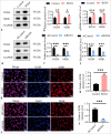
RGS4 upregulates MDK expression. (a and b) Stable overexpression of RGS4 in NCI–N87 cells, with immunoblot showing protein levels of MDK and RGS4. (c) The mRNA levels of RGS4 and MDK in NCI–N87 cells stabilized with overexpression of RGS4. (d and e) Knockdown of RGS4 in NCI–N87 cells, with immunoblot showing protein levels of MDK and RGS4. Scale bar: 50 μm. (f) The mRNA levels of RGS4 and MDK in NCI–N87 cells that silence RGS4. (g and h) Stable overexpression of RGS4 in NCI–N87 cells, with immunofluorescence, objective: 200×, showing MDK expression levels. Scale bar: 50 μm. (i and j) Knockdown of RGS4 in NCI–N87 cells, with immunofluorescence, objective: 200×, showing MDK expression levels. n = 3. P < 0.001. DAPI: 4’,6-Diamidino-2’-phenylindole, RGS4: Regulator of G protein signaling 4, MDK: Midkine.
Figure 5:
Figure 5:
RGS4 inhibits EMT in GC. (a and b) Immunofluorescence double staining, objective: 200×, of E-cadherin and N-cadherin in NCI– N87 cells after RSG4 overexpression. Scale bar: 50 μm. (c and d) Immunofluorescence double staining, objective: 200×, of E-cadherin and N-cadherin in NCI–N87 cells after silencing RSG4. Scale bar: 50 μm. n = 3. P < 0.01, P < 0.001. RGS4: Regulator of G protein signaling 4, GC: Gastric cancer.
Figure 6:
Figure 6:
RGS4 promotes angiogenesis in gastric cancer by upregulating MDK. (a and b) In NCI–N87 cells with RGS4 knockdown, transfection with MDK and control vectors, with immunoblot showing protein levels of MDK and RGS4. (c and d) Migration assay, objective: 200×, to analyze HUVEC migration and invasion capabilities. Scale bar: 50 μm. (e and f) Tube formation assay, objective: 200×, to analyze HUVEC angiogenesis. Scale bar: 50 μm. (g and h) Knockdown of MDK in NCI–N87 cells overexpressing RGS4, with immunoblot showing protein levels of MDK and RGS4. (i and j) Migration assay, objective: 200×, to analyze HUVEC migration and invasion capabilities. Scale bar: 50 μm. (k and l) Tube formation assay, objective: 200×, to analyze HUVEC angiogenesis. Scale bar: 50 μm. n = 3. ns: No significant, P < 0.01, P < 0.001. shMDK: MDK shRNA, RGS4: Regulator of G protein signaling 4, MDK: Midkine, HUVECs: Human umbilical vein endothelial cells.
Figure 7:
Figure 7:
RGS4 promotes gastric cancer tumor metastasis by upregulating MDK. (a) In NCI–N87 cells with RGS4 knockdown, transfection with MDK and control vectors, followed by detection of lung metastatic nodules in mice 6 weeks after tail vein injection. Scale bar: 50 or 20 μm. (b) Statistical analysis of the number of lung metastatic foci. (c and d) Immunohistochemical detection of CD31 in lung metastatic nodules. Scale bar: 50 or 20 μm. n = 3. P < 0.01, P < 0.001. RGS4: Regulator of G protein signaling 4, MDK: Midkine.
Figure 8:
Figure 8:
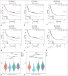
High expression of RGS4 and MDK predicts poor prognosis in gastric cancer patients. (a-f) Analysis of the correlation between RGS4 or MDK expression and patient outcomes (FP: First progression, OS: Overall survival, PPS: Post-progression survival) in gastric cancer using the KM database (http://kmplot.com/analysis/). (g and h) Correlation analysis of RSG4 expression levels with lung cancer stages. FP: First progression, OS: Overall survival, PPS: Post-progression survival, STAD: Stomach adenocarcinoma, RGS4: Regulator of G protein signaling 4, MDK: Midkine, KM: Kaplan–Meier.

Similar articles

References

    1. Smyth EC, Nilsson M, Grabsch HI, van Grieken NC, Lordick F. Gastric cancer. Lancet. 2020;396:635–48. doi: 10.1016/S0140-6736(20)31288-5. - DOI - PubMed
    1. Li GZ, Doherty GM, Wang J. Surgical management of gastric cancer: A review. JAMA Surg. 2022;157:446–54. doi: 10.1001/jamasurg.2022.0182. - DOI - PubMed
    1. Joshi SS, Badgwell BD. Current treatment and recent progress in gastric cancer. CA Cancer J Clin. 2021;71:264–79. doi: 10.3322/caac.21657. - DOI - PMC - PubMed
    1. Thrift AP, Wenker TN, El-Serag HB. Global burden of gastric cancer: Epidemiological trends, risk factors, screening and prevention. Nat Rev Clin Oncol. 2023;20:338–49. doi: 10.1038/s41571-023-00747-0. - DOI - PubMed
    1. Alsina M, Arrazubi V, Diez M, Tabernero J. Current developments in gastric cancer: From molecular profiling to treatment strategy. Nat Rev Gastroenterol Hepatol. 2023;20:155–70. doi: 10.1038/s41575-022-00703-w. - DOI - PubMed

LinkOut - more resources