. 2025 Apr 7:19:1541347.
doi: 10.3389/fncel.2025.1541347. eCollection 2025.

Mitochonic acid 5 mitigates age-related hearing loss progression by targeting defective 2-methylthiolation in mitochondrial transfer RNAs

Affiliations

Mitochonic acid 5 mitigates age-related hearing loss progression by targeting defective 2-methylthiolation in mitochondrial transfer RNAs

Teppei Kouga et al. Front Cell Neurosci. .

Abstract

Introduction: Age-related hearing loss (ARHL) is linked to dementia, with mitochondrial dysfunction playing a key role in its progression. Deficient mitochondrial tRNA modifications impair protein synthesis and energy metabolism, accelerating ARHL. Mitochonic acid 5 (MA-5) has shown promise as a therapeutic candidate by improving mitochondrial function, reducing oxidative stress, and stabilizing membrane potential.

Methods: In this study, we investigated the effects of MA-5 on ARHL in cyclin-dependent kinase 5 regulatory subunit-associated protein 1 (Cdk5rap1) knockout (KO) mice, which exhibit early-onset ARHL due to abnormalities in mitochondrial transfer RNA (mt-tRNA) modifications.

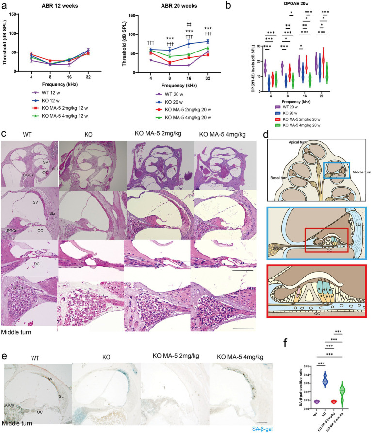
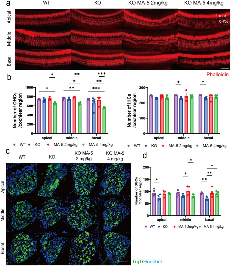
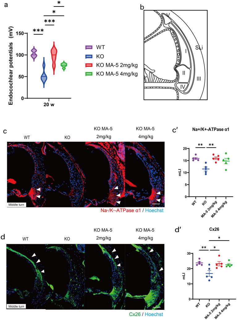
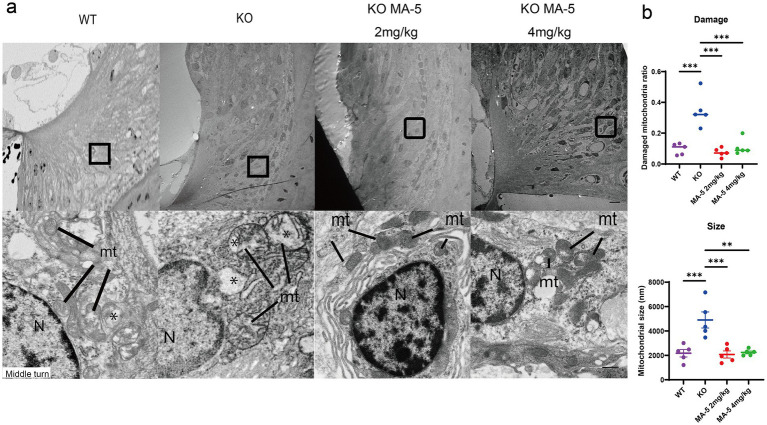
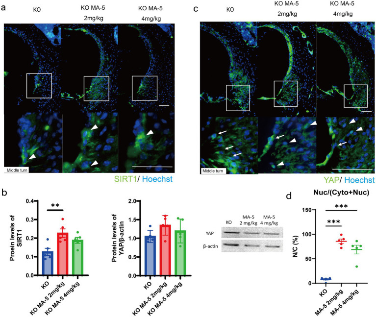
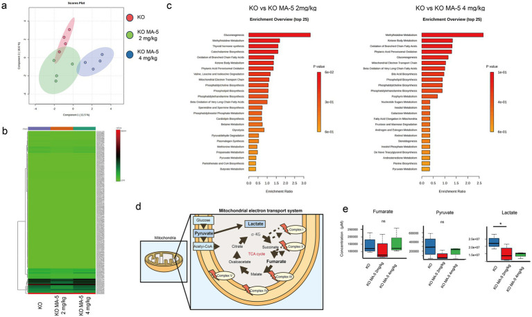
Results: MA-5 treatment effectively attenuated ARHL progression in Cdk5rap1-KO mice by improving auditory brainstem response thresholds and distortion product otoacoustic emissions. It also reduced spiral ganglion and outer hair cell loss, while preserving the cochlear structural integrity by preventing mitochondrial degeneration in spiral ligament fibrocytes. Mechanistically, MA-5 upregulated the expression of silent information regulator sirtuin 1 and promoted the nuclear translocation of yes-associated protein, both of which are involved in regulating mitochondrial function and cellular senescence. Metabolomics analysis further demonstrated that MA-5 restored mitochondrial metabolism, reduced lactate accumulation, and maintained mitochondrial integrity.

Conclusion: These findings suggest that MA-5 is a viable treatment option for ARHL and other age-related disorders associated with mitochondrial dysfunction.

Keywords: 2-methylthiolation; age-related hearing loss; cyclin-dependent kinase 5 regulatory subunit-associated protein 1; mitochondrial dysfunction; mitochonic acid 5.


Conflict of interest statement

The authors declare that the research was conducted in the absence of any commercial or financial relationships that could be construed as a potential conflict of interest.

Figures

Figure 1
Figure 1
MA-5 ameliorated ARHL and cochlear senescence in Cdk5rap1-KO mice. (a) ABR thresholds in Cdk5rap1-KO untreated mice and WT untreated mice at 12 and 20 weeks of age compared to those in MA-5-treated mice across various frequencies (n = 5 mice/group). Error bars represent the mean ± SE of five independent experiments. ††† indicates the significant differences (p < 0.001) between WT mice vs. untreated KO mice. *** indicates significant differences (p < 0.001) between untreated KO mice and MA-5 treated mice (2 mg/kg). ‡‡ indicates significant differences (p < 0.01) between untreated KO mice and MA-5 treated mice (4 mg/kg). (b) DPOAE levels in Cdk5rap1-KO untreated mice and WT untreated mice at 20 weeks of age compared to those in MA-5-treated mice across various frequencies (n = 5 mice/group). Error bars represent the mean ± SE of five independent experiments. *p < 0.05, **p < 0.01, ***p < 0.001. (c) Gross cochlear morphology, including the organ of Corti (OC), spiral ganglion cells (SGCs), stria vascularis (SV), and spiral ligament (SLi), at 20 weeks of age (n = 5 /group). (d) Cartoon diagram of cross-section of the mouse cochlea highlighting key structures. The blue and red boxes represent the middle turn of the cochlea and a zoomed-in view of the OC, respectively. (e) SA-β-gal staining of cochlear sections from the middle turn at 20 weeks of age, showing senescent cells in blue (n = 5/group). (f) Quantification of SA-β-gal-positive cells in the cochlea (n = 5/group). ***p < 0.001. Scale bars = 100 μm. MA-5, mitochonic acid 5; cdk5rap1, cyclin-dependent kinase (CDK)5 regulatory subunit-associated protein 1; KO, knockout; SA-β-gal, senescence-associated β-galactosidase; ARHL, age-related hearing loss; ABR, auditory brainstem-response; DPOAE, distortion product otoacoustic emissions; SLi, spiral ligament; SV, stria vascularis; SGCs; spiral ganglion cells; OC, Organ of Corti; SE, standard error of mean.
Figure 2
Figure 2
MA-5 partially prevented the loss of HCs and SGCs in Cdk5rap1-KO mice. (a) Phalloidin staining of IHCs and OHCs in untreated and MA-5-treated cochleae (n = 5/group). (b) Quantitative analysis of OHCs and IHCs (n = 5/group). (c) Anti-Tuj1 immunostaining of SGCs in cochleae from untreated and MA-5-treated mice (n = 5/group). (d) SGC counts in untreated and MA-5-treated mice (n = 5/group). *p < 0.05, **p < 0.01, ***p < 0.001. Scale bars = 100 μm. MA-5, mitochonic acid 5; cdk5rap1, cyclin-dependent kinase (CDK)5 regulatory subunit-associated protein 1; Tuj1, beta-tubulin III; KO, knockout; HC, hair cell; SGCs; spiral ganglion cells; IHCs, inner hair cells; OHCs, outer hair cells.
Figure 3
Figure 3
Endocochlear potential and immunohistological analysis of SLi tissues. (a) Endocochlear potential in Cdk5rap1-KO mice (untreated and MA-5-treated) and WT untreated mice at 20 weeks of age. (b) Schematic representation of the five different types of SLi fibrocytes. (c) Immunohistochemical staining of SLi tissues from untreated and MA-5-treated mice in the middle turn. Na+/K+-ATPase α1 expression (red) was detected as a marker of fibrocytes (types II, IV, and V). Arrowheads indicate Na+/K+-ATPase α1 expression. (c′) Quantitative analysis of Na+/K+-ATPase α1 expression in untreated and MA-5-treated mice, based on the mLI. (d) Immunohistochemical staining of SLi tissues showing Cx26 (green) expression in type-I fibrocytes in the middle turn. Arrowheads indicate Cx26 expression. (d′) mLI analysis of Cx26 expression in untreated and MA-5-treated mice. *p < 0.05, **p < 0.01, ***p < 0.001. Scale bars = 100 μm. MA-5, mitochonic acid 5; cdk5rap1, cyclin-dependent kinase (CDK)5 regulatory subunit-associated protein 1; KO, knockout; SLi, spiral ligament; mLI, modified labeling index; Cx26, connexin 26.
Figure 4
Figure 4
Restoration of type-II and type-IV fibrocytes in SLi mitochondria by MA-5 in Cdk5rap1-KO mice. (a) TEM images of type-II and type-IV fibrocytes from the middle turn of the cochlea. N = nucleus. Magnified views highlight mitochondrial cristae structures in Cdk5rap1-KO mice (untreated and MA-5-treated), with asterisks marking cristae loss. mt = mitochondria. Scale bars = 1 μm (full image) and 100 nm (magnified image). (b) Quantification of mitochondrial damage and size in untreated and MA-5-treated mice. **p < 0.01, ***p < 0.001. MA-5, mitochonic acid 5; cdk5rap1, cyclin-dependent kinase (CDK)5 regulatory subunit-associated protein 1; KO, knockout; SLi, spiral ligament; TEM, transmission electron microscopy.
Figure 5
Figure 5
MA-5 modulated key molecular regulators in the SLi. (a) Immunohistochemical staining for SIRT1 (green) in SLi tissues from untreated and MA-5-treated mice. Arrowheads indicate SIRT1 expressing cells. (b) Quantitative analysis of SIRT1 expression (ELISA) and YAP expression (western blot) in untreated and MA-5-treated mice. (c) Immunohistochemical staining for YAP (green) in SLi tissues from untreated and MA-5-treated mice. Arrowheads indicate nuclear YAP, while arrows indicate cytoplasmic YAP expression. (d) Comparison of the NCRs of YAP in untreated and MA-5-treated mice. **p < 0.01, ***p < 0.001. Scale bars = 100 μm. MA-5, mitochonic acid 5; cdk5rap1, cyclin-dependent kinase (CDK)5 regulatory subunit-associated protein 1; KO, knockout; SLi, spiral ligament; SIRT1, silent information regulator sirtuin; ELISA, enzyme-linked immunosorbent assay; YAP, yes-associated protein; NCR, nuclear-to-cytoplasmic ratio.
Figure 6
Figure 6
Mitochondrial metabolite changes in MA-5-treated Cdk5rap1-KO cochleae. (a) PCA of metabolome data (n = 3/group). (b) Heatmap of metabolite levels (n = 3/group), with differentially expressed metabolites plotted on the vertical axis and their expression levels represented in a green-to-red gradient. (c) Enrichment analysis (n = 3/group). The left panel shows significant associations of biological pathways/functions between KO and KO + MA-5 2 mg/kg, while the right panel shows significant associations between KO and KO + MA-5 4 mg/kg. (d) Schematic representation of the tricarboxylic acid (TCA) cycle and mitochondrial electron transport chain. (e) Comparison of fumarate, pyruvate, and lactate levels in untreated and MA-5-treated cochleae (n = 3/group). *p < 0.05. MA-5, mitochonic acid 5; cdk5rap1, cyclin-dependent kinase (CDK)5 regulatory subunit-associated protein 1; KO, knockout; PCA, principal component analysis.

References

    1. Arragain S., Handelman S. K., Forouhar F., Wei F.-Y., Tomizawa K., Hunt J. F., et al. . (2010). Identification of eukaryotic and prokaryotic methylthiotransferase for biosynthesis of 2-Methylthio-N6-threonylcarbamoyladenosine in TRNA. J. Biol. Chem. 285, 28425–28433. doi: 10.1074/jbc.m110.106831, PMID: - DOI - PMC - PubMed
    1. Dupont W. D., Plummer W. D. (1990). Power and sample size calculations. Control. Clin. Trials 11, 116–128. doi: 10.1016/0197-2456(90)90005-m, PMID: - DOI - PubMed
    1. Hequembourg S., Liberman M. C. (2001). Spiral ligament pathology: a major aspect of age-related cochlear degeneration in C57BL/6 mice. J. Assoc. Res. Otolaryngol. 2, 118–129. doi: 10.1007/s101620010075, PMID: - DOI - PMC - PubMed
    1. Karaiskos S., Grigoriev A. (2016). Dynamics of tRNA fragments and their targets in aging mammalian brain. F1000Research 5:2758. doi: 10.12688/f1000research.10116.1, PMID: - DOI - PMC - PubMed
    1. Kim M.-J., Haroon S., Chen G.-D., Ding D., Wanagat J., Liu L., et al. . (2019). Increased burden of mitochondrial DNA deletions and point mutations in early-onset age-related hearing loss in mitochondrial mutator mice. Exp. Gerontol. 125:110675. doi: 10.1016/j.exger.2019.110675, PMID: - DOI - PMC - PubMed

LinkOut - more resources