Review
. 2025 Apr 7:19:1552241.
doi: 10.3389/fncel.2025.1552241. eCollection 2025.

Physiological roles of embryonic microglia and their perturbation by maternal inflammation

Affiliations
Review

Physiological roles of embryonic microglia and their perturbation by maternal inflammation

Tsukasa Shimamura et al. Front Cell Neurosci. .

Abstract

The interplay between the nervous and immune systems is well documented in the context of adult physiology and disease. Recent advances in understanding immune cell development have highlighted a significant interaction between neural lineage cells and microglia, the resident brain macrophages, during developmental stages. Throughout development, particularly from the embryonic to postnatal stages, diverse neural lineage cells are sequentially generated, undergo fate determination, migrate dynamically to their appropriate locations while maturing, and establish connections with their surroundings to form neural circuits. Previous studies have demonstrated that microglia contribute to this highly orchestrated process, ensuring the proper organization of brain structure. These findings underscore the need to further investigate how microglia behave and function within a broader framework of neurodevelopment. Importantly, recent epidemiological studies have suggested that maternal immune activation (MIA), triggered by various factors, such as viral or bacterial infections, environmental stressors, or other external influences, can affect neurogenesis and neural circuit formation, increasing the risk of neurodevelopmental disorders (NDDs) in offspring. Notably, many studies have revealed that fetal microglia undergo significant changes in response to MIA. Given their essential roles in neurogenesis and vascular development, inappropriate activation or disruption of microglial function may impair these critical processes, potentially leading to abnormal neurodevelopment. This review highlights recent advances in rodent models and human studies that have shed light on the behaviors and multifaceted roles of microglia during brain development, with a particular focus on the embryonic stage. Furthermore, drawing on insights from rodent MIA models, this review explores how MIA disrupts microglial function and how such disturbances may impair brain development, ultimately contributing to the onset of NDDs.

Keywords: brain; development; maternal immune activation; maternal inflammation; microglia; neurodevelopmental disorder; neuron; psychiatric disorder.


Conflict of interest statement

The authors declare that the research was conducted in the absence of any commercial or financial relationships that could be construed as a potential conflict of interest.

Figures
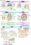
FIGURE 1
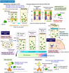
FIGURE 1
Ontology and distribution of “brain macrophages.” A schematic illustrates the process by which microglial progenitors colonize the brain both in humans and mice. Microglia and CAMs originate from the same progenitors, EMPs, which arise in the extraembryonic mesoderm of the yolk sac around PCW2–3 in humans and E7.5–8.5 in mice. Microglial progenitors differentiate from EMPs into microglia through the A1 and A2 states, and thereafter these cells migrate into the brain around at PCW3–4 in humans and around E9.5 in mice. A previous study reported that A2 progenitors can be divided into two distinct cell populations, characterized by the expression levels of CD206, a specific marker for CAMs (Utz et al., 2020). Another study using fate-mapping demonstrated that CD206+ progenitors retain the potential to differentiate into both CAMs and microglia, indicating that microglia are not exclusively derived from CD206 progenitors but also arise from early-committed CD206+ progenitors (Masuda et al., 2022a). Furthermore, it was revealed that a subset of microglia is derived from intraventricular CAMs, which frequently infiltrate the pallium at E12.5 in mice, indicating that microglia consist of at least two distinct populations with different colonization pathways (Hattori et al., 2023). The precise mechanisms governing when, where, and how these cells commit to their respective fates, as well as the factors influencing their migration into the brain parenchyma, remain to be fully elucidated. CAM, central nervous system-associated macrophage; E, embryonic day; EMP, erythromyeloid progenitor; P, postnatal day; PCW, postconceptional week.
FIGURE 2
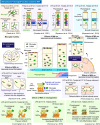
FIGURE 2
Multifaceted functions of microglia in the embryonic brain. The multifaceted functions of microglia in the embryonic brain are highlighted. A schematic illustrates how microglia alter their distribution in a stage-dependent manner during cerebral cortex development in mice and the roles they play in specific regions. During early embryonic stages, microglia are homogeneously distributed in the pallium. However, they transiently disappear from the CP between E15 and E16 before reentering around E17 (center panel). In regions where microglia are abundant, such as the VZ and the SVZ, they regulate neurogenesis and gliogenesis through cytokine and chemokine production (sections “5.1 Physiological and MIA-induced effects on neurogenesis” and “5.4 Physiological and MIA-induced effects on gliogenesis”). Conversely, the transient absence of microglia from the CP between E15 and E16 is crucial for the neuronal maturation, as microglia adjust their positioning to avoid interference (section “5.2 Physiological and MIA-induced effects on neuronal circuit formation”). In addition, microglia contribute to neuronal circuit formation by modulating the growth of dopaminergic axons and axon tracts in the corpus callosum (section “5.2 Physiological and MIA-induced effects on neuronal circuit formation”). They also play a role in interneuron positioning and migration (section “5.3 Physiological and MIA-induced effects on interneurons”). Furthermore, microglia support structural integrity in the CSA at E14 and CSB at E18 (section “3 Microglial distribution in the fetal brain”). CAM, central nervous system-associated macrophage; CCL2, C-C motif chemokine ligand 2; CP, cortical plate; CSA, cortico-striato-amygdalar boundary; CSB, cortico-septal boundary; CXCL10, C-X-C chemokine ligand 10; CXCL12, C-X-C chemokine ligand 12; CXCR4, C-X-C chemokine receptor 4; IFN-I, type I interferon; IL-6, interleukin-6; IP, intermediate progenitor; IZ, intermediate zone; LIF, leukemia inhibitory factor; MZ, marginal zone; NSC, neural stem cell; SP, subplate; SVZ, subventricular zone; VZ, ventricular zone.
FIGURE 3
FIGURE 3
MIA-induced effects on microglial function in the embryonic stage. A schematic summarizes previous studies investigating how MIA, induced by LPS or Poly(I:C), disrupts microglial function and its impact on surrounding neural lineage cells during embryonic development in rodent models. In the context of neurogenesis (section “5.1 Physiological and MIA-induced effects on neurogenesis”), MIA alters the state of microglia responsible for regulating the differentiation and number of NSCs and IPs, leading to a reduction in their population. Regarding its impacts on neuronal circuit formation (section “5.2 Physiological and MIA-induced effects on neuronal circuit formation”), MIA induces defasciculation of dorsal callosal axons in the corpus callosum and impairs dopaminergic axon extension in the subpallium. The effects of MIA on pyramidal neurons remain debated, with some studies reporting no significant changes, while another study has observed alterations in the number of CTIP2+ and TBR1+ neurons. In the process of interneuron migration guidance (section “5.3 Physiological and MIA-induced effects on interneurons”), MIA disrupts the positioning of LHX6+ interneurons in layer V of the CP, resulting in a broader dispersion of the LHX6+ population. Regarding microglial motility (section “5.5 Physiological and MIA-induced effects on microglial mobility and motility”), MIA has been reported to increase microglial process motility during the embryonic stage. CAM, central nervous system-associated macrophage; CP, cortical plate; CSA, cortico-striato-amygdalar boundary; CSB, cortico-septal boundary; CTIP2, COUP-TF-interacting protein 2; CXCL10, C-X-C chemokine ligand 10; CXCL12, C-X-C chemokine ligand 12; CXCR4, C-X-C chemokine receptor 4; IL-1β, interleukin-1 beta; iNOS, inducible nitric oxide synthase; IP, intermediate progenitor; IZ, intermediate zone; LHX6, LIM Homeobox 6; LPS, lipopolysaccharide; MIA, maternal immune activation; MZ, marginal zone; NSC, neural stem cell; PAX6, Paired box gene 6; Poly(I:C), polyinosinic-polycytidylic acid; SP, subplate; SVZ, subventricular zone; TBR1, T-box brain transcription factor 1; TBR2, T-box brain transcription factor 2; VZ, ventricular zone.

Similar articles

Cited by

References

    1. Ajami B., Samusik N., Wieghofer P., Ho P. P., Crotti A., Bjornson Z., et al. (2018). Single-cell mass cytometry reveals distinct populations of brain myeloid cells in mouse neuroinflammation and neurodegeneration models. Nat. Neurosci. 21 541–551. 10.1038/s41593-018-0100-x - DOI - PMC - PubMed
    1. Allen N. J., Lyons D. A. (2018). Glia as architects of central nervous system formation and function. Science 362 181–185. 10.1126/science.aat0473 - DOI - PMC - PubMed
    1. Alliot F., Godin I., Pessac B. (1999). Microglia derive from progenitors, originating from the yolk sac, and which proliferate in the brain. Brain Res. Dev. Brain Res. 117 145–152. 10.1016/s0165-3806(99)00113-3 - DOI - PubMed
    1. Andoh M., Shibata K., Okamoto K., Onodera J., Morishita K., Miura Y., et al. (2019). Exercise reverses behavioral and synaptic abnormalities after maternal inflammation. Cell Rep. 27 2817–2825.e5. 10.1016/j.celrep.2019.05.015 - DOI - PubMed
    1. Antony J. M., Paquin A., Nutt S. L., Kaplan D. R., Miller F. D. (2011). Endogenous microglia regulate development of embryonic cortical precursor cells. J. Neurosci. Res. 89 286–298. 10.1002/jnr.22533 - DOI - PubMed

LinkOut - more resources