. 2025 Apr 7:13:1565285.
doi: 10.3389/fcell.2025.1565285. eCollection 2025.

Lactoferrin-derived peptide PXL01 impacts nerve regeneration after sciatic nerve reconstruction in healthy and diabetic rats

Affiliations

Lactoferrin-derived peptide PXL01 impacts nerve regeneration after sciatic nerve reconstruction in healthy and diabetic rats

Derya Burcu Hazer Rosberg et al. Front Cell Dev Biol. .

Abstract

Introduction: Although advanced surgical techniques are available, satisfactory functional outcomes after peripheral nerve injuries are uncommon. Hence, immune-modulating factors such as PXL01, a lactoferrin-derived peptide that improves axonal outgrowth in injured human digital nerves, have gained attention. We previously reported a short-term immunosuppressive effect of PXL01 after the repair of transected rat sciatic nerves, but it had no effect on nerve regeneration. Here, we investigated the potential of PXL01 to improve nerve regeneration in healthy rats and in a rat model of type 2 diabetes (Goto-Kakizaki [GK] rats).

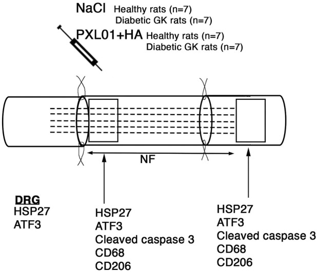
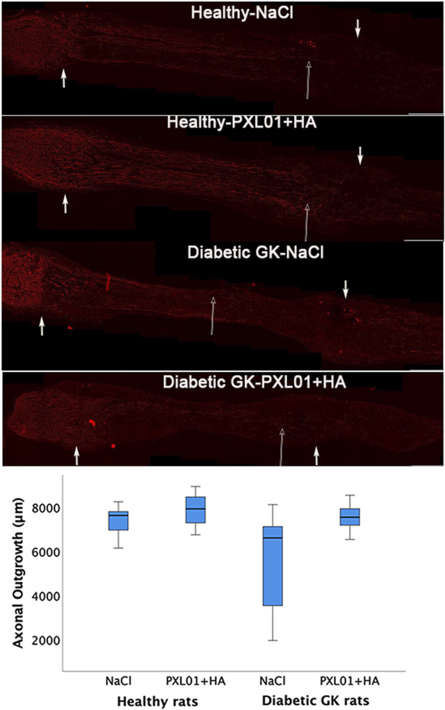
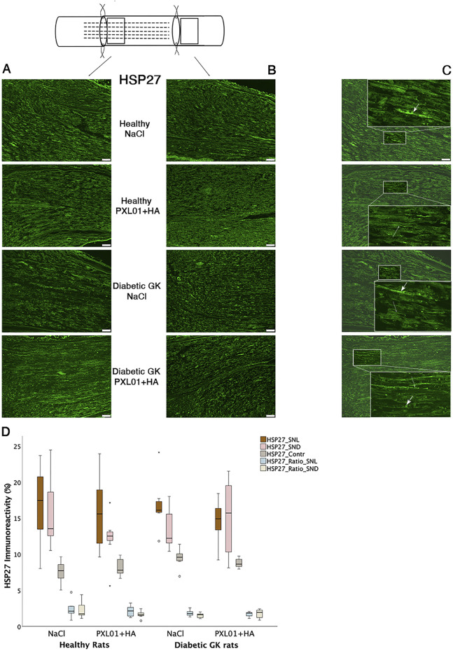
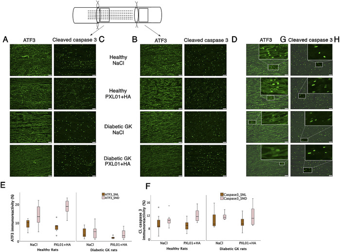
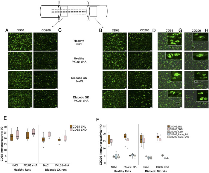
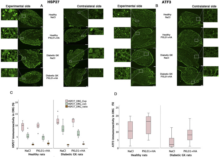
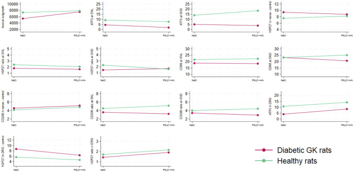
Methods: A 10-mm sciatic nerve defect was created in healthy (n = 14) and diabetic GK rats (n = 14) and reconstructed using nerve autografts. Immediately after surgery, PXL01 or sodium chloride (control, placebo) (n = 7 for each treatment) was administered around the autograft. On day 8, immunohistochemical staining of the sciatic nerve and dorsal root ganglia (DRGs) was performed to analyze axonal outgrowth (neurofilament staining); inflammation (CD68 and CD206 macrophage staining in nerve); Schwann cell and sensory neuron activation (transcription factor ATF3 staining in nerve and DRGs) and apoptosis (cleaved caspase 3 staining in nerve); and neuroprotection (heat shock protein [HSP27] staining in nerve and DRGs).

Results: PXL01 had no impact on the macrophage response in the autografts but increased axonal outgrowth and HSP27 expression in the DRGs of healthy and diabetic rats, despite a lower number of activated Schwann cells in the autograft. Diabetes affected axonal outgrowth, Schwann cell and macrophage responses, and HSP27 expression. These effects were observed in the sciatic nerve as well as the DRG.

Discussion: Application of PXL01, despite having no impact on macrophages, may improve axonal outgrowth and affects Schwann cell activation in autograft-reconstructed sciatic nerves, as well as conveys neuroprotection (HSP27 expression) in the DRGs of healthy and diabetic GK rats. Diabetes influenced nerve regeneration in such autografts. Therefore, PXL01 is a promising candidate to improve nerve regeneration.

Keywords: Goto-Kakizaki rat; PXL01; autograft; axonal outgrowth; diabetes; nerve reconstruction; nerve regeneration.


Conflict of interest statement

The authors declare that the research was conducted in the absence of any commercial or financial relationships that could be construed as a potential conflict of interest.

Figures

FIGURE 1
FIGURE 1
Schematic overview of the surgical method, the substances applied locally, and areas analyzed on the operated sciatic nerve. The presence of axonal outgrowth (length of neurofilament, NF; indicated by dashed lines), activated (ATF3-stained) and apoptotic (cleaved caspase 3-stained) Schwann cells, CD68 (activated, pan-macrophage)- and CD206 (pro-healing)-stained macrophages together with heat shock protein 27 (HSP27) expression at both the proximal lesion site (just distal to the proximal suture line) and at the distal nerve end (just distal to the distal suture line where there are no axons present at 8 days) were immunohistochemically analyzed at 8 days. In addition, immunoreactivity for HSP27 and ATF3 were analyzed in the bilateral dorsal root ganglia (DRGs).
FIGURE 2
FIGURE 2
Axonal outgrowth in nerve autografts 8 days after nerve reconstruction in healthy and diabetic Goto-Kakizaki (GK) rats with different treatments applied locally with sodium chloride (NaCl group) or PXL01 with sodium hyaluronate carrier (PXL01+HA group) is indicated by neurofilament staining. Scale bar = 1 mm. Small arrows in both ends show the suture sites. Long arrow shows the longest neurofilament staining. Box plots indicate the 25th and 75th percentiles (Tukey’s hinges) with the horizontal line in the middle indicating the median value. Error bars show minimum and maximum values.
FIGURE 3
FIGURE 3
HSP27 immunoreactivity at the proximal side of lesion (A) and at the distal nerve end (B) in autograft reconstructed sciatic nerves 8 days after the application of different treatment agents in Wistar and in diabetic GK rats. Scale bar = 100 μm. Box plots (D) show the HSP27 immunoreactivity in the operated sciatic nerve at the proximal side of lesion (SNL) and at distal distal nerve end (SND) as well as in the contralateral nerves (Cont). The groups are coded according to different treatment agents: sodium chloride treated group, NaCl; and PXL01 with sodium hyaluronate treated group, PXL01+HA. The images indicating the proximal site of lesion in SNL in all four experimental groups have a magnified insert indicating the Schwann cell expressing the HSP27 designated with short arrow, and the axonal expression of HSP27 with long arrow (C). Box plots indicate the 25th and 75th percentiles (Tukey’s hinges) with the horizontal line in the middle indicating the median value. Error bars show minimum and maximum values.
FIGURE 4
FIGURE 4
Activated (ATF3 stained) (A, B) and apoptotic (cleaved caspase 3-stained) Schwann cells (C, D) at the proximal side of lesion (SNL) and at the distal nerve end (SND) in autograft reconstructed sciatic nerve 8 days after the application of different treatment agents in healthy and diabetic GK rats. Scale bar = 50 μm. Box plots show the percentage of ATF3-stained (E) and cleaved caspase 3-stained Schwann cells (F) in the operated sciatic nerve. The groups are coded according to different treatment agents: sodium chloride treated group, NaCl; and PXL01 with sodium hyaluronate treated group, PXL01+HA. The magnification inserts showing ATF3 expressed (G) and cleaved caspase 3 (H) expressed Schwann cells are presented at the distal nerve end for each experimental group. Box plots indicate the 25th and 75th percentiles (Tukey’s hinges) with the horizontal line in the middle, indicating the median value. Error bars show minimum and maximum values.
FIGURE 5
FIGURE 5
Presence of CD68 and CD206 stained macrophages at the proximal side of lesion (A, C) and at the distal nerve end (B, D) in the autograft reconstructed sciatic nerve 8 days after applications of different treatment agents in healthy Wistar and in diabetic GK rats are presented. Bar = 50 μm. The magnification inserts showing CD68 expressed (G) and CD206 (H) expressed macrophages are presented at the distal nerve end for each experimental group. Box plots of percentage of CD68 stained (E) and CD206 stained macrophages (F) in the operated sciatic nerve (SNL = proximal side of lesion; SND = distal nerve end) and in the contralateral nerve (Cont) are shown. The groups are coded according to different treatment agents: sodium chloride treated group, NaCl; and PXL01 with sodium hyaluronate treated group, PXL01+HA. Box plots indicate the 25th and 75th percentiles (Tukey’s hinges) with the horizontal line in the middle, indicating the median value. Error bars show minimum and maximum values.
FIGURE 6
FIGURE 6
HSP27 immunoreactivity (A) and ATF3-stained sensory neurons (B) in the DRGs in operated (experimental) and contralateral side 8 days after application of different treatment agents in healthy and diabetic GK rats. Scale bar = 100 μm. Magnified inserts indicating the HSP27 expression in the sensory neuron (A) in the experimental side and in the contralateral side for each treatment modality and health status of the rats. Magnified inserts indicating the ATF3 expression in the sensory neuron (B) in the experimental side and in the contralateral side for each treatment modality and health status of the rats. Box plots show HSP27 immunoreactivity (C) and percentage of ATF3-stained sensory neurons (D) in DRGs. The groups are coded according to different treatment agents: sodium chloride treated group, NaCl; and PXL01 with sodium hyaluronate treated group, PXL01+HA. Exp: experimental side, con: contralateral side. Box plots indicate the 25th and 75th percentiles (Tukey’s hinges) with the horizontal line in the middle indicating the median value. Error bars show minimum and maximum values.
FIGURE 7
FIGURE 7
Interaction plots indicating the effects on the variables listed in Table 4 by health status (i.e., diabetic Goto-Kakizaki rats and healthy Wistar rats) and treatment (NaCl, saline; placebo and treatment with PLX01 in sodium hyaluronate carrier; PLX01+HA). For the location of the analyses, see Figure 1. SNL = at site of lesion. SND = at distal nerve end.

Similar articles

References

    1. Albers J. W., Pop-Busui R. (2014). Diabetic neuropathy: mechanisms, emerging treatments, and subtypes. Curr. Neurol. Neurosci. Rep. 14, 473. 10.1007/s11910-014-0473-5 - DOI - PMC - PubMed
    1. Bajic A., Andersson B., Ossinger A., Tavakoli S., Varghese O. P., Schizas N. (2024). Physically and chemically crosslinked hyaluronic acid-based hydrogels differentially promote axonal outgrowth from neural tissue cultures. Biomimetics (Basel) 9, 140. 10.3390/biomimetics9030140 - DOI - PMC - PubMed
    1. Bolandghamat S., Behnam-Rassouli M. (2020). Recent findings on the effects of pharmacological agents on the nerve regeneration after peripheral nerve injury. Curr. Neuropharmacol. 18, 1154–1163. 10.2174/1570159X18666200507084024 - DOI - PMC - PubMed
    1. Brownlee M. (2005). The pathobiology of diabetic complications: a unifying mechanism. Diabetes 54, 1615–1625. 10.2337/diabetes.54.6.1615 - DOI - PubMed
    1. Cattin A. L., Burden J. J., Van Emmenis L., Mackenzie F. E., Hoving J. J., Garcia Calavia N., et al. (2015). Macrophage-induced blood vessels guide Schwann cell-mediated regeneration of peripheral nerves. Cell 162, 1127–1139. 10.1016/j.cell.2015.07.021 - DOI - PMC - PubMed