. 2025 Apr 22;10(8):e178754.
doi: 10.1172/jci.insight.178754.

Glycine receptor activation promotes pancreatic islet cell proliferation via the PI3K/mTORC1/p70S6K pathway

Affiliations

Glycine receptor activation promotes pancreatic islet cell proliferation via the PI3K/mTORC1/p70S6K pathway

Ziyi Zhang et al. JCI Insight. .

Abstract

Glycine and β-alanine activate glycine receptors (GlyRs), with glycine known to enhance insulin secretion from pancreatic islet β cells, primarily through GlyR activation. However, the effects of GlyR activation on β cell proliferation have not been examined. Here, we aim to investigate the potential proliferative effects of glycine and β-alanine on islets. In vitro experiments on mouse and human islets revealed that glycine and β-alanine, via GlyR activation, stimulated the proliferation of β cells and α cells, without affecting insulin or glucagon secretion. Further analysis indicated the involvement of the PI3K/mTORC1/p70S6K signaling pathway in this process. Inhibition of GlyRs and PI3K/mTORC1/p70S6K signaling attenuated proliferative effects of glycine and β-alanine. In vivo and ex vivo studies supported these findings, showing increased β and α cell mass after 12 weeks of oral administration of glycine and β-alanine, with no changes in insulin secretion or glucose homeostasis under normal conditions. However, during an acute insulin resistance induced by insulin receptor antagonist S961, glycine and β-alanine enhanced insulin secretion and reduced blood glucose levels by increasing β cell secretory capacity. These findings demonstrate glycine and β-alanine in vivo and in vitro promote islet cell proliferation via GlyR activation and the PI3K/mTORC1/p70S6K pathway, potentially providing a target to enhance islet capacity.

Keywords: Beta cells; Diabetes; Endocrinology; Islet cells.


Conflict of interest statement

Conflict of interest: The authors have declared that no conflict of interest exists.

Figures

Figure 1
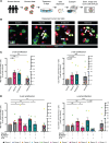
Figure 1. Glycine and β-alanine stimulate both β cell and α cell proliferation in mouse islets in vitro.
(A) Workflow for in vitro treatment of mouse islets. (B and C) Representative images of Ki67+ cells and islet cell proliferation measurement in mouse islets treated with vehicle, 1 mM glycine, or 1 mM β-alanine for 5 days in vitro. Harmine (10 μM) was used as a positive control for islet cell proliferation (n = 5 mice used for each treatment). We used 30 islets per mouse, with an average of 9,761 cells per sample to derive the data. Cell proliferation rate was calculated by normalizing Ki67+ β cell/α cell numbers to total β cell/α cell numbers on cytospin slides. Data are shown as mean ± SEM. Statistical significance was assessed using an unpaired t test, with Holm-Bonferroni correction applied for multiple comparisons. ***P < 0.001, **P < 0.01, compared with the control group. White arrows indicate Ki67-positive proliferating α cells and β cells.
Figure 2
Figure 2. Glycine and β-alanine stimulate both β cell and α cell proliferation in mouse islets via glycine receptor in vitro.
(A and B) Representative images of Ki67+ cells and islet cell proliferation in dispersed mouse islets treated with vehicle, 1 mM glycine, 1 mM β-alanine, or 10 μM Harmine, with or without 1 μM strychnine for 5 days (n = 5 mice used for each treatment). Data are depicted as mean ± SEM. Statistical significance was determined by using unpaired t test, with Holm-Bonferroni correction applied for multiple comparisons. ***P < 0.001, **P < 0.01, compared with the control group. ###P < 0.001, compared with the glycine or β-alanine treatment group. GlyR, glycine receptor; STR, strychnine. Scale bar: 50 μm. White arrows indicate Ki67+ proliferating α cells and β cells.
Figure 3
Figure 3. Glycine and β-alanine stimulate both β cell and α cell proliferation in human islets via glycine receptor in vitro.
(A) Workflow for in vitro treatment of human islets. (B and C) Representative images of Ki67+ cells and islet cell proliferation in human islets treated with vehicle, 1mM glycine, or 1 mM β-alanine for 5 days in vitro. Harmine (10 μM) was used as a positive control for islet cell proliferation. We used 30 islets per donor, with an average of 7,490 cells per sample to derive the data. Cell proliferation was calculated by normalizing Ki67+ β cell/α cell numbers to total β cell/α cell numbers on cytospin slides. Scale bar: 50 μm. White arrows indicate Ki67+ proliferating α cells and β cells. (D) Islet cell proliferative in primary human islets treated with vehicle, 1 mM glycine, or 1 mM β-alanine, with or without 1 μM strychnine for 5 days. Data are depicted as mean ± SEM. Statistical significance was determined by using Mann-Whitney U test or unpaired t test dependent on dataset normality test, with Holm-Bonferroni correction applied for multiple comparisons. *P < 0.05, **P < 0.01, ***P < 0.001, compared with the control group. #P < 0.05, compared with the glycine or β-alanine treatment group. STR, strychnine. We used islets from 11 donors, 7 of 11 donors have accessible HbA1c values, all falling within the normal range.
Figure 4
Figure 4. Glycine and β-alanine treatment had no effect on glucose-stimulated insulin secretion (GSIS) and total islet insulin content in mouse islets in vitro.
(A) Mouse islets were treated with vehicle, 1 mM glycine or 1 mM β-alanine for 5 days in vitro. Insulin secretion under the treatment of low glucose (2 mM), high glucose (11 mM), and high glucose + KCl was measured. (B) Total insulin content of 20 islets treated with vehicle, 1 mM glycine, or 1 mM β-alanine for 5 days in vitro. (C and D) Insulin secretion and total insulin content normalized to total DNA content. Data are depicted as mean ± SEM. n = 4–5 mice used for each treatment. Statistical significance was determined by using unpaired t test, with Holm-Bonferroni correction applied for multiple comparisons. There were no significant differences among the groups, with P > 0.05.
Figure 5
Figure 5. Glycine and β-alanine stimulate both β cell and α cell proliferation in mouse islets in vivo.
(A) Workflow for in vivo oral administration of 2% glycine and 2% β-alanine drinking water to mice and subsequent assessment. (B) Plasma glycine concentration. (C) Isolated islet size. Scale bars: 500 μm. (D) Total insulin content in 20 isolated mouse islets. (E and F) Representative images of Ki67+ cells and islet cell (both β cell and α cell) proliferation in mice treated with vehicle, 2% glycine, or 2% β-alanine for 12 weeks in vivo. We used 30 islets per mouse, with an average of 17,778 cells per sample to derive the data. Data are depicted as mean ± SEM. n = 7–9 in each group. Scale bar: 50 μm. White arrows indicate Ki67+ proliferating α cells and β cells. Statistical significance was determined by using unpaired t test, with Holm-Bonferroni correction applied for multiple comparisons where appropriate. *P < 0.05, **P < 0.01, ***P < 0.001, compared with the control group.
Figure 6
Figure 6. Glycine and β-alanine stimulate both β cell and α cell proliferation in mouse islets in vivo.
(A) Representative images of insulin-stained pancreatic sections from mice administered glycine or β-alanine for 12 weeks. Scale bar: 200 μm. (BG) Analysis of these sections were performed by calculating β cell mass (B), β cell number/area (C), average β cell size (D), α cell mass (E), α cell number/area (F), and average α cell size (G). n = 9–12 samples were analyzed in each group, with sections from 2 different levels in each sample stained and analyzed. Statistical significance was determined by using unpaired t test (B, C, E, and G) or Mann-Whitney U test (D and F) dependent on dataset normality test, with Holm-Bonferroni correction applied for multiple comparisons. *P < 0.05, compared with the control group.
Figure 7
Figure 7. Glycine and β-alanine administration did not influence glucose homeostasis in vivo upon glucose stimulation.
(A) Workflow for treatment and in vivo and in vitro assessment. (BH) Mice were treated with 2% glycine or 2% β-alanine through drinking water for 12 weeks and were then assessed for glucose homeostasis by evaluating body weight (B), fasting glucose (C), fasting insulin (D), fasting glucagon level (mice were treated with 2% glycine or normal drinking water for 5 weeks before glucagon assessment) (E), glucose changes during OGTT (F), insulin levels during OGTT (G), and glucose levels during ITT (H). n = 6–10 in each group. (I and J) glucose-stimulated insulin secretion in 20 islets isolated from mice treated with vehicle, 2% glycine or 2% β-alanine, with or without normalization by DNA content/cell number. n = 5–7 in each group. Data are depicted as mean ± SEM. Statistical significance was determined by using unpaired t test (B, D, E, and I), 1-way ANOVA (FH) or Mann-Whitney U test (C and J) dependent on dataset normality test, with Holm-Bonferroni correction applied for multiple comparisons.
Figure 8
Figure 8. Improved glucose tolerance and insulin secretion in S961-induced transient insulin resistance in glycine- and β-alanine–treated mice.
(A) Workflow for in vivo oral administration of 2% glycine and 2% β-alanine drinking water to mice. (B and C) Blood glucose levels and plasma insulin levels were measured hourly after S961 injection (30 nmol/kg). Data are depicted as mean ± SEM. n = 9–12 in each group. Statistical significance was determined by using unpaired t test, with Holm-Bonferroni correction applied for multiple comparisons. *P < 0.05, *P < 0.01, compared with the control group.
Figure 9
Figure 9. Glycine and β-alanine stimulate islet cell proliferation through PI3K/mTOR/p70S6K signaling pathway in mouse islets.
(A) Outline of the PI3K/mTOR/p70S6K signalling pathway and selective pathway inhibitors. (B and C) Mouse islets were exposed to various treatments for 5 days in vitro (n = 5 for each group). Cell proliferation was assessed by normalizing Ki67+ β cell/α cell numbers to total β cell/α cell numbers on cytospin slides. Glycine: 1 mM; β-alanine: 1 mM; Wortmannin (PI3K antagonist): 100 nM; PF-4708671 (p70S6K inhibitor): 10 μM; Rapamycin (mTORC1/2 inhibitor): 10 nM. Data are depicted as mean ± SEM. Statistical significance was determined by using unpaired t test, with Holm-Bonferroni correction applied for multiple comparisons. **P < 0.01, ***P < 0.001, compared with the control group. #P < 0.05, ##P < 0.01, ###P < 0.001, compared with the glycine or β-alanine treatment group. n = 4–5 in each group.

References

    1. Guillausseau PJ, et al. Abnormalities in insulin secretion in type 2 diabetes mellitus. Diabetes Metab. 2008;34(suppl 2):S43–S48. doi: 10.1016/S1262-3636(08)73394-9. - DOI - PubMed
    1. Gannon MC, et al. The metabolic response to ingested glycine. Am J Clin Nutr. 2002;76(6):1302–1307. doi: 10.1093/ajcn/76.6.1302. - DOI - PubMed
    1. Iverson JF, et al. Interaction of ingested leucine with glycine on insulin and glucose concentrations. J Amino Acids. 2014;2014:521941. doi: 10.1155/2014/521941. - DOI - PMC - PubMed
    1. Gonzalez-Ortiz M, et al. Effect of glycine on insulin secretion and action in healthy first-degree relatives of type 2 diabetes mellitus patients. Horm Metab Res. 2001;33(6):358–360. doi: 10.1055/s-2001-15421. - DOI - PubMed
    1. Yan-Do R, et al. A glycine-insulin autocrine feedback loop enhances insulin secretion from human β-cells and is impaired in type 2 diabetes. Diabetes. 2016;65(8):2311–2321. doi: 10.2337/db15-1272. - DOI - PubMed

MeSH terms

LinkOut - more resources