. 2025 May 20;10(5):e0173724.
doi: 10.1128/msystems.01737-24. Epub 2025 Apr 22.

Staphylococcus epidermidis uses the SrrAB regulatory system to modulate oxidative stress and intracellular survival in mouse macrophage cell line Ana-1

Affiliations

Staphylococcus epidermidis uses the SrrAB regulatory system to modulate oxidative stress and intracellular survival in mouse macrophage cell line Ana-1

Chunjing Zhao et al. mSystems. .

Abstract

The two-component system (TCS) SrrAB responds to oxidative stress in Staphylococcus epidermidis. A srrAB deletion mutant (∆srrAB) was constructed using S. epidermidis strain 1457 (SE1457) as the parent strain to study its regulatory function in oxidative stress. Compared to SE1457, the viable cell counts of the ∆srrAB mutant significantly decreased in the post-stationary phase culture, coinciding with a sharp increase in reactive oxidative species (ROS) accumulation. The impaired growth of the ∆srrAB mutant was partially restored by shifting the culture from oxic to microaerobic conditions. Consistently, growth of the ∆srrAB mutant in tryptone soy broth (TSB) medium containing H2O2 was notably inhibited compared to parent strain SE1457, and the mutant showed significantly decreased resistance (100- to 1,000-fold) to H2O2 and cumene hydroperoxide in both oxic and microaerobic conditions, which was fully rescued by the addition of ROS inhibitor 2,2-dipyridyl. Furthermore, the deletion of srrAB resulted in decreased intracellular survival in the Ana-1 macrophages, likely due to intracellular ROS accumulation. The complementation of srrAB in the ∆srrAB mutant restored ROS resistance and intracellular survival to wild-type levels. RNA-seq analysis revealed that srrAB deletion affected the transcription levels of 610 genes, including those involved in oxidative stress, respiratory and energy metabolism, and transition ion homeostasis. These findings were corroborated by quantitative real-time reverse transcription-PCR. In the ∆srrAB mutant, expressions of ROS-scavenging genes katA, ahpC, scdA, serp1797, and serp0483 were downregulated compared to SE1457. Electrophoretic mobility shift assay further demonstrated phosphorylated SrrA bound to the promoter regions of srrAB, katA, ahpC, scdA, serp1797, and serp0483 genes. This study elucidates that in S. epidermidis, SrrAB is the critical TCS to sense and respond to the oxidants, directly regulating transcription levels of the genes involved in ROS scavenging and ion homeostasis, thereby facilitating S. epidermidis detoxification of ROS and adaptation to the commensal environment.

Importance: Staphylococcus epidermidis in the human skin and mucous microbiome is a leading cause of hospital-acquired infection, whereas the mechanism by which it inhabits, adapts, and further results in infection is not well known. In this study, we found that the two-component regulatory system SrrAB directly regulates transcription levels of the genes involved in reactive oxidative species (ROS) scavenging and ion homeostasis in S. epidermidis, influencing ROS accumulation during growth, thereby facilitating detoxification of ROS and adaptation to the commensal environment. This work provides new molecular insight into the mechanisms of SrrAB in regulating resistance and intracellular viability against oxidative stress in S. epidermidis.

Keywords: ROS; Staphylococcal respiratory response; Staphylococcus epidermidis; macrophage; oxidative stress.


Conflict of interest statement

The authors declare no conflict of interest.

Figures

Fig 1
Fig 1
Transcriptional levels of srrA and srrB in SE1457 under oxidative stress. After culturing for 4 h, S. epidermidis strain 1457 was treated with different concentrations of H2O2 (A) or CHP (B) for 30 min of incubation. Staphylococcal cells were collected, and total RNA was extracted. The relative transcription levels of srrA and srrB were analyzed by qRT-PCR in comparison to the expression level of gyrB (housekeeping gene). The experiments were performed in triplicate and repeated at least three times. Data were represented as the means ± SDs; ***, P<0.001.
Fig 2
Fig 2
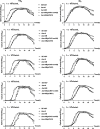
Growth curves of SE1457 srrAB isogenic mutants under oxic and microaerobic conditions. Overnight cultures of SE1457, ΔsrrAB, ΔsrrAB(pCN51-srrAB), and ΔsrrAB(pCN51) strains were diluted (1:200) in fresh TSB medium and incubated at 37°C for about 4 h with the OD600 reaching 0.8. After 10-fold serial dilutions, the bacterial suspension was inoculated (1:200) into a fresh TSB medium. Under oxic conditions (+O2), bacterial suspension was added to triplicate wells (200 µL/well) in a 96-well plate and placed into a heated microplate reader that allowed for free diffusion of gases. Under microaerobic conditions (−O2), the cultures were inoculated into a 96-well plate completely filled with the medium, all air bubbles were removed, and the plate was sealed with sealing film. The cultures were measured hourly at an OD600 for 24 h. The curves represented the results of the three independent experiments. Data were represented as the means  ±  SD of triplicate wells.
Fig 3
Fig 3
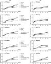
ROS accumulation of SE1457 srrAB isogenic mutants under oxic and microaerobic conditions. Overnight cultures were diluted (1:200) into fresh TSB medium and incubated at 37°C with aeration for 4 h (OD600 of 0.8). The bacterial suspension was adjusted to 1.0 × 10⁶ CFU/mL and serially diluted (10-fold), then pipetted into the microplate. At each time point, 100 µL of bacterial suspension was withdrawn, and 0.5 mL NBT (1 mg/mL) was added. The cultures were measured every 2 h at an OD575 for 24 h. The experiments were repeated at least three times, and one representative result was shown. Data were represented as means  ±  SD of triplicate wells.
Fig 4
Fig 4
Tolerance of the ∆srrAB mutant to oxidative stress under oxic and microaerobic conditions. Staphylococci grown to OD600 value of 0.8 at 37°C with aeration for about 4 h were serially diluted (1:10), and an aliquot (5 µL) was spotted onto the TSA plate containing different concentrations of H2O2 or CHP. For the detoxification of ROS, DIP was added. The plates were incubated at 37°C for 24 h under oxic conditions (+O2) or under microaerobic conditions (−O2). The image labeled “TSA” on the lower left in panel A or B was the duplicate serving as negative control from a set of assays. The results represented one of three independent experiments.
Fig 5
Fig 5
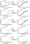
Growth curves of SE1457 srrAB isogenic mutants under oxidative stress. Staphylococci grown to OD600 value of 0.8 at 37°C with shaking were 10-fold serially diluted into TSB medium containing 1 mM H2O2. The incubation under oxic (+O2) and microaerobic (−O2) conditions were the same as described in the previous experiments. The cultures were measured hourly at an OD600 for 12 h. The curves represented the results of one of the three independent experiments. Data were represented as the means ± SD of triplicate wells.
Fig 6
Fig 6
Effect of srrAB deletion on the phagocytosis of S. epidermidis by macrophages. Ana-1 cells (1.5 × 10⁷ cells/mL, 1 mL) were co-incubated with S. epidermidis strains (1.5 × 10⁸ CFU/mL, 1 mL) at a multiplicity of infection of 10 in a six-well plate at 37°C with 5% CO2. At the time point of 6 h incubation, the extracellular bacterial cells unengulfed by macrophages were harvested, and the phagocytosis rate of S. epidermidis by macrophage was evaluated by CFU counting prior to Ana-1 cell lysis. Statistical significance was determined by one-way analysis of variance (ANOVA) analysis (P > 0.05) (A). The Ana-1 cells were treated with 100 µg/mL gentamycin and 20 µg/mL lysostaphin for 30 min to kill the extracellular and adherent bacterial cells and lysed with radioimmunoprecipitation assay buffer (RIPA) lysis buffer. The survival of S. epidermidis strains was assessed by CFU counting on TSA plate (B). The experiments were repeated at least three times, and the data were represented as the means ± SD. ***, P<0.001. The black square and circle represent the parent strain SE1457 and complementation strain ∆srrAB(pCN51-srrAB), respectively. The white circle represents the ∆srrA mutant. The white triangle and inverted triangle represent the ∆srrAB mutant and ∆srrAB(pCN51) vector control, respectively.
Fig 7
Fig 7
Effect of srrAB deletion on the ROS accumulation of macrophages infected by S. epidermidis. Mouse macrophage Ana-1 cells were co-incubated with S. epidermidis strains at a multiplicity of infection (MOI) of 10 in a six-well plate at 37°C for 6 h with 5% CO2. Samples were stained with DCFH-DA (10 µmol/L) at 37°C for 20 min in the dark, and the infected cells were washed three times with serum-free RPMI-1640 medium. The ROS-positive cells and fluorescence intensity were measured by flow cytometry (A) and a Microplate Reader (B), respectively. Rosup was designated as a positive control for the induction of ROS. The figures represent one of three independent experiments, and the data were represented as the means ± SD. **, P<0.01.
Fig 8
Fig 8
Transcriptional levels of ROS-scavenging genes in SE1457 srrAB isogenic mutants. S. epidermidis strains were inoculated into TSA medium challenged with or without 50 mM H2O2 and incubated at 37°C for 6 h. Bacterial cells were harvested, and total RNA was extracted. The relative expression levels of katA (A), ahpC (B), scdA (C), serp1797 (D), and serp0483 (E) were analyzed by qRT-PCR in comparison to the transcription level of gyrB (housekeeping gene). The measured values located on the bar were represented as the means of the relative transcript level. The experiments were performed in triplicate and repeated at least three times. Data were represented as the means ± SD.
Fig 9
Fig 9
EMSA analysis of S. epidermidis SrrA with the putative promoter regions. His-tagged SrrA was purified and phosphorylated (SrrA-P) by incubation with 50 mM acetyl phosphate. The putative promoter regions of srrA (A), katA (B), ahpC (C), scdA (D), SERP1797 (E), and SERP0483 (F) genes and negative control rpsJ gene (G) were PCR amplified, and the biotin-labeled DNA probes were purified. EMSAs were performed by incubating labeled probes with increasing amounts of SrrA-P. For each blot, lane 1 contained a no-protein control, and lanes 7 and 8 contained a 125-fold excess of the unlabeled specific probe (competitor control) and unlabeled nonspecific probe (DNA fragment within the gyrB coding region), respectively. Protein-DNA reactions were incubated at 25°C for 30 min, separated in a 6% nondenaturing polyacrylamide gel, and then blotted onto the nylon membrane. The biotin end-labeled DNA probe was detected using streptavidin conjugated to horseradish peroxidase (HRP) and a chemiluminescent substrate. Arrows indicate the position of the SrrA-DNA complex; triangles indicate the positions of free probes.
Fig 10
Fig 10
Schematic diagram of the SrrA promoter sequence in S. epidermidis. The suspected target genes directly regulated by SrrA were gathered, and a motif-based sequence analysis was performed at https://meme-suite.org/meme/. The SrrA binding motif (or box) was present in the proximal promoter region and almost located −4 bp to −18 bp upstream of the target genes at the transcription start site. The SrrA box with a high likelihood (p-value) was 8 nt in width (A). The SrrA binding box was shown as the standard pattern (B) or the reverse complement pattern (C). The conservative property of each base was indicated by the heights of each letter.

Similar articles

References

    1. Amer MA, Darwish MM, Soliman NS, Amin HM. 2024. Resistome, mobilome, and virulome explored in clinical isolates derived from acne patients in Egypt: unveiling unique traits of an emerging coagulase-negative Staphylococcus pathogen. Front Cell Infect Microbiol 14:1328390. doi:10.3389/fcimb.2024.1328390 - DOI - PMC - PubMed
    1. Severn MM, Horswill AR. 2023. Staphylococcus epidermidis and its dual lifestyle in skin health and infection. Nat Rev Microbiol 21:97–111. doi:10.1038/s41579-022-00780-3 - DOI - PMC - PubMed
    1. Maduka-Ezeh AN, Greenwood-Quaintance KE, Karau MJ, Berbari EF, Osmon DR, Hanssen AD, Steckelberg JM, Patel R. 2012. Antimicrobial susceptibility and biofilm formation of Staphylococcus epidermidis small colony variants associated with prosthetic joint infection. Diagn Microbiol Infect Dis 74:224–229. doi:10.1016/j.diagmicrobio.2012.06.029 - DOI - PubMed
    1. Mashruwala AA, Guchte A van de, Boyd JM. 2017. Impaired respiration elicits SrrAB-dependent programmed cell lysis and biofilm formation in Staphylococcus aureus. Elife 6:e23845. doi:10.7554/eLife.23845 - DOI - PMC - PubMed
    1. Carreau A, El Hafny-Rahbi B, Matejuk A, Grillon C, Kieda C. 2011. Why is the partial oxygen pressure of human tissues a crucial parameter? Small molecules and hypoxia. J Cell Mol Med 15:1239–1253. doi:10.1111/j.1582-4934.2011.01258.x - DOI - PMC - PubMed

LinkOut - more resources