. 2025 May 20;99(5):e0014325.
doi: 10.1128/jvi.00143-25. Epub 2025 Apr 22.

B cell lines fail to support efficient rhesus enteric calicivirus and human norovirus replication

Affiliations

B cell lines fail to support efficient rhesus enteric calicivirus and human norovirus replication

Tibor Farkas. J Virol. .

Abstract

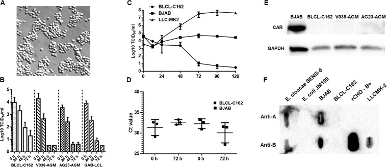
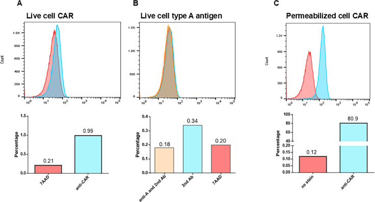
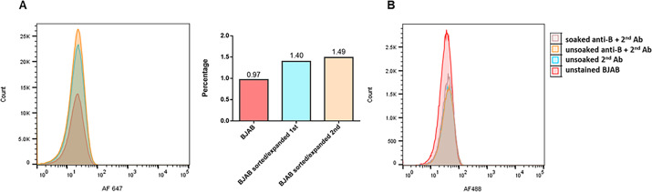
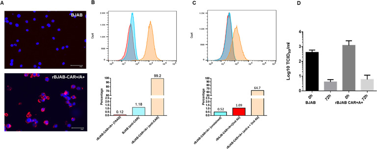
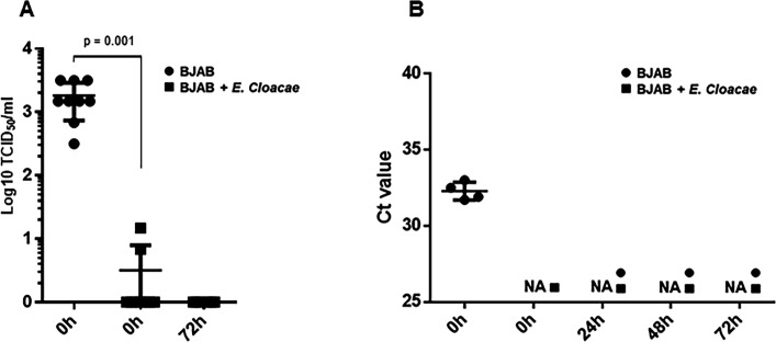
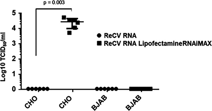
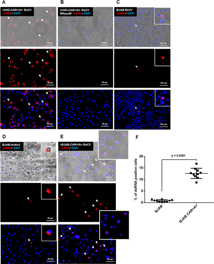
Analyses of intestinal biopsies of infected individuals and/or nonhuman primates (NHP) suggested the possible immune cell tropism of human noroviruses (HuNoV) and rhesus enteric caliciviruses (ReCV). Subsequently, the first HuNoV cell culture system using human B cell lines was reported. However, reproducibility issues raised questions about the validity and suitability of B cell cultures for HuNoV research. Histo-blood group antigens (HBGA) are known HuNoV susceptibility factors, but the full range of HuNoV susceptibility determinants remains unknown. In contrast, strain-specific ReCV susceptibility determinants have been recently characterized. Here, we evaluated NHP B cell lines and the human BJAB cell line for susceptibility to ReCV-FT285 infection, which is controlled by the Coxsackie and adenovirus receptor (CAR) and the type A or B HBGA. NHP B cell lines lacked CAR and HBGA expression and resisted infection. Inconsistent, low-level virus replication was detectable in BJAB cells, and expression of CAR and HBGAs was evident by Western blots. However, <1% of live, but >80% of fixed and permeabilized BJAB cells were CAR+, suggesting that CAR is mostly internalized. Co-transfection of BJAB cells with hCAR and A enzyme expression vectors led to substantial surface CAR and type A HBGA expression but not to an increase in ReCV titers. dsRNA staining revealed initial ReCV and HuNoV infection in a few cells that most likely became abortive. Based on both the similarities between ReCV and HuNoV replication profiles and the results obtained in the present study, considering BJAB cells an efficient culture system for HuNoV research is not justified.IMPORTANCERecently, two human norovirus (HuNoV) cell culture systems have been developed-the B cell culture system and the enteroid culture system. While the enteroid cell culture system became widely used in HuNoV research, mainly due to reproducibility issues, the B cell culture system did not. Here, we used HuNoV and rhesus enteric caliciviruses (ReCV) to evaluate enteric calicivirus B cell infections, in correlation to cell surface molecular determinants that control the susceptibility to infection. These are fully characterized for ReCVs, but not for HuNoVs. We found that only few BJAB cells express the cell surface molecules necessary for ReCV infection and support low-level, initial ReCV and HuNoV infection, but virus replication is most likely abortive, with minimal progeny virus release. Our findings and the poor reproducibility indicate that the B cell culture system in its current form is unsuitable for ReCV or HuNoV research and does not represent an efficient valid cell culture system.

Keywords: B cell; Coxsackie and adenovirus receptor; histo-blood group antigen; rhesus enteric calicivirus.


Conflict of interest statement

The author declares no conflict of interest.

Figures

Fig 1
Fig 1
BJAB cells but not the NHP B cell lines express CAR and support very low levels of ReCV replication. (A) Healthy B cell culture (BJAB cells are shown) with round cells displaying a sharp, shiny periphery and forming small clumps of 25–50 cells. (B) Titration of ReCV-FT285-infected (1 MOI) NHP B cell cultures in LLC-MK2 monolayers. No virus replication is indicated. (C) Titration of ReCV- FT285-infected (1 MOI) BLCL-C162 (rhesus) and BJAB (human) cell cultures in LLC-MK2 monolayers. A low level of virus replication is indicated in the BJAB but not in the BLCL-C162 cell line by the eclipse at 12 hpi and increasing titers. Note the different kinetics between the two cell lines. (D) qRT-PCR analysis of ReCV-infected BLCL-C162 and BJAB cell cultures. Ct values are shown. A low level of virus replication is indicated in the BJAB but not in the BLCL-C162 cell line by the decrease in Ct values at 72 hpi. (E) Western blot analysis for CAR expression. Only the BJAB cell lines but not the NHP B cell lines displayed detectable CAR expression. GAPDH was used as a loading control. (F) Anti-A and anti-B monoclonal antibodies detected the presence of both type A and type B HBGAs in slot blots of BJAB cell lysates and in the positive control rCHO-B +cells, but not in the NHP cell lines (BLCL-C162 is shown). One representative of three independent experiments using the BJAB-LSU cells is shown. BJAB-LSU and BJAB-CDC yielded similar results.
Fig 2
Fig 2
Flow cytometry analysis of BJAB cells indicated that CAR remains internalized. (A) Live cell staining revealed that <1% of cells display CAR on the cell surface. (B) No cell surface expression of HBGAs could be detected (data for the type B antigen are not shown). (C) Analysis of fixed and permeabilized BJAB cells revealed CAR expression in >80% of the cells. One representative of three independent experiments with BJAB-LSU is shown. BJAB-CDC yielded similar results.
Fig 3
Fig 3
The cell surface CAR- or HBGA-positive cell population did not increase after two rounds of sorting and expansion or incubating cells in human AB serum, respectively. (A) Live cell staining of the original or sorted and expanded CAR-positive cell populations with anti-CAR-FITC antibody. No enrichment of CAR-positive cells was achieved. (B) Live cell staining of the original (unsoaked) and human AB serum-soaked BJAB cell populations with anti-B mouse Mab and AF488-labeled secondary antibody. Soaking BJAB cells in human AB serum did not increase the number of HBGA-positive cells. One representative of three independent experiments with the BJAB-LSU cell clone is shown.
Fig 4
Fig 4
Cell surface expression of CAR and the type A HBGA in rBJAB-CAR+/A + cells. (A) Nonpermeabilized BJAB and rBJAB-CAR+/A + cells were stained by the anti-CAR antibody (RmcB) and AF-594 secondary antibody. Cell surface expression of CAR (white arrows) could be detected only in the rBJAB-CAR+/A + cultures. (B) Live cell staining with anti-CAR-FITC antibody and flow cytometry analysis revealed cell surface CAR on ~99% of the rBJAB-CAR+/A + cells, while only ~1% of the parental BJAB cells stained positive. (C) Live cell staining and flow cytometry analysis of the rBJAB-CAR+/A + cell population with anti-A mouse Mab- and AF488-labeled secondary antibody revealed the presence of cell surface type A HBGA in ~65% of the cells. One of three individual experiments with BJAB-LSU is shown. (D) BJAB and rBJAB CAR +A + cells were infected with ReCV-FT285 (1 MOI), and infectious virus titers were evaluated by titration on LLC-MK2 monolayers at 0 hpi and 72 hpi. The mean and SD of three independent experiments are shown.
Fig 5
Fig 5
Preincubation of ReCV-FT285 or HuNoV with inactivated E. cloacae SENG 6 significantly reduces viral load at 0 hour. (A) BJAB cells were inoculated with 1 MOI of ReCV-FT285 alone or with ReCV-FT285 preincubated with heat-inactivated E. cloacae SENG 6 for 1 hour pre-infection. After inoculation and incubation for 1 hour at 37°C, cells were washed three times in culture media by centrifugation for 5 minutes at 700 rpm and plated into 24-well tissue culture plates at a density of 3 × 10⁵ cells/mL. Cultures were harvested immediately after plating (0 hours) and at 72 hours post-infection and titrated on LLC-MK2 cells to determine infectious virus load. (B) BJAB cells were inoculated with BCM-HuNoVs with and without preincubation with E. cloacae SENG 6, as described in panel A. Samples were harvested immediately after plating (0 hours) and at 24, 48, and 72 hours post-infection and analyzed by qRT-PCR for genome copy number changes. Data shown represent the mean and SD of repeated experiments with the parental and recombinant BJAB-CDC cell clones. Except for the 0 hour time points, there was no difference in the virus yield between the E. cloacae SENG 6-treated and untreated samples. NA = no amplification signal was detected. Statistical significance was calculated by two-tailed, unpaired t-test using the GraphPad Prism software. A P-value ≤ 0.05 was considered statistically significant.
Fig 6
Fig 6
ReCV genomic RNA transfection and infectious virus recovery. BJAB and CHO cells were transfected with ReCV-FT285 genomic RNA and evaluated for the recovery of infectious virus at 72 hours after transfection by titration of cell lysates on LLC-MK2 cells. Cells receiving only viral RNA without the transfection reagent served as the negative control. Data shown represent experiment with BJAB-CDC. Statistical significance was calculated by two-tailed, unpaired t-test using the GraphPad Prism software. A P-value ≤ 0.05 was considered statistically significant.
Fig 7
Fig 7
Detection of replication intermediate dsRNA in ReCV- and HuNoV-infected cells. rCHO-CAR+/A + and BJAB cell cultures were infected with ReCV-FT285 (1 MOI) or GII.4 [P31]/BCM16-2 HuNoV (105 genome copy/well) and stained with dsRNA-specific J2 antibody at 6 hours post-infection. rCHO-CAR+/A + cells infected with ReCV-FT285 served as the positive control. The specificity of dsRNA staining was evaluated by treating the infected cells with RNase III before J2 antibody staining (only rCHO-CAR+/A + is shown) (panels A and B). A small number of ReCV- or HuNoV-infected parental BJAB cells (≤ 1 cell per view field) stained positive for dsRNA (panels C and D). The number of dsRNA-positive cells in the ReCV-FT285-infected rBJAB-CAR+/A + cultures was significantly higher (panels E and F). White arrows indicate dsRNA-positive cells. HuNoV BJAB cell infections were performed according to the protocol described by Jones et al. (8). A representative of three repeated experiments with BJAB-CDC is shown. Panel F was created based on 10 random images taken at the same magnification of three repeated infection/IFA staining experiments. Total and dsRNA-positive cell counts were determined by using the ImageJ software. Statistical significance was calculated by two-tailed, unpaired t-test using the GraphPad Prism software. A P-value ≤ 0.05 was considered statistically significant.

Similar articles

References

    1. Lopman BA, Steele D, Kirkwood CD, Parashar UD. 2016. The vast and varied global burden of norovirus: prospects for prevention and control. PLoS Med 13:e1001999. doi:10.1371/journal.pmed.1001999 - DOI - PMC - PubMed
    1. Burke RM, Mattison CP, Pindyck T, Dahl RM, Rudd J, Bi D, Curns AT, Parashar U, Hall AJ. 2021. Burden of norovirus in the United States, as estimated based on administrative data: updates for medically attended illness and mortality, 2001-2015. Clin Infect Dis 73:e1–e8. doi:10.1093/cid/ciaa438 - DOI - PMC - PubMed
    1. Wobus CE, Karst SM, Thackray LB, Chang KO, Sosnovtsev SV, Belliot G, Krug A, Mackenzie JM, Green KY, Virgin HW. 2004. Replication of Norovirus in cell culture reveals a tropism for dendritic cells and macrophages. PLoS Biol 2:e432. doi:10.1371/journal.pbio.0020432 - DOI - PMC - PubMed
    1. Souza M, Azevedo MSP, Jung K, Cheetham S, Saif LJ. 2008. Pathogenesis and immune responses in gnotobiotic calves after infection with the genogroup II.4-HS66 strain of human norovirus. J Virol 82:1777–1786. doi:10.1128/JVI.01347-07 - DOI - PMC - PubMed
    1. Cheetham S, Souza M, Meulia T, Grimes S, Han MG, Saif LJ. 2006. Pathogenesis of a genogroup II human norovirus in gnotobiotic pigs. J Virol 80:10372–10381. doi:10.1128/JVI.00809-06 - DOI - PMC - PubMed

MeSH terms

Substances

LinkOut - more resources