. 2025 May 20;99(5):e0042825.
doi: 10.1128/jvi.00428-25. Epub 2025 Apr 22.

Cholesterol 25-hydroxylase inhibits Newcastle disease virus replication by enzyme activity-dependent and direct interaction with nucleocapsid protein

Affiliations

Cholesterol 25-hydroxylase inhibits Newcastle disease virus replication by enzyme activity-dependent and direct interaction with nucleocapsid protein

Guangmei Zhu et al. J Virol. .

Abstract

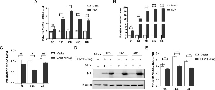
Newcastle disease virus (NDV) is a significant enveloped virus within the Paramyxoviridae family, posing a major threat to the global poultry industry. Increasing evidence suggests that cholesterol-25-hydroxylase (CH25H) and its enzymatic product, 25-hydroxycholesterol (25HC), exhibit broad-spectrum antiviral activity properties by modulating lipid metabolism and various signaling pathways. However, the specific role of CH25H in regulating NDV infection and replication remains unclear. In this study, we demonstrate that CH25H significantly inhibits NDV replication by blocking viral entry through its enzymatic product, 25HC. Notably, a catalytic mutant of CH25H (CH25H-M), which lacks hydroxylase activity, still retains partial ability to inhibit NDV replication, suggesting the involvement of an enzyme-independent antiviral mechanism. Furthermore, we found that CH25H interacts with the viral nucleoprotein (NP), leading to a reduction in NP expression and inhibition of viral ribonucleoprotein (RNP) complex activity. These findings reveal that CH25H exerts antiviral effects through both enzyme-dependent and -independent mechanisms, providing new insights into its role in host defense and offering potential targets for the development of antiviral therapies.IMPORTANCECholesterol 25-hydroxylase (CH25H) is a multifunctional host protein that has been implicated in regulating the life cycles of various viruses. As a prototype of paramyxovirus, Newcastle disease virus (NDV) poses a significant threat to the global poultry industry, causing substantial economic losses. Uncovering the mechanisms of NDV-host interactions is crucial for unraveling the viral pathogenesis and the host immune response, which can drive the development of effective antiviral therapies. Here, we demonstrate that CH25H, whose expression is induced upon NDV infection, plays a pivotal role in restricting viral replication. Specifically, CH25H interacts with the viral NP and inhibits the viral RNP activity. These findings expand our understanding of CH25H's antiviral functions and offer new insights into virus-host interactions, providing potential targets for the development of novel antiviral drugs against NDV and related pathogens.

Keywords: 25-hydroxycholesterol; Newcastle disease virus; cholesterol 25-hydroxylase; viral replication.


Conflict of interest statement

The authors declare no conflict of interest.

Figures

Fig 1
Fig 1
The CH25H was upregulated by NDV infection in DF-1 cells, and overexpressed CH25H inhibited replication of NDV. DF-1 cells were infected with NDV NA-1 strain at an MOI of 0.1. The mRNA levels of CH25H (A) and NP (B) were detected by qRT-PCR at 6, 12, 24, 36, and 48 hpi. DF-1 cells were transfected with either the Flag-tagged CH25H expressing plasmid or the empty vector pCAGGS for 24 h, followed by infection with NDV NA-1 at an MOI of 0.1. Cell samples and supernatants were harvested at 12, 24, and 48 hpi. NDV NP and mRNA levels were evaluated by qRT-PCR (C) and western blot analysis (D). The viral titers were quantified by TCID50 analysis (E). Results are presented as the mean ± SD from three independent experiments. *P < 0.05, **P < 0.01, ***P < 0.0001, ns (not significant), P > 0.05.
Fig 2
Fig 2
Knockdown of CH25H by siRNAs enhances NDV replication. DF-1 cells were transfected with three siRNAs (siRNA1, siRNA2, and siRNA3) or negative control (NC). At 48 hpt, the knockdown efficiency of CH25H was determined by qRT-PCR (A). DF-1 cells transfected with the three siRNAs were subsequently infected with the NDV NA-1 strain (MOI = 0.1) at 48 hpt. After 24 h of infection, cell samples were collected to assess the replication of NDV by qRT-PCR (B), western blot assays (C). Supernatants were collected for viral titer measurement by TCID50 analysis (D). Results are presented as mean ± SD from three independent experiments. *P < 0.05, **P < 0.01, ***P < 0.0001, ns (not significant), P > 0.05.
Fig 3
Fig 3
Construction and characterization of recombinant NDV expressing CH25H. (A) A schematic diagram illustrating the construction of recombinant NDV expressing CH25H. An artificial transcription cassette coding CH25H was inserted into the NDV genome between the P and M genes. (B) The insertion of the CH25H gene into rNDV-CH25H was verified by RT-PCR using a pair of primers spanning the insertion region of the CH25H gene. (C) DF-1 cells were infected with rNDV-CH25H at an MOI of 1. At 24 h post-infection, the cells were fixed and subjected to immunofluorescence assays to detect CH25H protein and NP. Primary antibodies (mouse anti-CH25H and rabbit anti-NP) were used, followed by secondary antibodies conjugated with AF488 (anti-mouse) and AF555 (anti-rabbit). (D) Multistep growth kinetics of the recombinant virus in DF-1 cells were analyzed. Cell monolayers were infected with rNDV-CH25H, rNDV-EGFP, or NDV NA-1 strain at an MOI of 1. Viruses were harvested at 12 h intervals, and virus titers were measured in DF-1 cells and expressed as TCID50/mL. (E) Biological characteristics of the recombinant viruses were evaluated. Data represent the mean values from triplicate experiments. *P < 0.05, **P < 0.01, ***P < 0.0001, ns (not significant, P > 0.05).
Fig 4
Fig 4
25HC inhibits NDV replication. DF-1 cells were treated with different concentrations of 25HC (0.25, 0.5, 1, 2, and 4 µM) or ethanol (ET) as a control for 24 h, and cell viability was assessed using the CCK-8 assay (A). To evaluate the effect of 25HC on NDV replication, DF-1 cells were pretreated with 1 µM 25HC for 24 h, and then infected with the NDV NA-1 strain at an MOI of 0.1. At 12, 24, and 48 hpi, cell samples and supernatants were collected and analyzed via qRT-PCR (B), western blot analysis (C), and TCID50 assay (D).
Fig 5
Fig 5
25HC blocks NDV penetration. For the attachment assay, DF-1 cells were pretreated with 1 µM 25HC or ET for 24 h at 37°C, then pre-chilled at 4°C for 1 h before infection with the NDV NA-1 strain (MOI = 10) for 2 h at 4°C. Cells were collected, and NDV NP mRNA levels were measured via qRT-PCR. For the penetration assay, DF-1 cells were pretreated with 25HC (1 µM) for 24 h, prechilled at 4°C for 1 h, then infected with NDV NA-1 strain (MOI = 10). After washing with PBS, the medium was replaced with DMEM containing 1 µM 25HC or ET, and cells were inoculated at 37°C for 2 h. Following three washes with PBS, cells were collected for qRT-PCR analysis of NP mRNA levels. For the replication assay, DF-1 cells were pretreated with 25HC (1 µM) at 37°C for 12 h, followed by NDV infection at 37°C. The infected cells were collected for qRT-PCR at 8 hpi. For the release assay, DF-1 cells were infected with NDV (MOI = 10). At 8 hpi, the virus-containing medium was replaced with fresh medium containing 1 µM 25HC or ET. After 2 h of 25HC treatment, cells were collected for qRT-PCR analysis (A). Supernatants and cell samples from the penetration assay were further analyzed via TCID50 (B) and western blot analysis (C). All results are presented as mean ± SD from three independent experiments, each performed in triplicate. *P < 0.05, **P < 0.01, ***P < 0.0001, ns: P > 0.05.
Fig 6
Fig 6
CH25H mutant lacking hydroxylase activity can suppress NDV replication. A catalytic mutant of CH25H (CH25H-M) was generated by substituting histidine residues at positions 242 and 243 with glutamine via site-directed mutagenesis (A). DF-1 cells were transfected with pCAGGS-CH25H, pCAGGS-CH25H-M, or the pCAGGS vector, then infected with the NDV NA-1 strain at an MOI of 0.1. After 24 h, cell samples and supernatants were collected for analysis by qRT-PCR (B), western blot analysis (C), and TCID50 assay (D). Data represent means ± SD from three independent experiments conducted in triplicate. *P < 0.05, **P < 0.01, ***P < 0.0001.
Fig 7
Fig 7
CH25H interacts with NDV NP and downregulates RNP activity. (A) DF-1 cells co-transfected with Flag-tagged CH25H and HA-tagged NP plasmids were fixed at 24 h post-transfection for immunofluorescence assays. A rabbit anti-Flag and mouse anti-HA antibodies were used as primary antibodies, followed by secondary antibodies conjugated with AF555 (anti-rabbit) and AF488 (anti-mouse). Nuclei were counterstained with DAPI. (B) DF-1 cells were co-transfected with recombinant plasmids encoding Flag-tagged CH25H and HA-tagged NP. At 24 h post-transfection, cell lysates were subjected to immunoprecipitation (IP) using an anti-Flag antibody. Whole-cell lysates and IP complexes were analyzed by western blot analysis with anti-Flag and anti-HA antibodies. (C) DF-1 were transfected with varying doses of CH25H and NP-expressing plasmids for 24 h. The replication of NDV was evaluated by Western blot analysis of NP expression. (D) DF-1 cells co-transfected with CH25H and NP-expressing plasmids were treated with specific inhibitors: MG132 (20 µM), 3-methyladenine (3-MA; 5 mM), or Ac-DEVD-CHO (25 µM) at 24 h post-transfection. After 12 h, cell lysates were analyzed by western blot analysis to detect the expression levels of NP, CH25H, and β-actin. (E) DF-1 cells were co-transfected with the NDV-MG minigenome reporter plasmid and helper plasmids encoding NP, P, and L proteins, along with either a CH25H expression plasmid or a control vector. At 48 hpost-transfection, EGFP-positive cells were imaged and quantified. Data shown are representative of three independent experiments.

Similar articles

References

    1. Wang C, Wang T, Duan L, Chen H, Hu R, Wang X, Jia Y, Chu Z, Liu H, Wang X, Zhang S, Xiao S, Wang J, Dang R, Yang Z. 2021. Evasion of host antiviral innate immunity by paramyxovirus accessory proteins. Front Microbiol 12:790191. doi:10.3389/fmicb.2021.790191 - DOI - PMC - PubMed
    1. Amarasinghe GK, Ayllón MA, Bào Y, Basler CF, Bavari S, Blasdell KR, Briese T, Brown PA, Bukreyev A, Balkema-Buschmann A, et al. . 2019. Taxonomy of the order mononegavirales: update 2019. Arch Virol 164:1967–1980. doi:10.1007/s00705-019-04247-4 - DOI - PMC - PubMed
    1. Ul-Rahman A, Ishaq HM, Raza MA, Shabbir MZ. 2022. Zoonotic potential of Newcastle disease virus: old and novel perspectives related to public health. Rev Med Virol 32:e2246. doi:10.1002/rmv.2246 - DOI - PubMed
    1. Yue H, Li D, Fu A, Ma L, Yang F, Tang C. 2008. shRNA-triggered RNAi inhibits expression of NDV NP gene in chicken embryo fibroblast. Front Biol China 3:433. doi:10.1007/s11515-008-0080-4 - DOI - PMC - PubMed
    1. El Najjar F, Schmitt AP, Dutch RE. 2014. Paramyxovirus glycoprotein incorporation, assembly and budding: a three way dance for infectious particle production. Viruses 6:3019–3054. doi:10.3390/v6083019 - DOI - PMC - PubMed

MeSH terms

LinkOut - more resources