

Bone marrow lesions related to bone marrow edema syndromes and osteonecrosis
- PMID: 40261331
- PMCID: PMC12037679
- DOI: 10.1007/s00132-025-04640-9
Bone marrow lesions related to bone marrow edema syndromes and osteonecrosis
Abstract
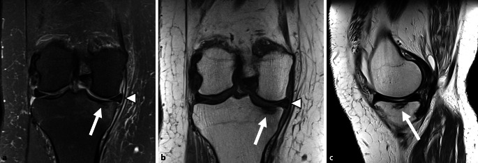
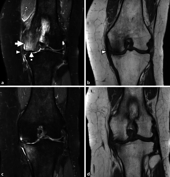
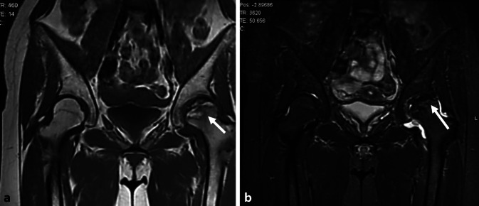
Bone marrow lesions (BML) are abnormalities in the bone marrow identified on magnetic resonance imaging (MRI) and can generally be classified as traumatic or atraumatic. This review focuses on atraumatic bone marrow edema syndromes (BMES) and their imaging evaluation. The MRI remains the modality of choice for assessing BMES, particularly using fluid-sensitive sequences although other sequences such as Dixon and T1-weighted imaging can be of further assistance. Emerging evidence supports dual-energy CT (DECT) as a reliable alternative, with high sensitivity and specificity for detecting bone marrow edema. The term BMES is a collective term for conditions, such as transient osteoporosis (TO) and regional migratory osteoporosis (RMO), predominantly affect weight-bearing bones in middle-aged individuals and pregnant or postpartum females. Subchondral insufficiency fractures of the knee (SIFK) are a key subset of BMES. These fractures most commonly involve the medial femoral condyle (MFC) and are associated with risk factors, such as meniscal root tears and extrusion of the meniscal body. The MRI findings typically include bone marrow edema-like signals and subchondral fracture lines, with additional features, such as secondary osteonecrosis in advanced cases. Prognostic indicators are crucial for stratifying patients and guiding management. Low-grade or reversible lesions often resolve with conservative treatment, whereas high-grade or irreversible lesions may require surgical intervention.Avascular necrosis, another atraumatic BML entity, differs from BMES by its association with systemic factors, such as steroid use or alcohol abuse. Accurate imaging, particularly in the early stages, is vital to distinguish between reversible and irreversible lesions, facilitating timely and appropriate management.
Knochenmarkläsionen (BML) sind Anomalien im Knochenmark, die in der Magnetresonanztomographie (MRT) festgestellt und generell als traumatisch oder atraumatisch klassifiziert werden können. Diese Übersicht konzentriert sich auf atraumatische Knochenmarködemsyndrome (BMES) und deren bildgebende Beurteilung. Die MRT ist nach wie vor das Mittel der Wahl für die Beurteilung von BMES, insbesondere unter Verwendung flüssigkeitsempfindlicher Sequenzen, wobei auch andere Sequenzen wie die Dixon- und T1-gewichtete Bildgebung von Nutzen sein können. Neue Erkenntnisse unterstützen die Dual-Energy-CT (DECT) als zuverlässige Alternative mit hoher Sensitivität und Spezifität für den Nachweis eines Knochenmarködems. Der Begriff BMES ist ein Sammelbegriff für Erkrankungen wie transiente Osteoporose und regionale Migrationsosteoporose, die vor allem gewichtstragende Knochen bei Personen mittleren Alters und schwangeren Frauen oder Frauen nach der Geburt betreffen. Subchondrale Insuffizienzfrakturen des Knies (SIFK) sind eine wichtige Untergruppe der BMES. Diese Frakturen betreffen am häufigsten den medialen Femurkondylus (MFC) und sind mit Risikofaktoren wie Meniskuswurzelrissen und Extrusion des Meniskuskörpers verbunden. Zu den MRT-Befunden gehören typischerweise Knochenmarködem-ähnliche Signale und subchondrale Bruchlinien sowie zusätzliche Merkmale wie sekundäre Osteonekrosen in fortgeschrittenen Fällen. Prognostische Indikatoren sind für die Stratifizierung der Patienten und die Steuerung der Behandlung von entscheidender Bedeutung. Geringgradige oder reversible Läsionen lassen sich häufig mit einer konservativen Behandlung beheben, während hochgradige oder irreversible Läsionen einen chirurgischen Eingriff erfordern können. Die avaskuläre Nekrose, eine weitere atraumatische BML-Entität, unterscheidet sich von der BMES durch ihren Zusammenhang mit systemischen Faktoren wie Steroidkonsum oder Alkoholmissbrauch. Eine genaue Bildgebung, insbesondere in den frühen Stadien, ist entscheidend für die Unterscheidung zwischen reversiblen und irreversiblen Läsionen und erleichtert eine rechtzeitige und angemessene Behandlung.
Keywords: Avascular necrosis; Dual-energy computed tomography; Magnetic resonance imaging; Sensitivity and specificity; Subchondral insufficiency fracture of the knee.
© 2025. The Author(s).
Conflict of interest statement
Declarations. Conflict of interest: G. Shabshin and N. Shabshin declare that they have no competing interests. Ethical standards: For this article no studies with human participants or animals were performed by any of the authors. All studies mentioned were in accordance with the ethical standards indicated in each case.
Figures


Similar articles
-
Is bone marrow edema syndrome a precursor of hip or knee osteonecrosis? Results of 49 patients and review of the literature.Diagn Interv Radiol. 2020 Jul;26(4):355-362. doi: 10.5152/dir.2020.19188. Diagn Interv Radiol. 2020. PMID: 32558648 Free PMC article. Review.
-
Bone marrow lesions and subchondral bone pathology of the knee.Knee Surg Sports Traumatol Arthrosc. 2016 Jun;24(6):1797-814. doi: 10.1007/s00167-016-4113-2. Epub 2016 Apr 13. Knee Surg Sports Traumatol Arthrosc. 2016. PMID: 27075892 Review.
-
Histopathologic confirmation of subchondral fracture in symptomatic pre-collapse osteonecrosis of the femoral head with bone marrow edema on magnetic resonance imaging.Skeletal Radiol. 2025 Jun;54(6):1275-1281. doi: 10.1007/s00256-024-04846-6. Epub 2024 Nov 29. Skeletal Radiol. 2025. PMID: 39611965
-
Subchondral insufficiency fracture of the knee: review of current concepts and radiological differential diagnoses.Jpn J Radiol. 2022 May;40(5):443-457. doi: 10.1007/s11604-021-01224-3. Epub 2021 Nov 29. Jpn J Radiol. 2022. PMID: 34843043 Free PMC article. Review.
-
Early irreversible osteonecrosis versus transient lesions of the femoral condyles: prognostic value of subchondral bone and marrow changes on MR imaging.AJR Am J Roentgenol. 1998 Jan;170(1):71-7. doi: 10.2214/ajr.170.1.9423603. AJR Am J Roentgenol. 1998. PMID: 9423603
References
-
- Aglietti P, Insall JN, Buzzi R, Deschamps G (1983) Idiopathic osteonecrosis of the knee. Aetiology, prognosis and treatment. J Bone Joint Surg Br 65:588–597. 10.1302/0301-620X.65B5.6643563 - PubMed
-
- Alvarez RE, Macovski A (1976) Energy-selective reconstructions in X‑ray computerized tomography. Phys Med Biol 21:733–744 - PubMed
-
- Assouline-Dayan Y, Chang C, Greenspan A et al (2002) Pathogenesis and natural history of osteonecrosis. Semin Arthritis Rheum 32:94–124 - PubMed
-
- Chen Z, Chen Y, Zhang H et al (2022) Diagnostic accuracy of dual-energy computed tomography (DECT) to detect non-traumatic bone marrow edema: A systematic review and meta-analysis. Eur J Radiol 153:110359. 10.1016/j.ejrad.2022.110359 - PubMed
-
- Clark SC, Pareek A, Hevesi M et al (2024) High incidence of medial meniscus root/radial tears and extrusion in 253 patients with subchondral insufficiency fractures of the knee. Knee Surg Sports Traumatol Arthrosc 32:2755–2761. 10.1002/ksa.12271 - PubMed
Publication types
MeSH terms
LinkOut - more resources
Full Text Sources
Medical