

Structural biology of Nipah virus G and F glycoproteins: Insights into therapeutic and vaccine development
- PMID: 40261700
- PMCID: PMC12208181
- DOI: 10.1556/1886.2025.00017
Structural biology of Nipah virus G and F glycoproteins: Insights into therapeutic and vaccine development
Abstract
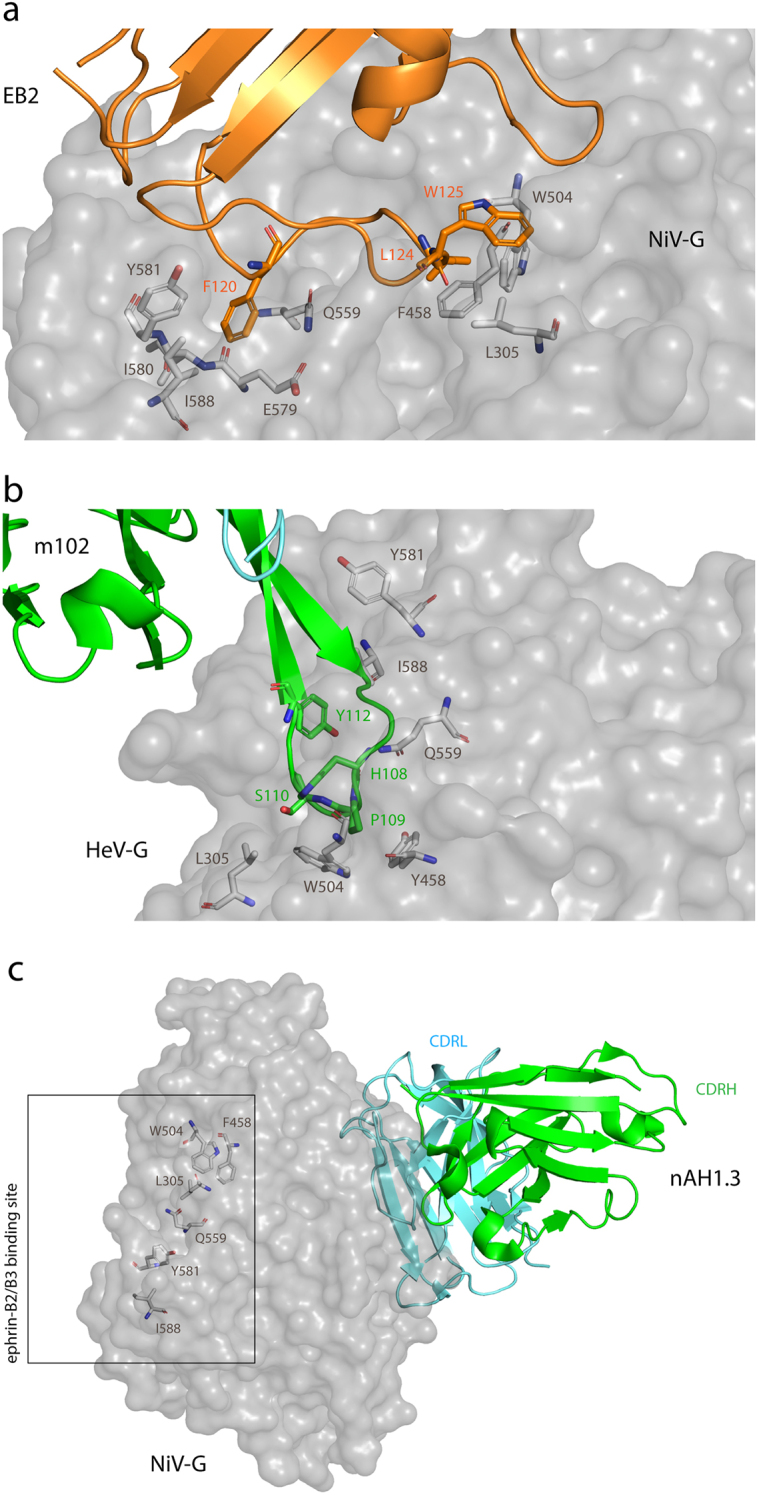
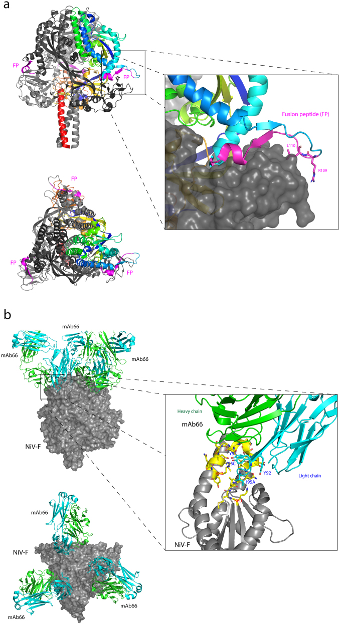
Nipah virus (NiV), a highly pathogenic zoonotic paramyxovirus, poses a significant public health threat due to its high mortality rate and potential for human-to-human transmission. The attachment (G) and fusion (F) glycoproteins play pivotal roles in viral entry and host-cell fusion, making them prime targets for therapeutic and vaccine development. Recent advances in structural biology have provided high-resolution insights into the molecular architecture and functional dynamics of these glycoproteins, revealing key epitopes and domains essential for neutralizing antibody responses. The G glycoprotein's head domain and the prefusion F ectodomain have emerged as focal points for vaccine design, with multivalent display strategies showing promise in enhancing immunogenicity and breadth of protection. Structural studies have also informed the development of monoclonal antibodies like m102.4, offering potential post-exposure therapies. Additionally, insights from cryo-electron microscopy and X-ray crystallography have facilitated the design of structure-based inhibitors and next-generation vaccines, including nanoparticle and multi-epitope formulations. This review highlights recent structural findings on the NiV G and F glycoproteins, their implications for therapeutic strategies, and the challenges in developing effective and targeted interventions. A deeper understanding of these glycoproteins will be crucial for advancing NiV-specific therapeutics and vaccines, ultimately enhancing global preparedness against future outbreaks.
Keywords: Nipah virus; attachment glycoprotein; fusion glycoprotein; therapeutic; vaccine.
Conflict of interest statement
Figures
Similar articles
-
In-silico molecular analysis and blocking of the viral G protein of Nipah virus interacting with ephrin B2 and B3 receptor by using peptide mass fingerprinting.Front Bioinform. 2025 Jun 25;5:1526566. doi: 10.3389/fbinf.2025.1526566. eCollection 2025. Front Bioinform. 2025. PMID: 40635997 Free PMC article.
-
A monoclonal antibody targeting conserved regions of pre-fusion protein cross-neutralizes Nipah and Hendra virus variants.Antiviral Res. 2025 Aug;240:106215. doi: 10.1016/j.antiviral.2025.106215. Epub 2025 Jun 18. Antiviral Res. 2025. PMID: 40541691
-
Immunogenicity and seroefficacy of pneumococcal conjugate vaccines: a systematic review and network meta-analysis.Health Technol Assess. 2024 Jul;28(34):1-109. doi: 10.3310/YWHA3079. Health Technol Assess. 2024. PMID: 39046101 Free PMC article.
-
Structural basis of different neutralization capabilities of monoclonal antibodies against H7N9 virus.J Virol. 2025 Jan 31;99(1):e0140024. doi: 10.1128/jvi.01400-24. Epub 2024 Dec 20. J Virol. 2025. PMID: 39704525 Free PMC article.
-
Systemic pharmacological treatments for chronic plaque psoriasis: a network meta-analysis.Cochrane Database Syst Rev. 2021 Apr 19;4(4):CD011535. doi: 10.1002/14651858.CD011535.pub4. Cochrane Database Syst Rev. 2021. Update in: Cochrane Database Syst Rev. 2022 May 23;5:CD011535. doi: 10.1002/14651858.CD011535.pub5. PMID: 33871055 Free PMC article. Updated.
References
-
- Arunkumar G, Chandni R, Mourya DT, Singh SK, Sadanandan R, Sudan P, et al. Outbreak investigation of Nipah virus disease in Kerala, India, 2018. J Infect Dis. 2019;219:1867–1878. - PubMed
Publication types
LinkOut - more resources
Full Text Sources