. 2025 Apr 22;10(11):e188146.
doi: 10.1172/jci.insight.188146. eCollection 2025 Jun 9.

New Pseudomonas infections drive Pf phage transmission in CF airways

Affiliations

New Pseudomonas infections drive Pf phage transmission in CF airways

Julie D Pourtois et al. JCI Insight. .

Abstract

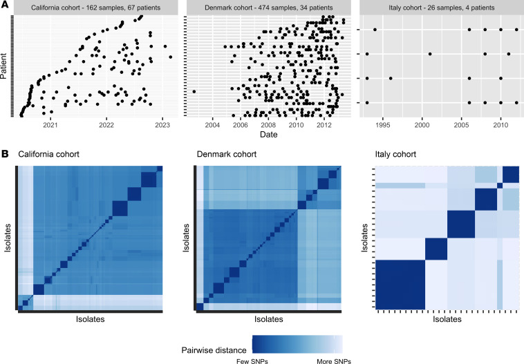
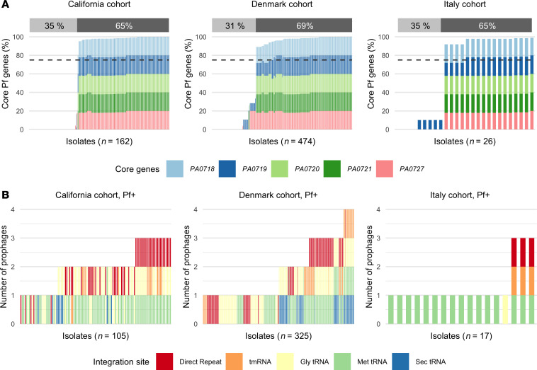
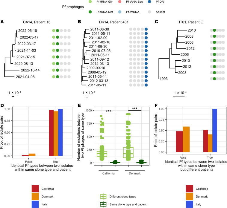
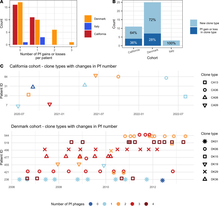
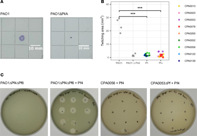
Pf bacteriophages, lysogenic viruses that infect Pseudomonas aeruginosa (Pa), are implicated in the pathogenesis of chronic Pa infections; phage-infected (Pf+) strains are known to predominate in people with cystic fibrosis (pwCF) who are older and have more severe disease. However, the transmission patterns of Pf underlying the progressive dominance of Pf+ strains are unclear. In particular, it is unknown whether phage transmission commonly occurs horizontally between bacteria via viral particles within the airway or whether Pf+ bacteria are mostly acquired via de novo Pseudomonas infections. Here, we studied Pa genomic sequences from 3 patient cohorts totaling 662 clinical isolates from 105 pwCF. We identified Pf+ isolates and analyzed transmission patterns of Pf within patients between genetically similar groups of bacteria called "clone types." We found that Pf was predominantly passed down vertically within Pa clone types and rarely via horizontal transfer between clone types within the airway. Conversely, we found extensive evidence of Pa de novo infection by a new, genetically distinct Pf+ Pa. Finally, we observed that clinical isolates showed reduced activity of type IV pili and reduced susceptibility to Pf in vitro. These results cast light on the transmission of virulence-associated phages in the clinical setting.

Keywords: Bacterial infections; Fibrosis; Infectious disease; Microbiology.


Figures

Figure 1
Figure 1. Overview of the 3 patient cohorts used in this study.
(A) Time series of sample collection from patients for each cohort. (B) Genetic distance matrix showing pairwise SNPs between each isolate for each cohort. All isolates were clustered using SNPs so that more similar isolates are located closer to each other on the x and y scales. The diagonal represents the comparison of each isolate to itself (0 SNPs). Clinical isolates cluster into groups of high genetic similarity (small dark blue squares) called clone types. In addition, larger clusters (medium blue squares) are observed. Genetic distance scale varies between cohorts.
Figure 2
Figure 2. Majority of Pa infected by Pf phages.
(A) Percentage of core Pf genes PA0718, PA0719, PA0720, PA0721, and PA0727 found in isolates for each patient cohort. The percentage of Pf+ isolates, defined with a minimum average coverage of 75% for these 5 core genes, is shown above each graph. (B) Number of prophages found in Pf+ isolates, as defined above, and integration site used by these Pf phages for each patient cohort. Integration sites are sequences in the bacterial genomes recognized by lysogenic phages to integrate into the bacterial chromosome. Different Pf lineages are able to use different integration sites, based on the presence of the corresponding integrase gene in the Pf genome.
Figure 3
Figure 3. Pf prophages are maintained within clone types within patients.
Presence or absence of 5 different Pf types, as defined by their integrase, for 1 clone type and patient in the California cohort (A), Denmark cohort (B), and Italy cohort (C). Sample dates are shown at the end of each branch. (D) Proportion of Pa isolate pairs with identical Pf infection patterns (same number and type of Pf phages, as determined by their integrase) for isolates of the same clone type and in the same patient. (E) Number of non-identical nucleotides (including SNPs and large insertions/deletions) for pairwise comparisons of Pf genomes of the same phage type (using the same integrase) for Pf infecting the same patient and Pa clone type, and for Pf infecting different clone types. Pf phages were more likely to share more of their genome if they infected the same clone type in the same patient. ***P < 0.001 by Wilcoxon’s rank-sum test. Boxes show the median (horizontal lines) and interquartile range (bounds of the boxes) and all data points are shown. (F) Proportion of Pa isolate pairs of the same clone type but in different patients with identical Pf infection patterns (same number and type of Pf phages, as determined by their integrase).
Figure 4
Figure 4. The majority of changes in the number of Pf phages in patients are associated with colonization or dominance of new clone types.
(A) Number of changes in the number of Pf phages per patient. (B) Percentage of the changes in the number of Pf phages that are due to a change in clone type or to infection of a clone type by Pf (horizontal transmission). (C) Time series of the number of phages and clone types for patients with at least one change in the number of phages in a clone type over time for the California cohort and for the Denmark cohort. No Pf change occurred within a clone type for any patient of the Italian cohort. Different shapes represent different clone types, while color indicates the number of Pf prophages found in that isolate.
Figure 5
Figure 5. Clinical isolates do not twitch and are less susceptible to Pf than PAO1.
Clinical isolates were selected from patients who were infected by both Pf and Pf+ isolates and that showed sufficient growth in vitro. (A) Positive (PAO1) and negative (PAO1ΔPilA) controls for twitching assays, showing motility of bacteria over agar after 24 hours. PAO1ΔPilA mutants do not have functional type IV pili and cannot twitch, resulting in a small stained area. (B) Twitching area for PAO1, PAO1ΔPilA, and Pf and Pf+ clinical isolates, with 4 replicates per isolate (mixed-effects model with isolate as random variable). ***P < 0.001 by t test. A lower twitching area indicates a lower twitching ability. (C) Plaque assays for Pf4 on PAO1ΔPf4ΔPf6 with negative control, on CPA0056 (Pf), and on CPA0053ΔPf, which are representative of results for all clinical isolates. Plaque assays for other Pf+ and Pf clinical isolates tested are shown in Supplemental Figure 5, for a total of 12 clinical isolates.

Similar articles

References

    1. Twort FW. An investigation on the nature of ultra-microscopic viruses. Lancet. 1915;186(4814):1241–1243. doi: 10.1016/S0140-6736(01)20383-3. - DOI - PMC - PubMed
    1. Strathdee SA, et al. Phage therapy: from biological mechanisms to future directions. Cell. 2023;186(1):17–31. doi: 10.1016/j.cell.2022.11.017. - DOI - PMC - PubMed
    1. Ackermann HW, DuBow MS. Viruses of Prokaryotes: General Properties of Bacteriophages. CRC Press; 1987.
    1. Howard-Varona C, et al. Lysogeny in nature: mechanisms, impact and ecology of temperate phages. ISME J. 2017;11(7):1511–1520. doi: 10.1038/ismej.2017.16. - DOI - PMC - PubMed
    1. Hay ID, Lithgow T. Filamentous phages: masters of a microbial sharing economy. EMBO Rep. 2019;20(6):e47427. doi: 10.15252/embr.201847427. - DOI - PMC - PubMed

MeSH terms