. 2025 Apr 22;135(12):e185430.
doi: 10.1172/JCI185430. eCollection 2025 Jun 16.

Antimicrobial peptide developed with machine learning sequence optimization targets drug resistant Staphylococcus aureus in mice

Affiliations

Antimicrobial peptide developed with machine learning sequence optimization targets drug resistant Staphylococcus aureus in mice

Biswajit Mishra et al. J Clin Invest. .

Abstract

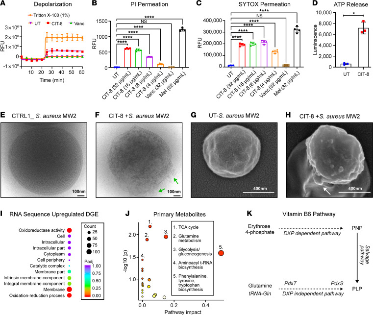
As antimicrobial resistance rises, new antibacterial candidates are urgently needed. Using sequence space information from over 14,743 functional antimicrobial peptides (AMPs), we improved the antimicrobial properties of citropin 1.1, an AMP with weak antimethicillin resistant Staphylococcus aureus (MRSA) activity, producing a short and potent antistaphylococcal peptide, CIT-8 (13 residues). At 40 μg/mL, CIT-8 eradicated 1 × 10⁸ drug-resistant MRSA and vancomycin resistant S. aureus (VRSA) persister cells within 30 minutes of exposure and reduced the number of viable biofilm cells of MRSA and VRSA by 3 log10 and 4 log10 in established biofilms, respectively. CIT-8 (at 32 μg/mL) depolarized and permeated the S. aureus MW2 membrane. In a mouse model of MRSA skin infection, CIT-8 (2% w/w in petroleum jelly) significantly reduced the bacterial burden by 2.3 log10 (P < 0.0001). Our methodology accelerated AMP design by combining traditional peptide design strategies, such as truncation, substitution, and structure-guided alteration, with machine learning-backed sequence optimization.

Keywords: Bacterial infections; Infectious disease; Microbiology.


Conflict of interest statement

Conflict of interest: The authors have declared that no conflict of interest exists.

Figures

Figure 1
Figure 1. CIT-based peptide design and optimization strategy.
Figure 2
Figure 2. Exploration of AMP sequence space.
(A) t-SNE transformed physicochemical property space, grouped by k-means clustering. (BE) Comparison of the physicochemical parameters of the 4 clusters of AMPs in our dataset based on (B) GRAVY, (C) helicity, (D) TPSA, and (E) hydrophobic moment. (F) Graph of amino acid group occurrence patterns (AGO). Each node represents a peptide fragment sequence, while the connected nodes indicate the most common parental fragment sequences with corresponding NOO (number of occurrences) values annotated on the edges. (G) Heat-map showing the percentage of each amino acid (APO) in all 13-mer helical AMP fragments in our dataset.
Figure 3
Figure 3. Antibiofilm and antipersister activity of CIT-8.
(A and B) Killing kinetics of CIT-8 against S. aureus MW2 in (A) exponential phase and (B) gentamicin-induced persister cells at concentrations of 4 and 40 μg/mL, compared with untreated bacterial (BC) control, colony forming units (CFU) counts were monitored for 120 minutes (n = 2, replicated thrice). (C and D) Killing kinetics of CIT-8 against S. aureus strain VRS1 in (C) exponential phase and (D) gentamicin-induced persister cells at 4 and 40 μg/mL, CFU counts were monitored for 120 minutes (n = 2, replicated thrice). We included ciprofloxacin (cipro) (at 10 μg/mL) and linezolid (at 100 μg/mL) as antibiotic controls and bithionol (at 10 μg/mL) as a positive control. (E and F) Disruption of 24 hour established biofilms of (E) MRSA (S. aureus MW2), and (F) VRSA (S. aureus VRS1) by CIT-8, measured as log reductions in bacterial loads on solid membranes treated with 4 and 40 μg/mL of CIT-8 (n = 6, *P < 0.05 by 1-way ANOVA followed by Dunnett’s multiple comparison test). We included 10 μg/mL vancomycin (Vanc) as control. (G and H) Inhibition of S. aureus MW2 biofilm formation by CIT-8 at concentrations ranging from 4–32 μg/mL after 24 hours of treatment, assessed using (G) live-cell viability (XTT assay), and (H) biomass quantification (crystal violet staining) (n = 3, replicated twice). (I and J) Disruption of 24 hours S. aureus MW2 established biofilms by CIT-8 at 4–32 μg/mL, evaluated by (I) reductions in live-cell viability (XTT assay) and (J) biomass loss (crystal violet staining) (n = 3, replicated twice). (K and L) Fluorescence microscopy images (10×) of 24 hour-established S. aureus MW2 biofilms stained with live/dead staining, (K) untreated control, and (L) biofilms treated with 32 μg/ml of CIT-8. Scale bars: 0.05 mm.
Figure 4
Figure 4. Structural insights to membrane targeting by CIT-8.
(A) Natural abundance 2D- 13C-HSQC spectrum of CIT-8. (B) 2D-NOESY spectrum and summary of important NOESY distance restraints used in the CIT-8 structure calculation. (C) CIT-8 NMR solution structure ensemble. (D) Ribbon representation of the first conformer in the ensemble. (E) Two surface representations obtained by 180º rotation along the x-axis showing the distribution of hydrophobic (yellow) and charged (blue) residues. All structure figures were prepared in Pymol using the YRB script. (F) Snapshot of an all-atom MD simulation of peptide CIT-8 in the presence of DOPC:DOPG (7:3) mimetic membrane model showing complete peptide insertion at 500 ns. Blue, charged residues; brown, hydrophobic residues. (G) Changes in membrane lipid density upon CIT-8-induced water perturbation. (H) Changes in membrane thickness upon CIT-8 interaction with model membrane.
Figure 5
Figure 5. Mechanism of action of CIT-8 and associated stress response by MRSA.
(A) Fluorescence-based, DIBAC4(3)-assisted S. aureus MW2 membrane depolarization caused by CIT-8 peptide (at 32 μg/mL) monitored for 40 minutes after peptide exposure (n = 3). (B and C) Fluorescence-based membrane permeability of S. aureus MW2 treated with CIT-8 (4–32 μg/mL), untreated bacteria (UT), vancomycin (Vanc), and melittin (Mel) at 32 μg/mL after 60 minutes, assessed using (B) PI and (C) SYTOX Green fluorescence (n = 4, ****P < 0.0001 by 1-way ANOVA followed by Dunnett’s multiple comparison test). (D) ATP release from S. aureus MW2 upon CIT-8 (at 32 μg/mL) interaction for 30 minutes (* denotes P < 0.05 by Student’s t-test, unpaired 2-tailed). (E) Cryo-EM image of control S. aureus MW2. (F) Cryo-EM image of S. aureus MW2 treated with CIT-8 at 80 μg/mL for 60 minutes (green arrows indicate membrane perturbation). (G) SEM image of control S. aureus MW2. (H) SEM image of S. aureus MW2 treated with CIT-8 at 40 μg/mL for 60 minutes (white arrows indicate membrane blebbing). (I) RNA-seq–derived differential gene expression (DGE) of significantly upregulated genes (n = 2 samples, P < 0.05, calculated using DESeq2 (76) in S. aureus MW2 by CIT-8 (at 2 μg/mL) treated for 30 minutes. (J) Pathway analysis of the targeted metabolome of S. aureus MW2 treated with peptide CIT-8 at 4 μg/mL for 30 minutes, revealing significant alterations in key stress and metabolic pathways (n = 3, significant metabolite in pathways were determined by their P < 0.05 obtained by Student’s t test, unpaired, 2-tailed). (K) Stress responsive vitamin B6 pathway in S. aureus MW2, indicating key regulatory genes (pdxT and pdxS revelated by our RNA-seq analysis) and metabolite (Erythrose 4-phosphate, identified by our targeted metabolomics analysis) positions in the pathway. Scale bars: 100 nm (E and F); 400 nm (G and H).
Figure 6
Figure 6. In vivo efficacy of CIT-8 in a skin-abraded murine infection model infected with S. aureus MW2.
(A) Schematic representation of the skin-abraded murine model representing both prophylactic and established models. (B) Quantified bacterial load from skin specimens collected from mice infected with exponential phase S. aureus MW2 and treated after 10 minutes (representing a prophylactic model) with CIT-8 (2% w/w), CIT-8 (1% w/w), and mupirocin (2% w/w) ointments compared with vehicle control (n = 12, ****P < 0.0001, **P < 0.01, *P < 0.05 calculated by 1-way ANOVA followed by Dunnett’s multiple comparison test). (C) Quantified bacterial load from skin specimens collected from mice infected with exponential phase S. aureus MW2 and treated after 24 hours (representing an established infection model) with CIT-8 (2% w/w), CIT-8 (1% w/w), and mupirocin (2% w/w) ointments compared with vehicle control (n = 8, **P < 0.01, *P < 0.05 calculated by 1-way ANOVA followed by Dunnett’s multiple comparison test). Cytokine estimations for (D) TNFA, (E) IL6, and (F) MCP1 (n = 6, *P < 0.05, calculated by Student’s t test, unpaired, 2-tailed) in murine skin treated with CIT-8 (2% w/w) after 10 minutes of bacterial infection in a prophylactic model.

Similar articles

References

    1. Prestinaci F, et al. Antimicrobial resistance: a global multifaceted phenomenon. Pathog Glob Health. 2015;109(7):309–318. doi: 10.1179/2047773215Y.0000000030. - DOI - PMC - PubMed
    1. Antimicrobial Resistance Collaborators. Global burden of bacterial antimicrobial resistance in 2019: a systematic analysis. Lancet. 2022;399(10325):629–655. doi: 10.1016/S0140-6736(21)02724-0. - DOI - PMC - PubMed
    1. Mishra B, et al. Host defense antimicrobial peptides as antibiotics: design and application strategies. Curr Opin Chem Biol. 2017;38:87–96. doi: 10.1016/j.cbpa.2017.03.014. - DOI - PMC - PubMed
    1. Ganesan N, et al. Antimicrobial peptides and small molecules targeting the cell membrane of Staphylococcus aureus. Microbiol Mol Biol Rev. 2023;87(2):e0003722. doi: 10.1128/mmbr.00037-22. - DOI - PMC - PubMed
    1. Lei M, et al. Engineering selectively targeting antimicrobial peptides. Annu Rev Biomed Eng. 2021;23:339–357. doi: 10.1146/annurev-bioeng-010220-095711. - DOI - PMC - PubMed

MeSH terms

Substances