. 2025 Apr 29;122(17):e2418409122.
doi: 10.1073/pnas.2418409122. Epub 2025 Apr 22.

NAT10 exacerbates acute renal inflammation by enhancing N4-acetylcytidine modification of the CCL2/CXCL1 axis

Affiliations

NAT10 exacerbates acute renal inflammation by enhancing N4-acetylcytidine modification of the CCL2/CXCL1 axis

Jia-Nan Wang et al. Proc Natl Acad Sci U S A. .

Abstract

Inflammation plays an essential role in eliminating microbial pathogens and repairing tissues, while sustained inflammation accelerates kidney damage and disease progression. Therefore, understanding the mechanisms of the inflammatory response is vital for developing therapies for inflammatory kidney diseases like acute kidney injury (AKI), which currently lacks effective treatment. Here, we identified N-acetyltransferase 10 (NAT10) as an important regulator for acute inflammation. NAT10, the only known "writer" protein for N4-acetylcytidine (ac4C) acetylation, is elevated in renal tubules across various AKI models, human biopsies, and cultured tubular epithelial cells (TECs). Conditional knockout (cKO) of NAT10 in mouse kidneys attenuates renal dysfunction, inflammation, and infiltration of macrophages and neutrophils, whereas its conditional knock-in (cKI) exacerbates these effects. Mechanistically, our findings from ac4C-RIP-seq and RNA-seq analyses revealed that NAT10-mediated ac4C acetylation enhances the mRNA stability of a range of key chemokines, including C-C motif chemokine ligand 2 (CCL2) and C-X-C motif chemokine ligand 1(CXCL1), promoting macrophage and neutrophil recruitment and accelerating renal inflammation. Additionally, CCL2 and CXCL1 neutralizing antibodies or their receptor inhibitors, abrogated renal inflammation in NAT10-overexpression TECs or NAT10-cKI mice. Importantly, inhibiting NAT10, either through Adeno-associated virus 9 (AAV9)-mediated silencing or pharmacologically with our found inhibitor Cpd-155, significantly reduces renal inflammation and injury. Thus, targeting the NAT10/CCL2/CXCL1 axis presents a promising therapeutic strategy for treating inflammatory kidney diseases.

Keywords: CCL2; CXCL1; NAT10; ac4C; renal inflammation.


Conflict of interest statement

Competing interests statement:The authors declare no competing interest.

Figures
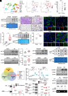
Fig. 1.
Fig. 1.
ac4C modifications increase in human biopsy, mouse models, and TECs in response to proinflammatory stimuli via a CREB1-dependent mechanism. (A) Uniform manifold approximation and projection (UMAP) of cell type clustering in human kidneys. Cell types include proximal tubule segment 1 (PT S1), proximal tubule segment 2 (PT S2), proximal tubule segment 3 (PT S3), thick ascending limb cell (TAL), descending thin limb cell (DTL), distal convoluted tubule cell (DCT), connecting tubule cell (CNT), principal cell (PC), intercalated cell (IC), podocyte (Pod), endothelial cell (EC), Immune cells (IMM). (B) UMAP plots displaying expression levels of NAT10 in each dataset group. (C) The expression levels of NAT10 in renal tubule cells of health and patients with AKI in single-cell data from Lake et al. (21). (D) The ac4C level in the human kidneys were determined using ac4C dot blot assay (n = 6 biological replicates). (E) Photomicrographs of PAS staining and NAT10 staining in kidney sections of patients with AKI. (Scale bar, 50 μm and 20 μm.) The red arrow indicates protein casts, the yellow triangle indicates tubular dilation, the black arrow indicates preserved proximal tubule with brush border, and the asterisks indicate shedding tubule cells. (F) NAT10 and LTL expression in kidney biopsies from patients with AKI was detected using immunofluorescence staining. LTL was used to stain the proximal tubules. (Scale bars, 50 μm.) (G) Quantifications showing NAT10 expression in the kidney from patients with AKI (n = 6 for normal subjects, n = 12 for patients with AKI, two-tailed unpaired Student’s t test). (H) The ac4C level in the I/R mouse model after 24 h were determined using ac4C dot blot assay. (I) The ac4C level in the IRI mouse model were determined using LC–MS/MS analysis (n = 3 biological replicates of mice, two-tailed unpaired Student’s t test). (J) Representative immunofluorescence staining for NAT10 and LTL in I/R-induced AKI after 24 h. (Scale bars, 50 μm.) (K and L) Western blotting (K) and real-time PCR (L) analyses of NAT10 in I/R (24 h)- and cisplatin (3 d)-induced AKI mouse models (n = 6 biological replicates of mice, two-tailed unpaired Student’s t test). (M) NAT10 was upregulated in response to different stimuli, including H/R (hypoxia 12 h and reoxygenation 6 h) and cisplatin (24 h) in HK2 cells. (N) The ac4C level in H/R- and cisplatin-induced HK2 cells were determined using ac4C dot blot assays. (O) Venn diagram showing the overlap of transcription factors (TFs) of NAT10 predicted by JASPAR, PROMO, and ChIPBase. (P) mRNA expression levels of NAT10 and CREB1 in HK2 cells were determined by real-time PCR analysis (n = 4 biological replicates, one-way ANOVA with Tukey’s multiple comparisons test). (Q) Western blotting of pCREB1 and CREB1 protein expression in H/R-induced HK2 cells. (R) Protein abundance of NAT10 in H/R-treated HK2 cells with CREB1 knockdown. (S) Luciferase activity of full-length or truncated NAT10 promoter constructs co-transfected with control or CREB1-overexpressing plasmids (n = 3 biological replicates, two-tailed unpaired Student’s t test). (T) Luciferase activity of NAT10 promoter constructs containing WT or mutated sites (Mut 1, 2, and 3) when co-transfected with control or CREB1 knockdown in HEK293T cells under H/R stimulation (n = 3 biological replicates, two-tailed unpaired Student’s t test). (U) ChIP assays of the binding of CREB1 to the NAT10 promoter in the presence or absence of H/R stimulation. Data represent the mean ± SEM. *P < 0.05, **P < 0.01, ***P < 0.001.
Fig. 2.
Fig. 2.
NAT10 deficiency protects against renal dysfunction, injury, and inflammation in I/R-induced AKI mice. (A) Schematic illustrating experiment groups. (B) Serum BUN and serum creatinine concentrations in control and NAT10-cKO mice exposed to I/R-induced injury after 24 h (n = 6 biological replicates of mice, two-way ANOVA with Tukey’s multiple comparisons test). (C) PAS score in NAT10 cKO mice with I/R-induced AKI (n = 6 biological replicates of mice, two-way ANOVA with Tukey’s multiple comparisons test). (D) PAS staining and immunohistochemistry staining of F4/80+ and Ly6G. (Scale bars, 100 and 50 μm.) The red arrow indicates protein casts, the yellow triangle indicates tubular dilation, the black arrow indicates preserved proximal tubule with brush border, and the asterisks indicate shedding tubule cells. (E) Western blotting of KIM-1, pp65, and p65 after NAT10 deficiency in I/R-induced AKI after 24 h. (F) ELISA analysis of the production of KIM-1 and NGAL in I/R-induced AKI with NAT10 knockout (n = 6 biological replicates, two-way ANOVA with Tukey’s multiple comparisons test). (G) Relative mRNA expressions of CCL2 were determined by real-time PCR (n = 6 biological replicates of mice, two-way ANOVA with Tukey’s multiple comparisons test). (H) The proportions of infiltrated Ly6Chi MDMs and Ly6G+ neutrophils in the kidney were detected by flow cytometry. Data represent the mean ± SEM. *P < 0.05, **P < 0.01, ***P < 0.001.
Fig. 3.
Fig. 3.
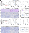
NAT10 deletion in renal tubular cells relieved cisplatin- and CLP-induced renal dysfunction, injury, and inflammation. (A) Schematic illustrating experiment groups. (B) Serum BUN and serum creatinine concentrations in control and NAT10-cKO mice exposed to cisplatin-induced injury sustained for 3 d (n = 6 biological replicates of mice, two-way ANOVA with Tukey’s multiple comparisons test). (C) PAS score in NAT10-cKO mice with cisplatin-induced AKI (n = 6 biological replicates of mice, two-way ANOVA with Tukey’s multiple comparisons test). (D) PAS staining and immunohistochemistry staining of F4/80+ and Ly6G. (Scale bars, 100 and 50 μm.) The red arrow indicates protein casts, the yellow triangle indicates tubular dilation, the black arrow indicates preserved proximal tubule with brush border, and the asterisks indicate shedding tubule cells. (E) The proportions of infiltrated Ly6Chi MDMs and Ly6G+ neutrophils in the kidney were detected by flow cytometry. (F) Schematic illustrating experiment groups. (G) Serum BUN and creatinine concentrations 24 h after in control and NAT10-cKO mice exposed to CLP-induced injury (n = 6 biological replicates of mice, two-way ANOVA with Tukey’s multiple comparisons test). (H) PAS score in NAT10-cKO mice with CLP-induced AKI (n = 6 biological replicates of mice, two-way ANOVA with Tukey’s multiple comparisons test). (I) Immunohistochemistry staining of F4/80+ and Ly6G. (Scale bars, 50 μm.) (J) The proportions of kidney Ly6Chi MDMs and Ly6G+ neutrophils were detected by flow cytometry. Data represent the mean ± SEM. ***P < 0.001.
Fig. 4.
Fig. 4.
Kidney tubular cell knock-in of NAT10 aggravates renal injury and inflammation in I/R-induced AKI. (A) Schematic illustrating experiment groups. (B) Serum BUN and creatinine concentrations in NAT10-cKI mice with I/R-induced AKI after 24 h (n = 6 biological replicates of mice, two-way ANOVA with Tukey’s multiple comparisons test). (C) Quantification of PAS staining in NAT10-cKI mice with I/R-induced AKI (n = 6 biological replicates of mice, two-way ANOVA with Tukey’s multiple comparisons test). (D) PAS staining and immunohistochemistry staining of F4/80+ and Ly6G. (Scale bars, 100 and 50 μm.) The red arrow indicates protein casts, the yellow triangle indicates tubular dilation, the black arrow indicates preserved proximal tubule with brush border, and the asterisks indicate shedding tubule cells. (E) Western blotting of KIM-1, pp65, and p65 in NAT10 overexpression on I/R-induced AKI (n = 6 biological replicates of mice, two-way ANOVA with Tukey’s multiple comparisons test). (F) ELISA analysis of the production of KIM-1 and NGAL in I/R-induced AKI with NAT10 overexpression (n = 6 biological replicates, two-way ANOVA with Tukey’s multiple comparisons test). (G and H) Flow cytometry analysis and quantitative data (n = 4 biological replicates of mice, two-way ANOVA with Tukey’s multiple comparisons test). Data represent the mean ± SEM. *P < 0.05, **P < 0.01, ***P < 0.001.
Fig. 5.
Fig. 5.
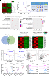
Characterization of ac4C modification and identification of NAT10 downstream targets through ac4C-RIP-seq and RNA-seq in H/R-treated with or without NAT10 knockdown. (A) Heatmap of ac4C-RIP-seq analysis showing differentially acetylated genes in H/R-induced HK2 cells with or without NAT10 knockdown. (B) Density distribution of ac4C-containing peaks across the mRNA transcripts. (C) The ac4C consensus motif in H/R-exposed cells with or without NAT10 knockdown was identified using HOMER. (D) KEGG enrichment analysis identified the top 10 pathways of hypoacetylated (Left) and downregulated genes (Right) following NAT10 knockdown. (E) Overlapping potential downstream targets of NAT10 in hypoacetylated and downregulated genes. (F) Heatmap showed the hypoacetylated genes (Left) and downregulated genes (Right) in the TNF-α pathway. (G) RNA decay assays with actinomycin D treatment were performed to detect the degradation rates of CXCL1 and CCL2 mRNA in NAT10 knockdown cells. (H) Correlation analysis between CCL2 and NAT10 (Left) and CXCL1 and NAT10 (Right) from single-cell data (GSE274819) from Li et al. (22) in I/R 24 h mice. (I) ELISA analysis of CXCL1 and CCL2 production in I/R-induced AKI with NAT10 knockout and NAT10 overexpression (n = 6 biological replicates of mice, two-way ANOVA with Tukey’s multiple comparisons test). (J) The levels of CCR2 and CXCR2 in myeloid cells were detected by flow cytometry. Data represent the mean ± SEM. *P < 0.05, **P < 0.01, ***P < 0.001.
Fig. 6.
Fig. 6.
CCL2 and CXCL1 are potential downstream target genes of NAT10. (A and B) Attenuation of NAT10 diminished ac4C modification of CCL2 (A) and CXCL1 (B) mRNA in H/R-treated HK2 cells. (C and D) ac4C-RIP-qPCR (C) and RIP-qPCR (D) analysis of alterations in the ac4C modifications of CCL2 and CXCL1 genes in H/R-treated HK2 cells with or without NAT10 knockdown (n = 4 biological replicates, two-tailed unpaired Student’s t test). (E) Wild-type and mutant CCL2 were inserted into pmirGLO reporter vectors. (F) Luciferase reporter assay measured the luciferase activities of CCL2-CDS WT or CCL2-CDS Mut in H/R-treated HK2 cells with or without NAT10 knockdown (n = 4 biological replicates, two-tailed unpaired Student’s t test). (G) Wild-type and mutant CXCL1 were inserted into pmirGLO reporter vectors. (H) Luciferase reporter assay measured the luciferase activities of CXCL1-CDS WT or CXCL1-CDS Mut in H/R-treated HK2 cells with or without NAT10 knockdown (n = 4 biological replicates, two-tailed unpaired Student’s t test). (I) Schematic illustrating the mutation site in the NAT10-G641E. (J) The total RNA ac4C level in wild-type NAT10 and mutant NAT10 was determined using anti-ac4C dot blot. (K and L) Western blotting (K) and real-time PCR (L) analysis of NAT expression in wild-type NAT10 and mutant NAT10 (n = 4 biological replicates, two-way ANOVA with Tukey’s multiple comparisons test). (M) ELISA analysis of the production of CCL2 and CXCL1 when NAT10-wt or NAT10-G641E treatment (n = 4 biological replicates, two-way ANOVA with Tukey’s multiple comparisons test). (N) RNA decay assays with actinomycin D treatment were performed to detect the degradation rate of CCL2 and CXCL1 mRNA in NAT10-wt or NAT10-G641E treatment. Data represent the mean ± SEM. *P < 0.05, **P < 0.01, ***P < 0.001.
Fig. 7.
Fig. 7.
CXCL1 and CCL2 neutralizing antibody or CCR2 and CXCR2 inhibitor rescues NAT10 overexpression-induced renal inflammation. (A) Western blotting analysis of pp65 and p65 protein expression. (B) Real-time PCR analysis of CXCL1 and CCL2 mRNA levels (n = 4 biological replicates, two-way ANOVA with Tukey’s multiple comparisons test). (C) Schematic of in vitro co-culture experiments. (D) Representative images of CXCL1 and CCL2 neutralizing antibody treatment on macrophage migration by transwell assay. (E) Schematic illustrating experiment groups and treatment in four groups. (F) Serum BUN and creatinine concentrations 24 h later in I/R-induced AKI mice (n = 6 biological replicates of mice, two-way ANOVA with Tukey’s multiple comparisons test). (G) Western blotting analysis of KIM-1 after CCR2 and CXCR2 inhibitor treatment. (H) ELISA analysis of CXCL1 and CCL2 levels (n = 6 biological replicates of mice, two-way ANOVA with Tukey’s multiple comparisons test). (I) Quantification of PAS staining in NAT10-cKI mice with CCR2 and CXCR2 inhibitor treatment (n = 6 biological replicates of mice, two-way ANOVA with Tukey’s multiple comparisons test). (J) PAS staining and immunohistochemistry staining of F4/80+ and Ly6G. (Scale bars, 100 and 50 μm.) Data represent the mean ± SEM. *P < 0.05, **P < 0.01, ***P < 0.001.
Fig. 8.
Fig. 8.
Therapy targeting NAT10 significantly attenuates renal dysfunction, injury, and inflammation. (A) Schematic illustrating the treatment of AAV9-mediated NAT10 knockdown in I/R-induced AKI. (B) Serum BUN and creatinine concentrations 24 h after in mice with I/R-induced nephropathy and knockdown of NAT10 (n = 6 biological replicates of mice, two-way ANOVA with Tukey’s multiple comparisons test). (C) PAS staining and immunohistochemistry staining of F4/80+ and Ly6G. (Scale bars, 100 and 50 μm.) The red arrow indicates protein casts, the yellow triangle indicates tubular dilation, the black arrow indicates preserved proximal tubule with brush border, and the asterisks indicate shedding tubule cells. (D) Western blot of KIM-1, pp65, and p65 after NAT10 knockdown in I/R-induced AKI. (E) Serum CXCL1 and CCL2 levels detected by ELISA (n = 6 biological replicates of mice, two-way ANOVA with Tukey’s multiple comparisons test). (F) Workflow of the hybrid virtual screening strategy. (G) Molecular docking demonstrated that Cpd-155 physically bound to the catalytic domain of NAT10. (H) RMSD of Cpd-155 during MD simulations. (I) The stabilization of NAT10 in vitro with or without Cpd-155 treatment detected by CETSA analysis. (J) Dot blot assay showing the effect of Cpd-155 treatment on ac4C abundance in H/R-treated HK2 cells. (K) Western blot analysis of KIM-1, pp65, and p65 after Cpd-155 treatment in H/R-induced HK2 cells. (L) Schematic illustrating administration of Cpd-155 before and after induction of I/R-induced AKI. ip, intraperitoneally. (M) Serum BUN and creatinine concentrations 24 h after I/R-induced AKI with Cpd-155 treatment (n = 6 biological replicates of mice, two-way ANOVA with Tukey’s multiple comparisons test). (N) PAS staining in I/R-induced AKI with Cpd-155 treatment. (Scale bars, 100 μm.) (O) ELISA analysis of CXCL1 and CCL2 levels (n = 6 biological replicates of mice, two-way ANOVA with Tukey’s multiple comparisons test). Data represent the mean ± SEM. ***P < 0.001.

References

    1. Ronco C., Bellomo R., Kellum J. A., Acute kidney injury. Lancet 394, 1949–1964 (2019). - PubMed
    1. Haines R. W., Fowler A. J., Kirwan C. J., Prowle J. R., The incidence and associations of acute kidney injury in trauma patients admitted to critical care: A systematic review and meta-analysis. J. Trauma Acute Care Surg. 86, 141–147 (2019). - PubMed
    1. Rabb H., et al. , Inflammation in AKI: Current understanding, key questions, and knowledge gaps. J. Am. Soc. Nephrol. 27, 371–379 (2016). - PMC - PubMed
    1. Ferenbach D. A., Bonventre J. V., Mechanisms of maladaptive repair after AKI leading to accelerated kidney ageing and CKD. Nat. Rev. Nephrol. 11, 264–276 (2015). - PMC - PubMed
    1. Song H., et al. , METTL3-mediated m(6)A RNA methylation promotes the anti-tumour immunity of natural killer cells. Nat. Commun. 12, 5522 (2021). - PMC - PubMed

MeSH terms

LinkOut - more resources