. 2025 Apr 22;8(7):e202402837.
doi: 10.26508/lsa.202402837. Print 2025 Jul.

Human iPS cell-derived respiratory organoids as a model for respiratory syncytial virus infection

Affiliations

Human iPS cell-derived respiratory organoids as a model for respiratory syncytial virus infection

Rina Hashimoto et al. Life Sci Alliance. .

Abstract

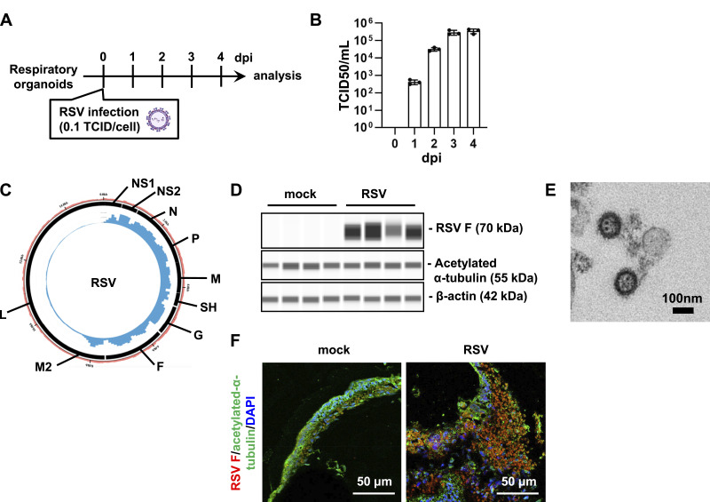
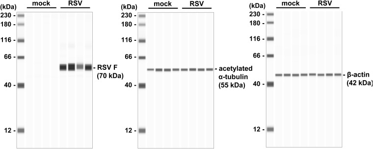
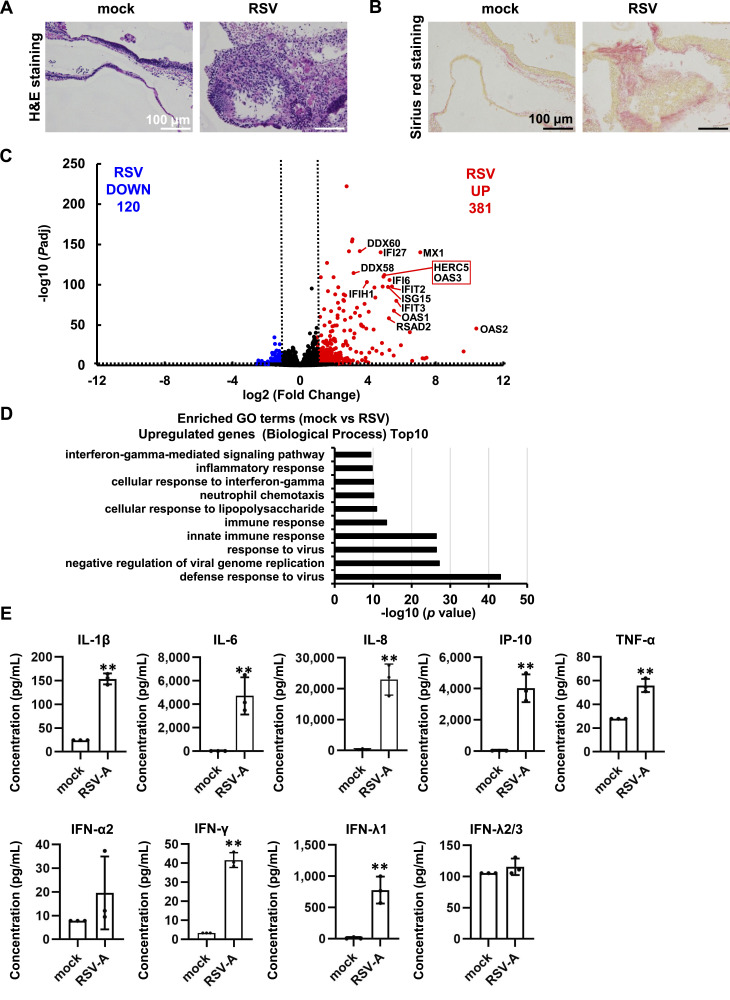
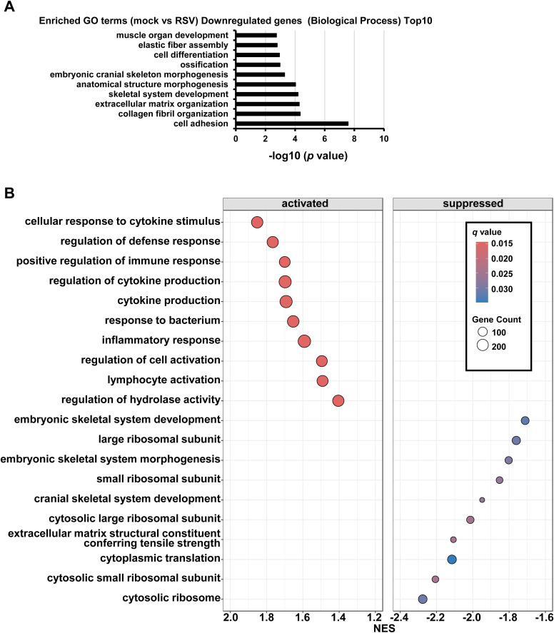
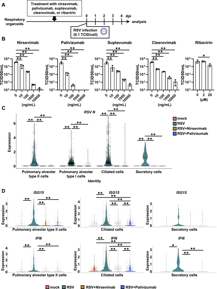
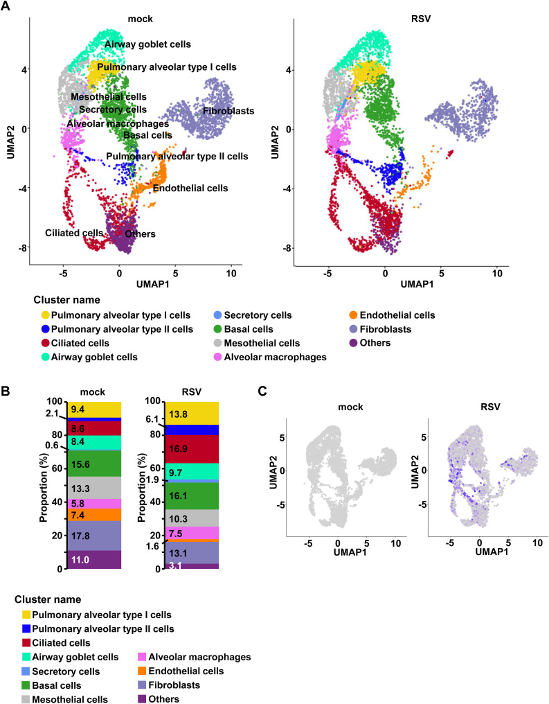
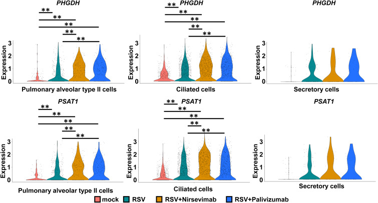
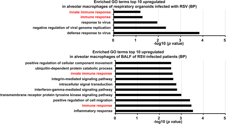
Respiratory syncytial virus (RSV) is a seasonal respiratory pathogen that primarily affects young children, potentially causing severe lower respiratory tract disease. Despite the high disease burden, understanding of RSV pathophysiology remains limited. To address this, advanced RSV infection models are needed. Whereas HEp-2 cells are widely used because of their high susceptibility to RSV, they do not accurately reflect the host response of the human respiratory tract. In this study, we evaluated human-induced pluripotent stem cell-derived respiratory organoids, which contain respiratory epithelial cells, immune cells, fibroblasts, and vascular endothelial cells, for their potential to model RSV infection and support pharmaceutical research. RSV-infected organoids exhibited high viral genome and protein expression, epithelial layer destruction, and increased collagen accumulation. Pro-inflammatory cytokine levels in culture supernatants also increased post-infection. Furthermore, RSV infection was significantly inhibited by monoclonal antibodies (nirsevimab, palivizumab, suptavumab, or clesrovimab), although ribavirin showed limited efficacy. These findings highlight the utility of respiratory organoids for RSV research.


Conflict of interest statement

The authors declare that they have no conflict of interest.

Figures

Figure 1.
Figure 1.. RSV infects human iPS cell–derived respiratory organoids.
(A) Human iPS cell–derived respiratory organoids were infected with 0.1 TCID/cell RSV (8 × 10⁴ TCID/well) and cultured for 4 d. (B) TCID50 assay was temporally performed in the RSV-infected respiratory organoids. (C) RNA sequencing was performed for RSV-infected respiratory organoids. Circos plot showing the distribution of reads along the RSV genome. The inner blue circles indicate viral RNA-seq reads obtained from RSV-infected respiratory organoids. The outer black blocks show the coding regions of RSV. The outer pale red bar plot signifies the percentage of GC content. The Circos plot was created using Circleator (v 1.0.2) (Crabtree et al, 2014). (D) Expression levels of RSV F, acetylated α-tubulin, and β-actin were measured by a capillary-based immunoassay (Jess analysis). (E) TEM image of RSV. (F) Immunofluorescence analysis of acetylated α-tubulin (green) and RSV F (red) in the respiratory organoids.
Figure S1.
Figure S1.. Protein expression analysis in RSV-infected respiratory organoids.
Human iPS cell–derived respiratory organoids were infected with 0.1 TCID/cell (8 × 10⁴ TCID/well) RSV and cultured for 4 d. Expression levels of RSV F, acetylated α-tubulin, and β-actin were measured by a capillary-based immunoassay (Jess analysis).
Figure 2.
Figure 2.. Analysis of host cell response in RSV-infected respiratory organoids.
Human iPS cell–derived respiratory organoids were infected with 0.1 TCID/cell RSV (8 × 10⁴ TCID/well) and cultured for 4 d. (A) H&E staining of respiratory organoids. (B) Sirius red staining of respiratory organoids. (C) A volcano plot of differentially expressed genes in uninfected and infected respiratory organoids (Log2 fold-change > 1, adjusted P-value [Padj] < 0.01). Red and blue dots represent up-regulated and down-regulated genes, respectively. (D) DAVID-based GO analysis of RSV-infected respiratory organoids was performed. The top 10 significantly enriched GO terms of up-regulated genes (Biological Process) in RSV-infected samples compared with uninfected samples are shown. (E) The concentration (pg/ml) of 9 cytokines in the cell culture supernatant of the RSV-infected respiratory organoids, as measured by bead-based immunoassays. Data information: data are shown as means ± SD (n = 3). Two-tailed t test (**P < 0.01).
Figure S2.
Figure S2.. Analysis of host cell response in RSV-infected respiratory organoids.
(A) Database for Annotation, Visualization and Integrated Discovery (DAVID)-based gene ontology (GO) analysis of RSV-infected respiratory organoids was performed. The top 10 significantly enriched GO terms of down-regulated genes (Biological Process) in RSV-infected samples compared with uninfected samples are shown. (B) Dot plot of the top 10 significantly enhanced (normalized enrichment score > 1.2 and the false discovery rate [FDR, qvalue of gseGO results] < 0.03) and decreased (normalized enrichment score < −1.6 and FDR < 0.03) gene sets from Gene Set Enrichment Analysis of RSV-infected respiratory organoids compared with uninfected respiratory organoids. Enrichment of gene ontology (GO) biological process terms in differentially expressed gene clusters was evaluated using the clusterProfiler package (v4.4.4; R-version 4.2.1) (Wu et al, 2021). The complete set of expressed genes was used as background for the analysis. GO terms enriched with an FDR < 0.25 were considered significant. gseGO with the following settings: ont = “BP,” nPerm 10,000, minGSSize = 3, maxGSSize = 800, pvalueCutoff = 0.05, orgDB = “org. Hs. e.g., db.” Dot color indicates FDR (qvalue of gseGO results), and dot size represents the fraction of genes annotated with each term.
Figure 3.
Figure 3.. Evaluation of anti-RSV F antibodies and ribavirin using human iPS cell–derived respiratory organoids.
(A) Human iPS cell–derived respiratory organoids were infected with 0.1 TCID/cell (8 × 10⁴ TCID/well) RSV-A and cultured with medium containing nirsevimab, suptavumab, clesrovimab, palivizumab, or ribavirin for 4 d. (B) At 4 dpi, the cell culture supernatant of infected respiratory organoids was collected and TCID50 assay was performed. One-way ANOVA followed by the Dunnet post hoc test (*P < 0.05, **P < 0.01, compared with 0 ng/ml or 0 μM). Data are shown as mean ± SD (n = 3). (C) Violin plot displaying the gene expression of RSV-A N in each cluster of epithelial cells (alveolar type II cells, alveolar type I cells, ciliated cells, and secretory cells). (D) Violin plot displaying the gene expression of interferon stimulated gene (ISG) 15 and interferon alpha inducible protein (IFI) 6 in alveolar type II cells, ciliated cells, and secretory cells of uninfected and RSV-infected respiratory organoids treated with or without antibodies (nirsevimab or palivizumab). Data information: Wilcoxon rank-sum test with the Bonferroni correction. (**P < 0.01).
Figure S3.
Figure S3.. Analysis of host cell response in RSV-infected respiratory organoids.
Human iPS cell–derived respiratory organoids were infected with 0.1 TCID/cell (8 × 10⁴ TCID/well) RSV and cultured for 4 d. (A) UMAP plot of single-cell RNA-seq data of respiratory organoids (mock and RSV). Each dot represents a single cell, and cells were computationally clustered based on transcriptional similarities. (B) Cellular composition of respiratory organoids (mock and RSV). The percentage of each cell cluster is indicated in figure. (C) Feature plots corresponding to the uninfected or RSV-infected respiratory organoids and displaying RSV N expression.
Figure S4.
Figure S4.. Analysis of host cell response in anti-RSV antibody-treated infected respiratory organoids.
Human iPS cell–derived respiratory organoids were infected with 0.1 TCID/cell (8 × 10⁴ TCID/well) RSV and cultured with medium containing 1 μg/ml nirsevimab or palivizumab for 4 d. Violin plot displaying the gene expression of phosphoglycerate dehydrogenase (PHGDH) and phosphoserine aminotransferase 1 (PSAT1) in alveolar type II cells, ciliated cells, and secretory cells of uninfected and RSV-infected respiratory organoids treated with or without antibodies (nirsevimab or palivizumab).
Figure S5.
Figure S5.. Analysis of host cell response in alveolar macrophages of RSV-infected respiratory organoids.
Human iPS cell–derived respiratory organoids were infected with 0.1 TCID/cell (8 × 10⁴ TCID/well) RSV and cultured for 4 d. DAVID-based GO analysis in alveolar macrophages was performed using scRNA-seq data obtained from RSV-infected respiratory organoids (top) and bronchoalveolar lavage fluid of RSV-infected patients (bottom). Genes uniquely up-regulated by RSV infection were extracted and submitted for DAVID-based analysis. The top 10 significantly enriched GO terms in RSV-infected samples compared with uninfected samples are shown.

References

    1. Alcorn JL, Stark JM, Chiappetta CL, Jenkins G, Colasurdo GN (2005) Effects of rsv infection on pulmonary surfactant protein sp-a in cultured human type ii cells: Contrasting consequences on sp-a mrna and protein. Am J Physiol Lung Cell Mol Physiol 289: L1113–L1122. 10.1152/ajplung.00436.2004 - DOI - PubMed
    1. Anders S, Pyl PT, Huber W (2015) HTSeq-a Python framework to work with high-throughput sequencing data. Bioinformatics 31: 166–169. 10.1093/bioinformatics/btu638 - DOI - PMC - PubMed
    1. Bodie NM, Hashimoto R, Connolly D, Chu J, Takayama K, Uhal BD (2023) Design of a chimeric ace-2/fc-silent fusion protein with ultrahigh affinity and neutralizing capacity for sars-cov-2 variants. Antib Ther 6: 59–74. 10.1093/abt/tbad001 - DOI - PMC - PubMed
    1. Chen YW, Huang SX, de Carvalho A, Ho SH, Islam MN, Volpi S, Notarangelo LD, Ciancanelli M, Casanova JL, Bhattacharya J, et al. (2017) A three-dimensional model of human lung development and disease from pluripotent stem cells. Nat Cell Biol 19: 542–549. 10.1038/ncb3510 - DOI - PMC - PubMed
    1. Cone RA (2009) Barrier properties of mucus. Adv Drug Deliv Rev 61: 75–85. 10.1016/j.addr.2008.09.008 - DOI - PubMed

MeSH terms