

FAPI-Targeted Molecular Imaging: Transforming Insights into Post-Ischemic Myocardial Remodeling?
- PMID: 40263181
- PMCID: PMC12227512
- DOI: 10.1007/s40291-025-00778-6
FAPI-Targeted Molecular Imaging: Transforming Insights into Post-Ischemic Myocardial Remodeling?
Abstract
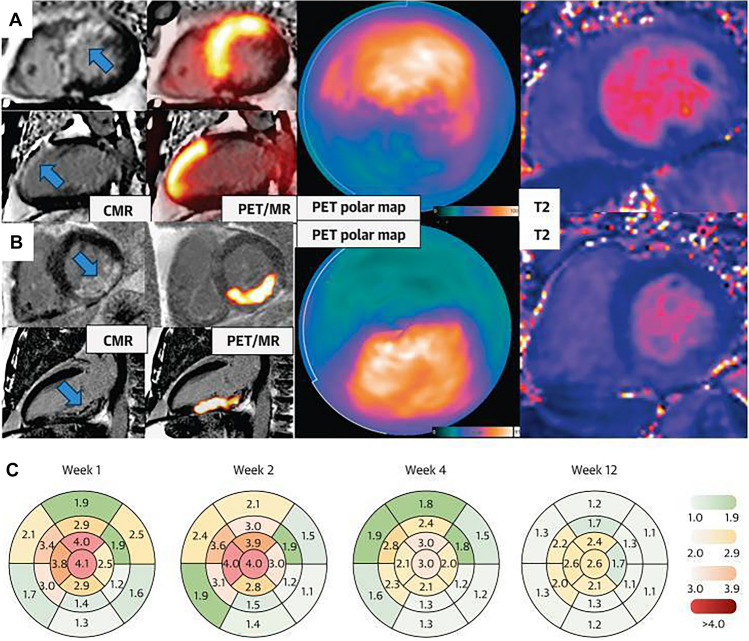
Post-ischemic myocardial remodeling significantly impacts clinical outcomes after acute myocardial infarction (MI), involving structural and functional changes such as ventricular dilation, infarct wall thinning, and fibrosis development. These processes, driven by inflammatory cascades, neurohormonal activation, and extracellular matrix remodeling, result in impaired cardiac output and an increased risk of heart failure. Imaging with fibroblast activation protein inhibitors (FAPI) has emerged as a promising non-invasive tool for assessing myocardial fibrosis via positron emission tomography (PET) or single-photon emission computed tomography (SPECT), targeting activated fibroblasts; the mediators of reparative and fibrotic processes. This innovative approach enables precise visualization and quantification of fibrosis dynamics, surpassing traditional imaging modalities. Preclinical studies using [68Ga]Ga-FAPI PET/computed tomography (CT) demonstrated the tracer's specificity for fibroblast activation and its peak uptake in the infarct border zone at day 6 post-MI. These findings, corroborated by histology and autoradiography, highlight its potential for tracking reparative fibrosis. Clinical translation of FAPI imaging was recently achieved with [68Ga]Ga-FAPI-46 PET/magnetic resonance imaging (MRI), showing persistent fibroblast activity beyond infarct zones and strong correlations with myocardial injury markers. Complementary research on [99mTc]Tc-HFAPi SPECT imaging in patients post-MI established its predictive value for left ventricular remodeling, emphasizing its cost-effectiveness and accessibility compared with PET. These advancements underscore FAPI-based imaging's potential to transform risk stratification and therapeutic guidance in post-MI care.
© 2025. The Author(s).
Conflict of interest statement
Declarations. Funding: Open access funding provided by Università degli Studi di Roma Tor Vergata within the CRUI-CARE Agreement. Conflict of interest: L.F., M.A.P., and O.S. have no conflicts of interest that are directly relevant to the content of this article. Consent (participation and publication): Not applicable. Availability of data and material: Data sharing is not applicable to this article as no datasets were generated or analyzed. Code availability: Not applicable. Author contributions: Conceptualization: L.F. and O.S.; writing first draft: L.F. and M.A.P.; editing and revision: L.F., M.A.P., and O.S. All authors read and approved the final version of the manuscript. Ethical approval: Not applicable.
Figures
Similar articles
-
18F-FAPI PET/CT for Early Detection and Severity Assessment of Intestinal Fibrosis in a Mouse Model.Inflamm Bowel Dis. 2025 Jul 7;31(7):2019-2026. doi: 10.1093/ibd/izaf086. Inflamm Bowel Dis. 2025. PMID: 40349210
-
Enhanced Detection of Early Pulmonary Fibrosis Disease Using 68Ga-FAPI-LM3 PET.Mol Pharm. 2024 Jul 1;21(7):3684-3692. doi: 10.1021/acs.molpharmaceut.4c00405. Epub 2024 Jun 20. Mol Pharm. 2024. PMID: 38899595 Free PMC article.
-
Molecular Imaging of Fibroblast Activity After Myocardial Infarction Using a 68Ga-Labeled Fibroblast Activation Protein Inhibitor, FAPI-04.J Nucl Med. 2019 Dec;60(12):1743-1749. doi: 10.2967/jnumed.119.226993. Epub 2019 Aug 12. J Nucl Med. 2019. PMID: 31405922 Free PMC article.
-
The Value of FAPI PET/CT in Cholangiocarcinoma and Pancreatic Cancer: An Update.Semin Nucl Med. 2025 Sep;55(5):701-709. doi: 10.1053/j.semnuclmed.2025.06.011. Epub 2025 Jul 16. Semin Nucl Med. 2025. PMID: 40675899 Review.
-
Targeting fibroblast activation protein in rheumatoid arthritis: from molecular imaging to precision therapeutics.Front Immunol. 2025 Jun 18;16:1616618. doi: 10.3389/fimmu.2025.1616618. eCollection 2025. Front Immunol. 2025. PMID: 40607439 Free PMC article. Review.
References
-
- Hinderer S, Schenke-Layland K. Cardiac fibrosis—a short review of causes and therapeutic strategies. Adv Drug Deliv Rev. 2019;146:77–82. - PubMed
-
- Yap J, Irei J, Lozano-Gerona J, Vanapruks S, Bishop T, Boisvert WA. Macrophages in cardiac remodelling after myocardial infarction. Nat Rev Cardiol. 2023;20:373–85. - PubMed
Publication types
MeSH terms
Substances
LinkOut - more resources
Full Text Sources
