. 2025 Jul;68(7):1476-1491.
doi: 10.1007/s00125-025-06424-4. Epub 2025 Apr 23.

RFX3 is essential for the generation of functional human pancreatic islets from stem cells

Affiliations

RFX3 is essential for the generation of functional human pancreatic islets from stem cells

Bushra Memon et al. Diabetologia. 2025 Jul.

Abstract

Aims/hypothesis: The role of regulatory factor X 3 (RFX3) in human pancreatic islet development has not been explored. This study aims to investigate the function of RFX3 in human pancreatic islet development using human islet organoids derived from induced pluripotent stem cells (iPSCs), hypothesising that RFX3 regulates human islet cell differentiation.

Methods: We generated RFX3 knockout (RFX3 KO) iPSC lines using CRISPR/Cas9 and differentiated them into pancreatic islet organoids. Various techniques were employed to assess gene expression, cell markers, apoptosis, proliferation and glucose-stimulated insulin secretion. Single-cell RNA-seq datasets from human embryonic stem cell-derived pancreatic islet differentiation were re-analysed to investigate RFX3 expression in specific cell populations at various developmental stages. Furthermore, bulk RNA-seq was conducted to further assess transcriptomic changes. RFX3 overexpression was implemented to reverse dysregulated gene expression.

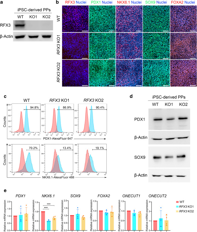
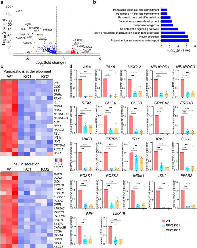
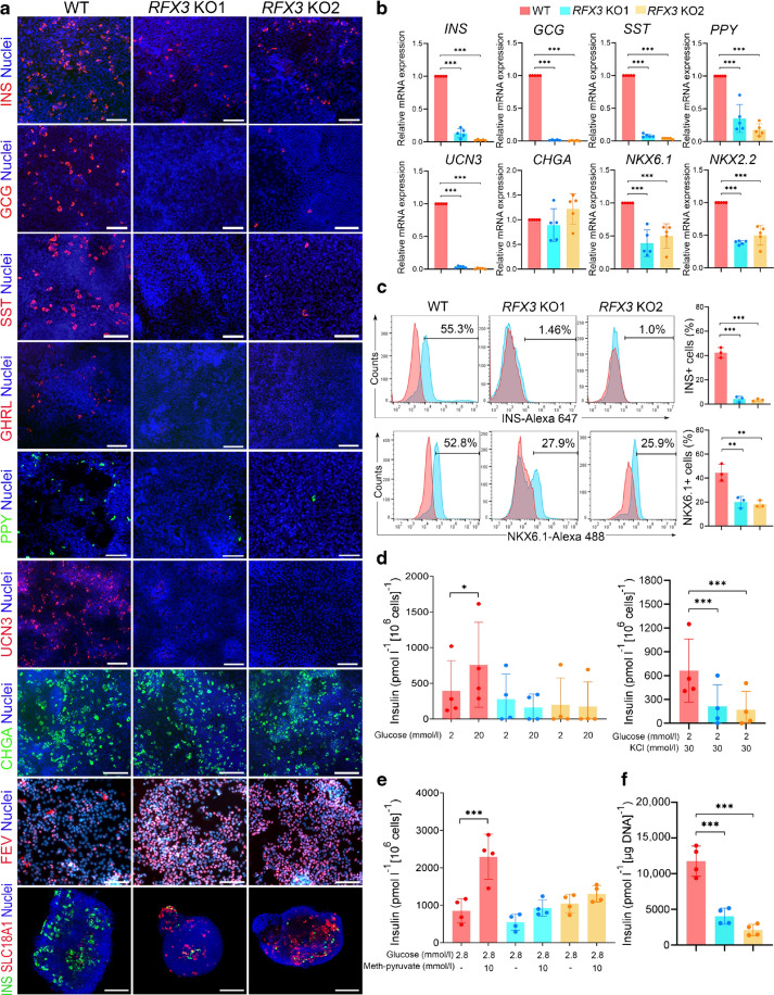
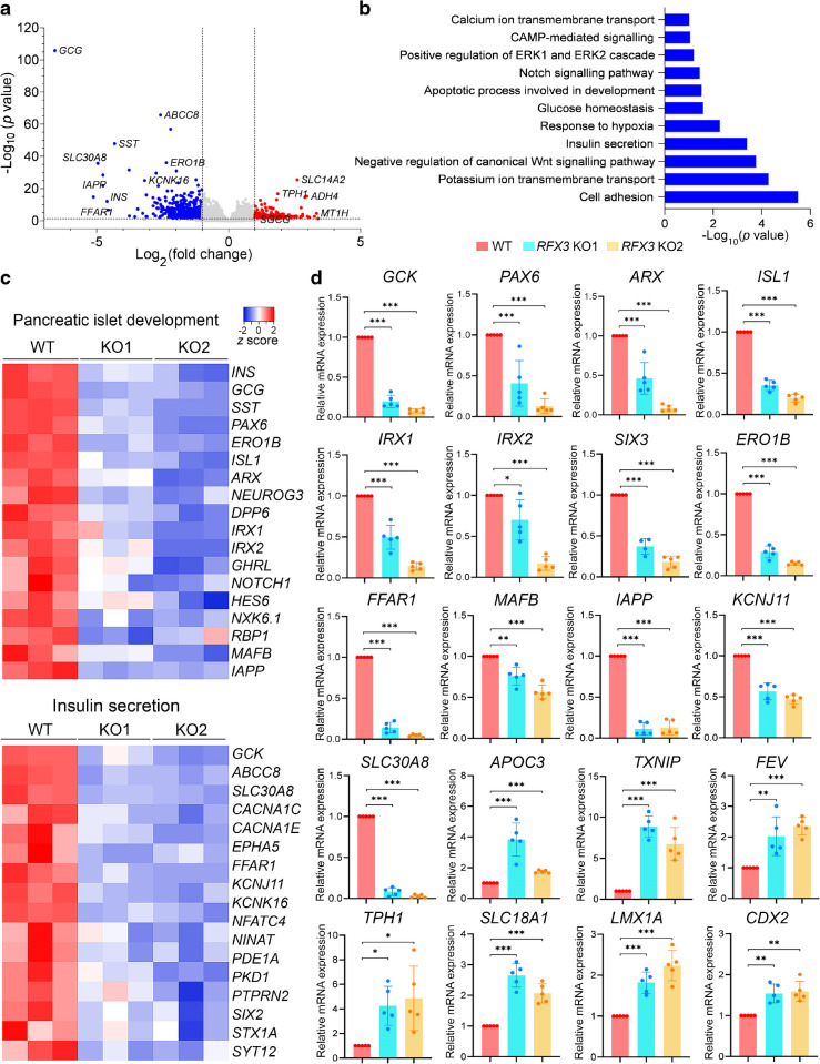
Results: RFX3 was found to be highly expressed in pancreatic endocrine cell populations within pancreatic progenitors (PPs), endocrine progenitors (EPs) and mature islet stages derived from iPSCs. Single-cell RNA-seq further confirmed RFX3 expression across different endocrine cell clusters during differentiation. The loss of RFX3 disrupted pancreatic endocrine gene regulation, reduced the number of hormone-secreting islet cells and impaired beta cell function and insulin secretion. Despite a significant reduction in the expression levels of pancreatic islet hormones, the pan-endocrine marker chromogranin A remained unchanged at both EP and islet stages, likely due to an increase in the abundance of enterochromaffin cells (ECs). This was supported by our findings of high EC marker expression levels in RFX3 KO EPs and islets. In addition, RFX3 loss led to smaller islet organoids, elevated thioredoxin-interacting protein levels and increased apoptosis in EPs and islets. Furthermore, RFX3 overexpression rescued the expression of dysregulated genes in RFX3 KO at the PP and EP stages.

Conclusions/interpretation: These findings underscore the crucial role of RFX3 in regulating human islet cell differentiation and its role in suppressing EC specification. These insights into RFX3 function have implications for understanding islet biology and potential diabetes susceptibility.

Data availability: The RNA-seq datasets have been submitted to the Zenodo repository and can be accessed via the following links: DOI https://doi.org/10.5281/zenodo.13647651 (PPs); and DOI https://doi.org/10.5281/zenodo.13762055 (SC-islets).

Keywords: Differentiation; Endocrine pancreas; Enterochromaffin cells; Transcription factor; iPSC model.


Conflict of interest statement

Data availability: The RNA-seq datasets have been submitted to the Zenodo repository and can be accessed via the following links: DOI https://doi.org/10.5281/zenodo.13647651 (PPs); and DOI https://doi.org/10.5281/zenodo.13762055 (SC-islets). Funding: Open Access funding provided by the Qatar National Library. This work was funded by grants from Sidra Medicine (SDR400217) and QBRI (QBRI-HSCI Project 1). NA, the co-first author of this article, is a PhD student with a scholarship funded by QRDI (GSRA9-L-1-0511-22008). Authors’ relationships and activities: SH is a co-founder and shareholder of Sequantrix GmbH and has received research funding from Novo Nordisk and Askbio. The authors declare that there are no other relationships or activities that might bias, or be perceived to bias, their work. Contribution statement: BM, NA, AKE performed the experiments and analysed the data. NA and AKE conducted all revision experiments. SI and SH analysed the sequencing data. BM and EMA wrote the manuscript. EMA conceived and designed the study, supervised the project, and analysed and interpreted the data. All authors critically reviewed the article and approved the final version of the manuscript. EMA is the guarantor of this work.

Figures

Fig. 1
Fig. 1
RFX3 is predominantly expressed in pancreatic endocrine lineages during differentiation of iPSCs into islets. (a) Immunofluorescence images showing stage-specific expression pattern of RFX3 in pancreatic cells (images representative of at least three biological experiments). Note the absence of RFX3 expression in PDX1+ and NKX6.1+ cells at the PP stage. RFX3 was co-expressed with NKX2.2 and NEUROG3 in iPSC-derived EPs and with INS and GCG in iPSC-derived islets. (b) Timeline RT-qPCR analysis for RFX3 mRNA expression during pancreatic islet cell differentiation (n=4), from definitive endoderm (S1) to primitive gut tube (S2), posterior foregut (S3), PP (S4) EP (S5) and islet stages (S6). (c) Immunofluorescence images showing the co-expression of RFX3 and RFX6 in PPs, EPs and islets derived from hESC-H9 RFX6 HA-tagged cell line. (d) Dot plots demonstrating gene expression in various cell clusters determined by single-cell RNA-seq. The expression level in each cluster (C0–C5) is scaled based on the percentages of cells expressing RFX3 (dot size) and mean expression (colour intensity) of the gene. Dot plots are presented for day 11 (D11), day 14 (D14), day 21 (D21) and day 39 (D39) of hESC differentiation. (e) Violin plots depicting expression pattern of key islet and EC markers across various cell clusters (as shown in d) at day 39 (mature islets). Data are presented as means±SD. ***p<0.001. Scale bar, 50 µm
Fig. 2
Fig. 2
Impact of RFX3 deletion on iPSC-derived PPs. (a) Western blotting analysis validating the loss of RFX3 protein expression in RFX3 KO PPs. (b) Immunofluorescence images showing expression of RFX3, PDX1, NKX6.1, SOX9 and FOXA2 in RFX3 KO PPs and WT PPs (images representative of at least three biological experiments). (c) Representative flow cytometry histograms showing the expression of PDX1 and NKX6.1 in RFX3 KO PPs and WT PPs. (d) Representative western blots showing the expression levels of PDX1 and SOX9 proteins in RFX3 KO PPs and WT PPs. (e) RT-qPCR analysis for PP developmental markers (n=4). Relative mRNA expression calculated as fold change vs WT (set as 1). Data are presented as means±SD. ***p<0.001. Scale bar, 100 µm
Fig. 3
Fig. 3
Abolished endocrine specification in iPSC-derived PPs lacking RFX3. DEGs and pathways were identified by bulk RNA-seq analysis of RFX3 KO PPs and WT PPs (n=3). (a) Volcano plot showing upregulated (red) and downregulated (blue) DEGs. (b) GO of significantly enriched biological processes in downregulated DEGs. (c) Heatmaps depicting z scores of significantly downregulated DEGs regulating pancreatic islet development and insulin secretion. (d) RT-qPCR analysis of key DEGs affected by RFX3 deletion in iPSC-derived PPs (n=4). Relative mRNA expression calculated as fold change vs WT (set as 1). Data are presented as means±SD. *p<0.05, **p<0.01, ***p<0.001
Fig. 4
Fig. 4
Effect of RFX3 deletion on EPs derived from iPSCs. (a, b) Representative immunofluorescence images (n=3) (a) and RT-qPCR analysis (n=4) (b) showing the expression of the EP markers CHGA, NEUROG3, NKX2.2 and NKX6.1, in EPs derived from WT and RFX3 KO iPSCs. (c) RT-qPCR analysis showing reduced expression of key markers regulating endocrine pancreas differentiation, along with increased expression of EC markers in EPs derived from RFX3 KO iPSCs compared with WT iPSCs (n=4). Relative mRNA expression calculated as fold change vs WT (set as 1). (d) Representative immunofluorescence images showing the co-expression of the EC marker SLC18A1 with CHGA, NKX6.1 and NKX2.2 in RFX3 KO EPs compared with WT EPs (n=3). (e) Western blotting analysis showing an increase in the expression levels of CDX2 in RFX3 KO EPs compared with WT EPs (n=3). Data are presented as means±SD. *p<0.05, **p<0.01, ***p<0.001. Scale bar, 100 µm
Fig. 5
Fig. 5
Impaired generation of hormone-secreting islet cells from RFX3 KO iPSCs. (a) Representative immunofluorescence images showing reduced expression of islet hormones (INS, GCG, SST, GHRL and PPY) and UCN3 in islet cells differentiated from RFX3 KO iPSCs. In contrast, CHGA expression remained unchanged, while FEV and SLC18A1 expression increased (n=4). (b) RT-qPCR analysis for key islet markers in WT islets and RFX3 KO islets (n=5). (c) Flow cytometry analysis and quantification of the expression of INS and NKX6.1 in islets differentiated from RFX3 KO iPSCs compared with WT controls (n=3). (d) Glucose-stimulated insulin secretion assay illustrating diminished beta cell functionality and reduced insulin release upon treatment with high glucose (20 mmol/l, n=4) and KCl (30 mmol/l, n=4), respectively. (e) Insulin release in response to 10 mmol/l methyl pyruvate from RFX3 KO islets compared with WT controls. (f) Total insulin content was measured from lysed islet organoids derived from RFX3 KO iPSCs compared with WT controls using acid-ethanol (n=4). Data are presented as means±SD. *p<0.05, **p<0.01, ***p<0.001. Scale bar, 100 µm
Fig. 6
Fig. 6
Transcriptome changes in pancreatic islets lacking the RFX3 gene. (a) Volcano plot depicting DEGs identified by transcriptome analysis of islet cells derived from RFX3 KO iPSCs and WT iPSCs (n=3). Significantly upregulated DEGs are shown in red; downregulated DEGs are shown in blue. (b) GO of downregulated biological processes in RFX3 KO islets compared with WT islets. (c) Heatmaps for key DEGs involved in pancreatic islet development and insulin secretion depicting their z scores. (d) RT-qPCR analysis showing downregulation of key islet markers and upregulation of genes associated with ECs in iPSC-derived islets lacking RFX3 (n=5). Relative mRNA expression calculated as fold change vs WT (set as 1). Data are presented as means±SD. *p<0.05, **p<0.01, ***p<0.001
Fig. 7
Fig. 7
RFX3 loss leads to increased apoptosis and disruption of iPSC-derived islet organoids. (a) Representative phase contrast images showing morphological changes of islet organoids during stages 5 and 6 (EPs and islets, respectively) derived from RFX3 KO iPSCs and WT iPSCs (n=3). Note the reduced size of organoids derived from RFX3 KO iPSCs compared with WT controls. ‘S’ denotes stage; ‘D’ denotes day. (b) Flow cytometry quantification of apoptotic (Annexin V+) cells in stages 4 (PPs) and 5 (EPs) indicating increased apoptosis in RFX3-deficient cells. (c) Immunostaining images showing an increase in the number of cells expressing the apoptosis marker Annexin V and the EC marker SLC18A1 in RFX3-deficient EPs, with no co-localisation observed between the two markers (n=2). (d) Immunostaining images showing no change in the expression of the proliferation marker Ki67 and an increase in the expression of SLC18A1 in RFX3-deficient EPs, with no co-localisation observed between the two markers (n=2). (e) Flow cytometry analysis of BrdU incorporation showing no significant changes in cell proliferation (BrdU+ cells) in PPs and EPs derived from RFX3 KO iPSC lines compared with those from WT iPSCs. (f) Western blotting analysis showing an increase in the expression levels of TXNIP in EPs and islets derived from RFX3 KO iPSCs compared with WT controls (representative of n=3). Scale bars, 100 µm in (a) and 200 µm in (c, d)

Similar articles

References

    1. Lutz TA (2023) Mammalian models of diabetes mellitus, with a focus on type 2 diabetes mellitus. Nat Rev Endocrinol 19(6):350–360. 10.1038/s41574-023-00818-3 - PubMed
    1. Memon B, Abdelalim EM (2022) Toward precision medicine with human pluripotent stem cells for diabetes. Stem Cells Transl Med 11(7):704–714. 10.1093/stcltm/szac030 - PMC - PubMed
    1. Abdelalim EM (2021) Modeling different types of diabetes using human pluripotent stem cells. Cell Mol Life Sci 78(6):2459–2483. 10.1007/s00018-020-03710-9 - PMC - PubMed
    1. Ait-Lounis A, Baas D, Barras E et al (2007) Novel function of the ciliogenic transcription factor RFX3 in development of the endocrine pancreas. Diabetes 56(4):950–959. 10.2337/db06-1187 - PubMed
    1. Ait-Lounis A, Bonal C, Seguin-Estevez Q et al (2010) The transcription factor Rfx3 regulates beta-cell differentiation, function, and glucokinase expression. Diabetes 59(7):1674–1685. 10.2337/db09-0986 - PMC - PubMed

MeSH terms

Substances

LinkOut - more resources